Floral Structure of Kirkia (Kirkiaceae) and its ... - Annals of Botany
Floral Structure of Kirkia (Kirkiaceae) and its ... - Annals of Botany
Floral Structure of Kirkia (Kirkiaceae) and its ... - Annals of Botany

You also want an ePaper? Increase the reach of your titles
YUMPU automatically turns print PDFs into web optimized ePapers that Google loves.
544<br />
A<br />
B<br />
s<br />
F G H s<br />
s<br />
I<br />
s<br />
s<br />
Bachelier <strong>and</strong> Endress — Flowers <strong>of</strong> <strong>Kirkia</strong> <strong>and</strong> Position in Sapindales<br />
C D E<br />
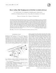
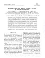
F IG.6. <strong>Kirkia</strong> wilmsii. (A) Longitudinal section (LS) <strong>of</strong> uniseriate multicellular papillae <strong>of</strong> the stigma before anthesis. (B) LS <strong>of</strong> the hemispherical protrusion<br />
in the floral centre above the ovary (compare with Fig. 5A). (C) LS <strong>of</strong> the slightly campylotropous ovule filling the locule at anthesis, with the<br />
exp<strong>and</strong>ed large-celled distal parts <strong>of</strong> the two integuments forming a long micropyle. (D) Transverse section (TS) <strong>of</strong> base <strong>of</strong> sepals, petal <strong>and</strong> stamens<br />
before anthesis, showing carpet <strong>of</strong> secretory hairs (with short multiseriate stalk <strong>and</strong> large multicellular head) on the inner side <strong>of</strong> the petal base, <strong>and</strong><br />
petal base exp<strong>and</strong>ing dorsally between the free margins <strong>of</strong> two sepal bases. (E) LS <strong>of</strong> sepal base showing the epidermal <strong>and</strong> sub-epidermal special mucilage<br />
cells. (F–I) TS <strong>of</strong> a preanthetic gynoecium <strong>and</strong> corresponding enlarged micrographs, showing the inner angle <strong>of</strong> a fertile locule <strong>and</strong> centre <strong>of</strong> the<br />
gynoecium (locule dorsal side oriented downwards; compare with Fig. 5K); arrows point to placentae; arrowhead points to the second reduced locule<br />
developing on the same radius as the fertile one (compare with Fig. 7A); morphological surfaces drawn with thick continuous lines; postgenitally<br />
fused morphological surfaces drawn with dashed lines; vascular bundles drawn with thin continuous lines; ‘s’: synlateral vascular bundle; dash rectangles<br />
in (F) <strong>and</strong> (H) show location <strong>of</strong> (G) <strong>and</strong> (I). (F, G) In the upper part <strong>of</strong> the locule, the endocarp differentiation begins laterally <strong>and</strong> the ventral inner surface<br />
<strong>of</strong> the carpel is S-shaped (arrowhead). (H, I) Lower down, the endocarp encompasses the inner angle <strong>of</strong> the locule <strong>and</strong> the second locule is isolated (arrowhead).<br />
Scale bars: A ¼ 30 mm; B, C, D ¼ 200 mm; E, F, G, H, I ¼ 100 mm.<br />
Endress, 1994), <strong>and</strong> slightly campylotropous with only the<br />
very base <strong>of</strong> the nucellus <strong>and</strong> embryo sac curved (Fig. 5A).<br />
The two integuments surround the nucellus <strong>and</strong>, although<br />
both appear to be <strong>of</strong> the same thickness, the inner integument<br />
comprises three or four cell layers but the outer<br />
only two or three cell layers (Figs 5A <strong>and</strong> 6C). Above the<br />
nucellus the integuments are elongate <strong>and</strong> thickened. At<br />
anthesis, the inner integument is about twice as long as<br />
the nucellus <strong>and</strong> the outer even two <strong>and</strong> a half times<br />
(Figs 5A <strong>and</strong> 6C). Thus, the micropyle is unusually long<br />
<strong>and</strong> comprises two distinct zones. The proximal zone is a<br />
straight tubular canal formed by the inner integument,<br />
s<br />
s<br />
whereas the distal zone is not tubular <strong>and</strong> is somewhat<br />
wavy, <strong>and</strong> is formed by the second integument (Figs 5A<br />
<strong>and</strong> 6C). The extended part <strong>of</strong> the integuments above the<br />
nucellus comes about by cell enlargement (Fig. 6C). In isolated<br />
ovules studied with the SEM, these enlarged cells tend<br />
to collapse (Fig. 4 N). The ovule fills the locule <strong>and</strong> the<br />
micropyle is contiguous with the placenta (Fig. 5A).<br />
Anatomy<br />
Sepals have one median <strong>and</strong> two lateral main vascular<br />
bundles, which extend almost through their whole length,