Morphology and distribution of some marine diatoms, family ... - Algae
Morphology and distribution of some marine diatoms, family ... - Algae
Morphology and distribution of some marine diatoms, family ... - Algae

Create successful ePaper yourself
Turn your PDF publications into a flip-book with our unique Google optimized e-Paper software.
Research Article<br />
<strong>Algae</strong> 2011, 26(4): 299-315<br />
http://dx.doi.org/10.4490/algae.2011.26.4.299<br />
Open Access<br />
<strong>Morphology</strong> <strong>and</strong> <strong>distribution</strong> <strong>of</strong> <strong>some</strong> <strong>marine</strong> <strong>diatoms</strong>, <strong>family</strong><br />
Rhizosoleniaceae, genus Proboscia, Neocalyptrella, Pseudosolenia,<br />
Guinardia, <strong>and</strong> Dactyliosolen in Korean coastal waters<br />
Suk Min Yun 1,a <strong>and</strong> Jin Hwan Lee 1, *<br />
1 Department <strong>of</strong> Life Science, Sangmyung University, Seoul 110-743, Korea<br />
The morphology, taxonomy, <strong>and</strong> <strong>distribution</strong> <strong>of</strong> species belonging to the diatom <strong>family</strong> Rhizosoleniaceae were studied<br />
from the <strong>marine</strong> coastal waters <strong>of</strong> Korea. Rhizosolenid diatom taxa were collected at 30 sites from September 2008<br />
to February 2010 <strong>and</strong> were analyzed by light <strong>and</strong> scanning electron microscopy. We identified 6 rhizosolenid genera,<br />
including Rhizosolenia, Proboscia, Pseudosolenia, Neocalyptrella, Guinardia, <strong>and</strong> Dactyliosolen. We describe 5 genera in<br />
this study, except Rhizosolenia. Five genera were compared in detail with congeneric species. Six genera within the <strong>family</strong><br />
Rhizosoleniaceae were divided into two groups based on morphological diagnostic characters including valve shape,<br />
areolae pattern, the shape <strong>of</strong> external process, <strong>and</strong> girdle segments in the column. The first group had a conoidal valve<br />
<strong>and</strong> loculate areolae, which comprised Rhizosolenia, Proboscia, Pseudosolenia, <strong>and</strong> Neocalyptrella, <strong>and</strong> the second group<br />
<strong>of</strong> Guinardia <strong>and</strong> Dactyliosolen showed a flat or rounded valve <strong>and</strong> poroid areolae. Important key diagnostic characters<br />
were based on valve shape, areolae pattern on the segment, external process, position <strong>of</strong> the tube, <strong>and</strong> the valve margin.<br />
D. phuketensis was new to Korean coastal waters.<br />
Key Words: Dactyliosolen; <strong>diatoms</strong>; <strong>distribution</strong>; Guinardia; morphology; Neocalyptrella; Proboscia; Pseudosolenia;<br />
Rhizosolenia<br />
INTRODUCTION<br />
Peragallo (1892) regarded the genera Dactyliosolen<br />
Castracane, Lauderia Cleve, Attheya T. West, Guinardia<br />
H. Peragallo, <strong>and</strong> Rhizosolenia Brightwell as members<br />
<strong>of</strong> Rhizosoléniées. Thereafter, Hustedt (1930) suggested<br />
that 23 <strong>marine</strong> <strong>and</strong> 4 freshwater species belonged to this<br />
<strong>family</strong>. He synonymized several species, as several earlier<br />
studies described variations <strong>of</strong> seemingly the same<br />
species.<br />
Sundström (1986) suggested that only those species<br />
with valves bearing an external process, otaria, claspers,<br />
<strong>and</strong> copulae perforated by loculate areolae should be<br />
included in the genus Rhizosolenia. According to Sund-<br />
This is an Open Access article distributed under the terms <strong>of</strong> the<br />
Creative Commons Attribution Non-Commercial License (http://creativecommons.org/licenses/by-nc/3.0/)<br />
which permits unrestricted<br />
non-commercial use, <strong>distribution</strong>, <strong>and</strong> reproduction in any medium,<br />
provided the original work is properly cited.<br />
ström (1986), the genus Proboscia Sundström <strong>and</strong> Pseudosolenia<br />
Sundström were separated from Rhizosolenia<br />
because they have external processes. The two species<br />
<strong>of</strong> R. calcar-avis Schultze <strong>and</strong> R. alata Brightwell were<br />
subsequently transferred to Pseudosolenia calcar-avis<br />
Sundström <strong>and</strong> Proboscia alata Sundström, respectively<br />
(Sundström 1986). R. robusta Norman was also transferred<br />
to Neocalyptrella robusta Hernández-Becerril <strong>and</strong><br />
Meave (Hernández-Becerril <strong>and</strong> Meave del Castillo 1996,<br />
1997).<br />
More recently, the <strong>family</strong> Rhizosoleniaceae included<br />
Neocalyptrella, Pseudosolenia, Proboscia, <strong>and</strong> Urosolenia.<br />
Received September 10, 2011, Accepted November 5, 2011<br />
*Corresponding Author<br />
E-mail: jhlee@smu.ac.kr<br />
Tel: +82-2-2287-5152, Fax: +82-2-2287-0098<br />
a Present address: Laboratory <strong>of</strong> Plankton Ecology, Korea Institute<br />
<strong>of</strong> Coastal Ecology, Inc., Bucheon 421-808, Korea<br />
Copyright © The Korean Society <strong>of</strong> Phycology 299 http://e-algae.kr pISSN: 1226-2617 eISSN: 2093-0860
<strong>Algae</strong> 2011, 26(4): 299-315<br />
Representatives <strong>of</strong> these genera are commonly found as<br />
solitary cells in <strong>marine</strong> environments, except Urosolenia,<br />
which is restricted to freshwater (Edlund <strong>and</strong> Stoermer<br />
1993, Rott et al. 2006, Li et al. 2009). The genera Guinardia<br />
<strong>and</strong> Dactyliosolen have been allocated to the <strong>family</strong> Rhizosoleniaceae.<br />
In Korea, the genera Rhizosolenia, Guinardia, <strong>and</strong> Dactyliosolen<br />
were recorded by Shim (1994). Many authors<br />
(Moon <strong>and</strong> Choi 1991, Yoon et al. 1992, Chang <strong>and</strong> Shim<br />
1993, Kim et al. 1993, Yoon <strong>and</strong> Koh 1994, 1995) <strong>and</strong> Lee<br />
(1995) added Pseudosolenia <strong>and</strong> Proboscia to his checklist.<br />
Additional studies on the <strong>family</strong> Rhizosoleniaceae in<br />
Korea have been conducted sporadically (Yun <strong>and</strong> Lee<br />
2010, Yun et al. 2011), but species identification, synonymies,<br />
<strong>and</strong> the phylogeny the <strong>family</strong> Rhizosoleniaceae<br />
have been insufficiently investigated. The present study<br />
provides a detailed survey <strong>of</strong> <strong>marine</strong> <strong>diatoms</strong> belonging<br />
to the genera Proboscia, Pseudosolenia, Neocalyptrella,<br />
Guinardia, <strong>and</strong> Dactyliosolen from the coastal waters <strong>of</strong><br />
Korea. This survey provides detailed light <strong>and</strong> scanning<br />
electron microscopy illustrations <strong>and</strong> a critical review <strong>of</strong><br />
the taxonomical <strong>and</strong> <strong>distribution</strong>al data.<br />
MATERIALS AND METHODS<br />
Field samples were collected in Korean coastal waters<br />
from September 2008 to February 2010 (Table 1). Phytoplankton<br />
was collected using a 20 μm mesh-sized net by<br />
vertical towing. Samples were immediately fixed in neutralized<br />
formalin (final concentration 4%), glutaraldehyde<br />
(final concentration 2%), <strong>and</strong> Lugol’s solution. Organic<br />
material in the samples was removed using the methods<br />
<strong>of</strong> Hasle <strong>and</strong> Fryxell (1970) <strong>and</strong> Simonsen (1974). The materials<br />
were examined under a light microscope (Axioskop<br />
40; Carl Zeiss, Jena, Germany), photographed with a<br />
MRc5 camera (Carl Zeiss) <strong>and</strong> a scanning electron microscope<br />
(JSM-5600LV; Jeol, Tokyo, Japan). Sizes <strong>of</strong> cells were<br />
measured using image calculation s<strong>of</strong>tware (AxioVision<br />
AC v. 4.5; Carl Zeiss).<br />
Terminology was from that recommended in the first<br />
report <strong>of</strong> the working Committee on Diatom Terminology<br />
(Anonymous 1975) from the third Symposium on<br />
Recent <strong>and</strong> Fossil Marine Diatoms, Kiel. Other terminology<br />
follows Ross et al. (1979), Sundström (1986), Round<br />
et al. (1990), Hernández-Becerril (1995), <strong>and</strong> Hasle <strong>and</strong><br />
Syvertsen (1996).<br />
RESULTS<br />
http://dx.doi.org/10.4490/algae.2011.26.4.299 300<br />
We identified 6 rhizosolenid genera, including Rhizosolenia,<br />
Proboscia, Pseudosolenia, Neocalyptrella, Guinardia,<br />
<strong>and</strong> Dactyliosolen. We described five <strong>of</strong> these genera<br />
except Rhizosolenia. The morphological characters observed<br />
in the genera Proboscia, Neocalyptrella, Pseudosolenia,<br />
Dactyliosolen, <strong>and</strong> Guinardia species are shown in<br />
Tables 2-4. According to the system suggested by Sundström<br />
(1986), 9 phytoplanktonic diatom taxa representing<br />
1 order, 1 suborder, 1 <strong>family</strong>, 5 genera, <strong>and</strong> 9 species<br />
were identified in this study. The systematic accounts are<br />
as follows:<br />
Class Bacillariophyceae Haeckel 1878<br />
Order Centrales Hustedt 1930<br />
Suborder Rhizosoleniineae Simonsen 1979<br />
Family Rhizosoleniaceae De Toni 1890<br />
Genus Proboscia Sundström 1986<br />
Proboscia alata (Brightwell) Sundström<br />
1986<br />
Proboscia indica (H. Peragallo)<br />
Hernández-Becerril 1995<br />
Genus Neocalyptrella (Norman)<br />
Hernández-Becerril & Meave 1996<br />
Neocalyptrella robusta Hernández-<br />
Becerril & Meave 1996<br />
Genus Pseudosolenia Sundström 1986<br />
Pseudosolenia calcar-avis (Schultze)<br />
Sundström 1986<br />
Genus Guinardia H. Peragallo 1892<br />
Guinardia delicatula (Cleve) Hasle 1995<br />
Guinardia flaccida (Castracane)<br />
H. Peragallo 1892<br />
Guinardia striata (Stolterforth) Hasle 1995<br />
Genus Dactyliosolen Castracane 1886<br />
Dactyliosolen fragilissimus (Bergon)<br />
Hasle 1995<br />
Dactyliosolen phuketensis (Sundström)<br />
Hasle 1995<br />
Proboscia alata (Brightwell) Sundström 1986<br />
(Fig. 1, A-H)<br />
Brightwell 1858, p. 95, Pl. 5, Fig. 8; Peragallo 1892, p.<br />
115, Pl. 18, Figs 11-20; Hustedt 1920, Pl. 317; Hustedt<br />
1930, p. 600, Fig. 345; Cupp 1943, p. 90, Fig. 52A & B; Okuno<br />
1952, p. 353, Pl. 2, Figs 5 & 6; Okuno 1960, p. 310, Pl.<br />
1, Fig. 1; Hendey 1964, p. 146, Pl. 2, Fig. 2; Drebes 1974,<br />
p. 57, Fig. 39c & d; Navarro 1981, p. 430, Figs 33 & 34 as<br />
R. alata; Sundström 1986, p. 99, Figs 258-266; Jordan et<br />
al. 1991, p. 65, Figs 1-9; Takahashi et al. 1994, p. 413, Figs<br />
2-7; Hernández-Becerril 1995, p. 252, Figs 2-4; Hasle <strong>and</strong>
Syvertsen 1996, p. 159, Pl. 30; Sunesen <strong>and</strong> Sar 2007, p.<br />
639, Figs 82-88 & 98.<br />
Synonyms. Rhizosolenia alata Brightwell 1858, Rhizosolenia<br />
alata f. gracillima (Cleve) Gran 1905.<br />
Cells are solitary or in pairs, narrow cylindrical, bilaterally<br />
symmetrical, 3.3-13.3 μm in diameter, 270.0-485.7<br />
μm long. Valve is sub-conoidal, the ventral part longer<br />
than the dorsal part <strong>and</strong> proboscis structure is slightly<br />
curved, tapering towards the apical part <strong>of</strong> the valve, circular<br />
in cross section, 15.0-30.0 μm long. Apical surface<br />
<strong>of</strong> the proboscis is composed <strong>of</strong> variously sized spinules.<br />
Number <strong>of</strong> spinules is 7-16, 0.1-0.4 μm long. Contiguous<br />
area is convex towards the valve surface, distally limited<br />
by asymmetric claspers. The valve areolae are rounded,<br />
52-90 in 10 μm, arranged in longitudinal striae, converg-<br />
Yun & Lee <strong>Morphology</strong> <strong>and</strong> Distribution <strong>of</strong> Marine Diatoms<br />
ing towards the apex. Girdle segment areolae are loculate,<br />
arranged in columns, with the external velum perforated<br />
by central pores, <strong>and</strong> internal circular foramina, 25-62 in<br />
10 μm. Interlocular pores are commonly surrounded by<br />
six loculi. Segment horizontal axis <strong>and</strong> perpendicular axis<br />
are 3.3-13.3 <strong>and</strong> 10.0-26.7 μm long, respectively.<br />
Distribution. Proboscia alata has frequently been reported<br />
in the Argentine Sea (Ferrario <strong>and</strong> Galávan 1989<br />
as R. alata <strong>and</strong> R. alata f. gracillima). During this study,<br />
P. alata was frequently observed in September 2008 <strong>and</strong><br />
June 2009 at the Wolsung coast, Jeju Isl<strong>and</strong>, <strong>and</strong> the Korea<br />
Strait.<br />
Remarks. Sundström (1986) did not share the biogeographical<br />
limits <strong>of</strong> P. alata because synonyms were used<br />
for probably all taxa included in the genus. The specific<br />
Table 1. Sampling sites for the genera Proboscia, Neocalyptrella, Pseudosolenia, Guinardia, <strong>and</strong> Dactyliosolen <strong>of</strong> the <strong>family</strong> Rhizosoleniaceae<br />
Location Latitude (N) Longitude (E) Sampling date Species<br />
Korea Strait 34°44.965 128°50.083 Sep 10, 2008 Proboscia alata<br />
Wolsung coast 35°43.836 129°29.225 May 20, 2009<br />
Jeju Is. 33°33.442 126°47.706 Sep 15, 2009<br />
Yellow Sea 35°30.521 124°10.773 Sep 22, 2008 P. indica<br />
Daecheon Harbor 36°18.203 126°30.925 May 3, 2009<br />
Geoje Is. 34°59.588 128°40.519 Jun 25, 2009<br />
Daebu Is. 37°17.852 126°34.351 Jul 25, 2009<br />
Yangyang coast 38°07.411 128°37.961 Oct 15, 2009<br />
Yellow Sea 35°30.521 124°10.773 Sep 22, 2008 Neocalyptrella robusta<br />
Korea Strait 34°37.629 128°44.887 Feb 10, 2009<br />
Wolsung coast 35°43.836 129°29.225 May 20, 2009<br />
Geoje Is. 34°59.588 128°40.519 Jun 25, 2009<br />
Yellow Sea 35°30.521 124°10.773 Sep 22, 2008 Pseudosolenia calcar-avis<br />
Jeju Is. 33°12.578 126°15.557 Sep 15, 2009<br />
Jeju Is. 33°18.572 126°09.876 Sep 15, 2009<br />
Sacheon coast 37°20.731 126°41.181 Jul 25, 2009 Guinardia delicatula<br />
Incheon coast 37°22.125 126°33.193 Aug 17, 2009<br />
Mokpo Bay 34°46.843 126°22.884 Jan 12, 2010<br />
Yellow Sea 35°30.521 124°10.773 Sep 22, 2008 G. flaccida<br />
Geoje Is. 34°59.588 128°40.519 Jun 25, 2009<br />
Namhae Br. 34°54.986 128°01.956 Jul 19, 2009<br />
Sacheon coast 35°00.276 128°01.195 Aug 6, 2009<br />
Tongyeong coast 34°54.558 128°26.491 Aug 7, 2010<br />
Incheon coast 37°22.125 126°33.193 Aug 17, 2009<br />
Namhae Br. 34°54.986 128°01.956 Jul 19, 2009 G. striata<br />
Daebu Is. 37°17.852 126°34.351 Jul 25, 2009<br />
Incheon coast 37°22.125 126°33.193 Aug 17, 2009<br />
Yeongdeok coast 36°36.038 129°24.573 Oct 16, 2009<br />
Goseong coast 38°16.205 128°33.438 Feb 20, 2010 Dactyliosolen fragilissimus<br />
Geoje Is. 34°59.588 128°40.519 Jun 25, 2009 D. phuketensis<br />
Sacheon coast 35°00.276 128°01.195 Aug 6, 2009<br />
Tongyeong coast 34°54.545 128°18.851 Aug 6, 2009<br />
Yellow Sea 35°30.521 124°10.773 Sep 22, 2008<br />
Yeongduk coast 36°36.038 129°24.573 Oct 16, 2009<br />
301 http://e-algae.kr
<strong>Algae</strong> 2011, 26(4): 299-315<br />
Table 2. Morphological characteristics <strong>of</strong> the Proboscia species examined in this study<br />
Cell Segment Areolae<br />
Valve<br />
Valve in<br />
10 μm<br />
Segment in<br />
10 μm<br />
Perpendicular<br />
axis length<br />
(μm)<br />
Horizontal<br />
axis length<br />
(μm)<br />
Species<br />
Spinule in valve<br />
Pervalvar axis<br />
length<br />
(μm)<br />
Shape Diameter<br />
(μm)<br />
Number Length (μm)<br />
Shape Length<br />
(μm)<br />
25-62 52-90<br />
10.0-26.7<br />
(n = 22)<br />
3.3-13.3<br />
(n = 43)<br />
7-16 0.1-0.4<br />
(n = 21)<br />
15.0-30.0<br />
(n = 25)<br />
Tapering<br />
conical<br />
270.0-485.7<br />
(n = 11)<br />
3.3-13.3<br />
(n = 43)<br />
Proboscia alata Narrow<br />
cylindrical<br />
- 30-60<br />
10.6-16.0<br />
(n = 29)<br />
25.0-125.0<br />
(n = 32)<br />
8-13 0.2-0.5<br />
(n = 39)<br />
Sub-conical 39.0-83.3<br />
(n = 25)<br />
193.4-764.2<br />
(n = 20)<br />
25.0-125.0<br />
(n = 32)<br />
P. indica Largely<br />
cylindrical<br />
-, no data.<br />
Table 3. Morphological characteristics <strong>of</strong> the Pseudosolenia <strong>and</strong> Neocalyptrella species examined in this study<br />
http://dx.doi.org/10.4490/algae.2011.26.4.299 302<br />
Cell Segment Areolae<br />
Valve in<br />
10 μm<br />
Segment<br />
in 10 μm<br />
Striation Outline<br />
shape<br />
(velum)<br />
Perpendicular<br />
axis length<br />
(μm)<br />
External process Horizontal axis<br />
Pervalvar<br />
axis length<br />
(μm)<br />
Shape Diameter<br />
(μm)<br />
Species<br />
length<br />
(μm)<br />
Diameter<br />
(μm)<br />
Shape Length<br />
(μm)<br />
16-34<br />
(n = 5)<br />
Circular 21-38<br />
(n = 14)<br />
Regularly<br />
straight<br />
9.3-46.2<br />
(n = 43)<br />
9.3-90.0<br />
(n = 68)<br />
-<br />
10.7-51.4<br />
(n = 27)<br />
Claw,<br />
screw<br />
206.7-793.8<br />
(n = 12)<br />
9.3-90.0<br />
(n = 68)<br />
Elongated<br />
cylindrical<br />
Pseudosolenia<br />
calcar-avis<br />
13-16<br />
(n = 2)<br />
Narrow silt 17-22<br />
(n = 4)<br />
Regularly<br />
straight<br />
11.7-25.0<br />
(n = 17)<br />
108.3-190.6<br />
(n = 10)<br />
0.8-1.0<br />
(n = 2)<br />
6.7-7.3<br />
(n = 2)<br />
Short<br />
tube<br />
413.3<br />
(n = 1)<br />
108.3-190.6<br />
(n = 10)<br />
Sigmoid<br />
form<br />
Neocalyptrella<br />
robusta<br />
-, no data.
B<br />
E<br />
F<br />
Yun & Lee <strong>Morphology</strong> <strong>and</strong> Distribution <strong>of</strong> Marine Diatoms<br />
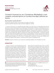
Fig. 1. Proboscia alata. (A) A complete cell, light microscopy (LM). (B) A complete cell, scanning electron microcopy (SEM). (C) Apical part <strong>of</strong> the<br />
valve, noticeable clasper (arrow), LM. (D) Apical part <strong>of</strong> valve, noticeable clasper (arrow), SEM. (E) Details <strong>of</strong> proboscis structure, varied spinule<br />
size, SEM. (F) Details <strong>of</strong> clasper (arrow) <strong>and</strong> contiguous area, SEM. (G) Girdle segments, LM. (H) Girdle segments, SEM. Scale bars represent: A & B,<br />
50 μm; C, 20 μm; D & H, 5 μm; E, 0.5 μm; F, 1 μm; G, 10 μm.<br />
A<br />
D<br />
H<br />
303 http://e-algae.kr<br />
C<br />
G
<strong>Algae</strong> 2011, 26(4): 299-315<br />
Table 4. Morphological characteristics <strong>of</strong> the Dactylisolen <strong>and</strong> Guinardia species examined in this study<br />
Species<br />
Dactylisolen<br />
fragilissimus<br />
Shape Diameter<br />
(μm)<br />
Straight 8.3-20.0<br />
(n = 24)<br />
D. phuketensis Curved 10.0-20.0<br />
(n = 42)<br />
Guinardia<br />
delicatula<br />
Straight 7.9-13.2<br />
(n = 41)<br />
G. flaccida Straight 23.3-42.5<br />
(n = 25)<br />
G. striata Curved 10.0-20.0<br />
(n = 24)<br />
-, no data.<br />
limits <strong>of</strong> the taxa including Proboscia have been determined<br />
by many authors (Jordan <strong>and</strong> Priddle 1991, Jordan<br />
et al. 1991, Takahashi et al. 1994, Jordan <strong>and</strong> Saito 1999,<br />
Jordan <strong>and</strong> Ito 2002, Jordan <strong>and</strong> Ligowski 2004, 2006).<br />
Takahashi et al. (1994) reported that the genus contains<br />
five modern species distributed from polar to temperate<br />
regions. Jordan <strong>and</strong> Ligowski (2004) stated that P. alata<br />
is not cosmopolitan, because it appears to be a complex<br />
cryptic species. Some P. alata representatives are<br />
commonly found in polar waters. However, Hernández-<br />
Becerril (1995) found that P. alata is distributed from<br />
tropical to subtropical waters.<br />
Proboscia indica (Peragallo) Hernández-Becerril<br />
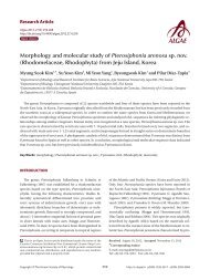
emend. Jordan & Ligowski 1995 (Fig. 2, A-F)<br />
Hustedt 1930, p. 602, Fig. 346; Cupp 1943, p. 93, Fig.<br />
52C; Hendey 1964, p. 147, Pl. 2, Fig. 4; Hernández-Becerril<br />
1995, p. 254, Figs 5 & 6; Moreno et al. 1996, p. 15, Pl. 29,<br />
Figs 6 & 7; Jordan <strong>and</strong> Ligowski 2004, p. 98, Pl. 4, Figs 5-7;<br />
Gómez <strong>and</strong> Souissi 2007, p. 287, Fig. 4g-h; Sunesen <strong>and</strong><br />
Sar 2007, p. 639, Figs 89-97 & 99.<br />
Synonyms. Rhizosolenia indica H. Peragallo 1892, Rhizosolenia<br />
alata f. indica (Peragallo) Gran 1905.<br />
Cells are solitary or in pairs, cylindrical, bilaterally<br />
symmetrical, 25.0-125.0 μm in diameter, 193.4-764.2 μm<br />
long. Valve is sub-conoidal, the ventral part longer than<br />
the dorsal part. Proboscis structure is strongly curved, tapered<br />
towards the apical part in the valve, circular in cross<br />
section, 39.0-83.3 μm long. Apical surface <strong>of</strong> the proboscis<br />
is composed <strong>of</strong> varied sized spinules <strong>and</strong> the slit is pore<br />
http://dx.doi.org/10.4490/algae.2011.26.4.299 304<br />
Cell Segment<br />
Pervalvar<br />
axis length<br />
(μm)<br />
25.0-33.4<br />
(n = 18)<br />
31.3-129.2<br />
(n = 41)<br />
24.9-30.0<br />
(n = 21)<br />
50.0-125.0<br />
(n = 16)<br />
50.0-120.0<br />
(n = 16)<br />
External process Horizontal<br />
Shape Length<br />
(μm)<br />
Oblique tube 1.1-4.3<br />
(n = 9)<br />
Short tube 1.3-6.7<br />
(n = 20)<br />
Narrow tube 2.1-5.0<br />
(n = 11)<br />
Short tube 1.5-1.8<br />
(n = 4)<br />
Slight hook 4.3-6.7<br />
(n = 12)<br />
axis length<br />
(μm)<br />
Perpendicular<br />
axis length<br />
(μm)<br />
- -<br />
10.0-20.0<br />
(n = 42)<br />
7.9-13.2<br />
(n = 41)<br />
23.3-42.5<br />
(n = 25)<br />
10.0-20.0<br />
(n = 24)<br />
1.3-6.6<br />
(n = 70)<br />
shape situated below the apex. Spinule number is 8-13<br />
<strong>and</strong> 0.2-0.5 μm long. Contiguous area is convex towards<br />
the valve surface, distally limited by asymmetric claspers.<br />
The valve areolae are rounded, 30-60 in 10 μm, arranged<br />
in longitudinal striae, converging towards the apex. Girdle<br />
segment areolae are loculate, arranged in columns,<br />
<strong>and</strong> the external velum is perforated by central pores <strong>and</strong><br />
internal circular foramina. Interlocular pores are commonly<br />
surrounded by four loculi. The horizontal axis <strong>of</strong><br />
the segments is 25.0-125.0 μm <strong>and</strong> the perpendicular axis<br />
is 10.6-16.0 μm.<br />
Distribution. Hendey (1964) reported that P. indica is<br />
common in temperate <strong>and</strong> sub-tropical seas as R. alata<br />
var. indica. This species has been reported from Buenos<br />
Aires <strong>marine</strong> waters (Marques Da Cunha <strong>and</strong> Da Fonseca<br />
1917, Balech 1964, 1971, 1979, Lange 1985 as R. alata var.<br />
indica). During the present study, P. indica was rare but<br />
distributed widely at Geoje Isl<strong>and</strong>, Daebu Isl<strong>and</strong>, Daecheon<br />
Harbor, the Yangyang coast, <strong>and</strong> the Yellow Sea<br />
from September 2008 to October 2009.<br />
Remarks. Proboscia indica <strong>and</strong> P. alata are fairly similar<br />
species. However, P. indica differs from P. alata by the<br />
larger diameter <strong>of</strong> the frustule <strong>and</strong> valve morphology. The<br />
valve shape <strong>of</strong> P. indica is sub-conical, round, <strong>and</strong> tapers<br />
into a strongly curved proboscis. Additionally, the pattern<br />
<strong>of</strong> the interlocular pores also differs between the two<br />
species, similar to a feature previously described by Hasle<br />
(1975) <strong>and</strong> Takano (1990). P. indica have interlocular<br />
pores surrounded by four loculae, but the P. alata interlocular<br />
pores are surrounded by six loculae, as observed<br />
by Sundström (1986).<br />
-<br />
1.9-5.0<br />
(n = 140)<br />
4.3-4.6<br />
(n = 2)
A B<br />
C<br />
Neocalyptrella robusta (Norman) Hernández-<br />
Becerril & Meave 1996 (Fig. 3, A-H)<br />
Pritchard 1861, p. 866, Pl. 8, Fig. 42; Peragallo 1892, p.<br />
109, Pl. 14, Fig. 1; Hustedt 1920, Pl. 320, Figs 1-3; Hustedt<br />
1930, p. 578, Fig. 330; Cupp 1943, p. 83, Fig. 46; Okuno<br />
1957, p. 105, Pl. 2, Fig. 1; Okuno 1968, Figs 1(6), 10A, 17 &<br />
18; Navarro 1981, p. 430, Figs 43 & 45; Sundström 1986, p.<br />
Yun & Lee <strong>Morphology</strong> <strong>and</strong> Distribution <strong>of</strong> Marine Diatoms<br />
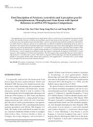
Fig. 2. Proboscia indica. (A) A complete cell, light microscopy (LM). (B) Apical part <strong>of</strong> valve, clasper (arrow), LM. (C) Apical part <strong>of</strong> the valve, clasper<br />
(arrow), scanning electron microcopy (SEM). (D) Details <strong>of</strong> Fig. 9, proboscis structure; longitudinal slit-like pore below the tip (arrow), SEM. (E) Details<br />
<strong>of</strong> the proboscis structure, varied spinule size, SEM. (F) Girdle segments, LM. Scale bars represent: A & B, 50 μm; C & F, 10 μm; D, 5 μm; E, 1 μm.<br />
D<br />
E<br />
F<br />
104, Figs 289 & 290 as R. robusta; Hernández-Becerril <strong>and</strong><br />
Meave del Castillo 1996, p. 199, Figs 1-20 as Calyptrella<br />
robusta; Hasle <strong>and</strong> Syvertsen 1996, p. 159, Pl. 30 as R. robusta;<br />
Gómez <strong>and</strong> Souissi 2007, p. 287, Fig. 4i; Sunesen<br />
<strong>and</strong> Sar 2007, p. 637, Figs 62-67.<br />
Synonyms. Rhizosolenia robusta Norman in Pritchard<br />
1861, Calyptrella robusta (Norman) Hernández-Becerril<br />
<strong>and</strong> Meave 1996.<br />
305 http://e-algae.kr
<strong>Algae</strong> 2011, 26(4): 299-315<br />
A B C<br />
D<br />
E<br />
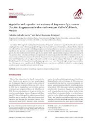
Fig. 3. Neocalyptrella robusta. (A) A complete cell, light microscopy (LM). (B) Apical part <strong>of</strong> valve, external tube at the valve apex (arrow), LM. (C)<br />
Apical part <strong>of</strong> valve, valve with part <strong>of</strong> the cingulum, LM. (D) Apical part <strong>of</strong> valve, valve with part <strong>of</strong> the cingulum, scanning electron microscopy (SEM).<br />
(E) Striation at valve apex, SEM. (F) Valve apex showing calyptra structure <strong>and</strong> external tube, SEM. (G) Detail <strong>of</strong> cingulum; cingulum ends in an obtuse<br />
straight line, SEM. (H) Details <strong>of</strong> Fig. 3E loculate areolae, SEM. Scale bars represent: A, 100 μm; B & C, 20 μm; D, 50 μm; E & G, 10 μm; F, 0.5 μm; H, 2 μm.<br />
http://dx.doi.org/10.4490/algae.2011.26.4.299 306<br />
F<br />
G<br />
H
Cells are solitary, large, bilaterally symmetrical, 108.3-<br />
190.6 μm in diameter, 413.3 μm long, elliptical in cross section,<br />
crescent shaped in lateral view <strong>and</strong> <strong>of</strong> sigmoid form<br />
in ventro-dorsal view. Valve is conoidal with a rounded or<br />
truncated apex <strong>and</strong> with longitudinal undulations. Process<br />
is a cylindrical external tube, straightened towards<br />
the distal part, merging with the calyptra structure <strong>and</strong><br />
circular pore in the distal part <strong>of</strong> the tip, 6.7-7.3 μm long,<br />
0.8-1.0 μm in diameter. Valve areolae, 13-16 in 10 μm, are<br />
arranged in regularly straight striations, with a secondary<br />
quincuncial pattern. Otaria, claspers, <strong>and</strong> contiguous areas<br />
are absent. Girdle segments are oriented in a straight<br />
line <strong>and</strong> arranged in two dorsiventral columns. Segment<br />
areolae, 17-22 in 10 μm, are arranged in regular, straight<br />
striations, with a secondary quincuncial pattern, loculate<br />
areolae, with the velum perforated by slit-like pores <strong>and</strong><br />
internal foramina, circular to subcircular. Horizontal axis<br />
<strong>and</strong> perpendicular axis <strong>of</strong> segments are 108.3-190.6 <strong>and</strong><br />
11.7-25.0 μm in length, respectively.<br />
Distribution. Neocalyptrella robusta is distributed<br />
from tropical to temperate waters (Hasle <strong>and</strong> Syvertsen<br />
1996, Hernández-Becerril <strong>and</strong> Meave del Castillo 1996).<br />
It has been reported to occur in littoral Argentinean waters<br />
(Ferrario <strong>and</strong> Galávan 1989, as Rhizosolenia robusta).<br />
During this study, N. robusta was rare but observed in<br />
September 2008 <strong>and</strong> June 2009 at Geoje Isl<strong>and</strong>, the Korea<br />
Straight, the Wolsung coast, <strong>and</strong> the Yellow Sea.<br />
Pseudosolenia calcar-avis (Schultze) Sundström<br />
1986 (Fig. 4, A-H)<br />
Schultze 1858, p. 339, Pl. 13, Figs 5-10; Peragallo 1892,<br />
p. 113, Pl. 17, Fig. 9; Hustedt 1930, p. 592, Fig. 339 as R. calcar-avis;<br />
Cupp 1943, p. 89, Fig. 51 as R. calar-avis; Navarro<br />
1981, p. 430, Figs 36 & 37 as R. calcar-avis; Sundström<br />
1986, p. 95, Figs 40-46 & 247-257; Hernández-Becerril<br />
1995, p. 254, Figs 7-10; Hasle <strong>and</strong> Syvertsen 1996, p. 160,<br />
Pl. 30; Sunesen <strong>and</strong> Sar 2007, p. 637, Figs 68-81.<br />
Synonym. Rhizosolenia calcar-avis Schultze 1858.<br />
Cells are usually solitary, elongated, <strong>of</strong> cylinder shape,<br />
bilaterally symmetrical, circular in cross section, 9.3-90.0<br />
μm in diameter, 206.7-793.8 μm long. Valve is sub-conical,<br />
asymmetrical, with the ventral part slightly longer than<br />
the dorsal part. Contiguous area is a narrow groove, sigmoid,<br />
extended from the basal part <strong>of</strong> the process to the<br />
margin in the ventral part <strong>of</strong> the valve. Process is claw or<br />
screw shaped, slightly or strongly curved, <strong>and</strong> tapered<br />
towards the distal part, 10.7-51.4 μm long. Otaria <strong>and</strong><br />
claspers are absent. Valve areolae are poroid, circular,<br />
16-34 in 10 μm. Striations are regular <strong>and</strong> straight, with<br />
Yun & Lee <strong>Morphology</strong> <strong>and</strong> Distribution <strong>of</strong> Marine Diatoms<br />
a secondary quincuncial pattern. Girdle segments are<br />
scale-shaped to rhomboidal, arranged in two or multiples<br />
<strong>of</strong> two columns, with a sub marginal seam-like structure<br />
close to the advalvar margin with entire hyaline edges.<br />
Horizontal axis <strong>and</strong> perpendicular axis <strong>of</strong> segments are<br />
9.3-90.0 <strong>and</strong> 9.3-46.2 μm long, respectively. Segmented<br />
areolae are 21-38 in 10 μm in a secondary quincuncial<br />
pattern.<br />
Distribution. Pseudosolenia calcar-avis is a circumglobally<br />
distributed species (Sundström 1986) <strong>and</strong> occurs<br />
in warm waters <strong>and</strong> occasionally in temperate waters<br />
(Hasle <strong>and</strong> Syvertsen 1996). It has been reported several<br />
times in both oceanic <strong>and</strong> near-shore waters along the<br />
coastline <strong>of</strong> Argentina (Ferrario <strong>and</strong> Galávan 1989, as<br />
Rhizosolenia calcar-avis). In the present study, this species<br />
was rarely observed in September 2008 to September<br />
2009 in the oceanic waters <strong>of</strong> Jeju Isl<strong>and</strong> <strong>and</strong> the Yellow<br />
Sea.<br />
Guinardia delicatula (Cleve) Hasle 1995<br />
(Fig. 5, A & B)<br />
Cleve 1900, p. 28, Fig. 11; Hustedt 1930, p. 577, Fig. 328;<br />
Cupp 1943, p. 83, Fig. 44; Hendey 1964, p. 147, Pl. 4, Fig. 2;<br />
Drebes 1974, p. 49, Fig. 35a; Sundström 1986, p. 103, Figs<br />
272 & 273.<br />
Basionym. Rhizosolenia delicatula Cleve 1900.<br />
Cells form fairly straight chains <strong>and</strong> are bilaterally symmetrical.<br />
Cells are 7.9-13.2 μm in diameter, 24.9-30.0 μm<br />
in length. Valve margins are round. External process is<br />
thin <strong>and</strong> short, <strong>and</strong> narrow, tube-shaped, <strong>and</strong> oblique to<br />
the pervalvar axis. External processes are 2.1-5.0 μm long.<br />
External process fits into a depression on the adjacent<br />
valve. Girdle segments are composed <strong>of</strong> open b<strong>and</strong>s, with<br />
poroid areolae, <strong>and</strong> are not noticeable. Segment horizontal<br />
axes are 7.9-13.2 μm long.<br />
Distribution. Hasle <strong>and</strong> Syvertsen (1996) reported that<br />
G. delicatula is a cosmopolitan species in temperate <strong>and</strong><br />
tropical waters. During the present study, this species was<br />
recorded in July 2009 <strong>and</strong> January 2010 in the coastal waters<br />
<strong>of</strong> Sacheon, Incheon, <strong>and</strong> Mokpo.<br />
Guinardia flaccida (Castracane) H. Peragallo 1892<br />
(Fig. 5, C-E)<br />
Castracane 1886, p. 74, Pl. 29, Fig. 4; Peragallo 1892,<br />
p. 107, Pl. 1, Figs 3-5; Bergon 1903, p. 78, Pl. 2, Figs 1-3;<br />
Hustedt 1930, p. 562, Fig. 322; Cupp 1943, p. 78, Fig. 40;<br />
Hendey 1964, p. 147, Pl. 5, Fig. 5; Drebes 1974, p. 58, Fig.<br />
43a; Hasle 1975, p. 116, Figs 64, 65 & 81-89; Navarro 1981,<br />
307 http://e-algae.kr
<strong>Algae</strong> 2011, 26(4): 299-315<br />
A B<br />
F<br />
G H<br />
Fig. 4. Pseudosolenia calcar-avis. (A) A complete cell, light microscopy (LM). (B) Complete cells, scanning electron microcopy (SEM). (C) Apical<br />
part <strong>of</strong> valve, internal structure <strong>of</strong> external process (arrow), LM. (D) Apical part <strong>of</strong> valve, claw or screw shaped external process (arrow), SEM. (E)<br />
Details <strong>of</strong> girdle segments, regularly straight striation, SEM. (F) Girdle segments, LM. (G) Apical part <strong>of</strong> valve, sigmoid contiguous area, SEM. (H)<br />
Screw shaped external process, SEM. Scale bars represent: A & B, 50 μm; C-E & H, 10 μm; F & G, 5 μm.<br />
http://dx.doi.org/10.4490/algae.2011.26.4.299 308<br />
E<br />
C<br />
D
A B<br />
C D E<br />
F G<br />
p. 430, Figs 31 & 32; Takano 1990, pp. 260-261.<br />
Basionym. Rhizosolenia flaccida Castracane 1886.<br />
Cells are solitary or form fairly straight chains, <strong>and</strong> are<br />
bilaterally symmetrical. Cells are 14.0-42.5 μm in diameter,<br />
50.0-125.0 μm in length. Valves are flat or slightly concave.<br />
External processes are short <strong>and</strong> tube-shaped. Short<br />
tube-shaped external processes are located on the external<br />
valve surface. External processes are 1.5-1.8 μm long.<br />
Yun & Lee <strong>Morphology</strong> <strong>and</strong> Distribution <strong>of</strong> Marine Diatoms<br />
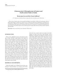
Fig. 5. Guinardia delicatula. (A) Chain formed four cells, light microscopy (LM). (B) External process in valve marginal part (arrow), LM. (C-E)<br />
Guinardia flaccid. (C) A complete cells, LM. (D) Detail <strong>of</strong> girdle segments, LM. (E) Apical part <strong>of</strong> valve (arrow), LM. (F-H) Guinardia striata. (F) Chain<br />
formed two cells, LM. (G) External part <strong>of</strong> process, LM. (H) Girdle b<strong>and</strong>s (arrow), LM. Scale bars represent: A-E, G & H, 10 μm; F, 20 μm.<br />
Girdle segments composed <strong>of</strong> open b<strong>and</strong>s with poroid<br />
areolae. The segment horizontal axis <strong>and</strong> perpendicular<br />
axis are 14.0-42.5 <strong>and</strong> 1.3-5.0 μm in length, respectively.<br />
Distribution. Guinardia flaccida shows a cosmopolitan<br />
<strong>distribution</strong> except the two polar bodies <strong>of</strong> water<br />
(Hasle <strong>and</strong> Syvertsen 1996). During the present study, G.<br />
flaccida was frequently observed in September 2008 <strong>and</strong><br />
August 2009 in the coastal waters <strong>of</strong> the Yellow Sea, Geoje<br />
309 http://e-algae.kr<br />
H
<strong>Algae</strong> 2011, 26(4): 299-315<br />
Isl<strong>and</strong>, Namhae, Sacheon, Tongyeang, <strong>and</strong> Incheon.<br />
Guinardia striata (Stolterforth) Hasle 1995<br />
(Fig. 5, F-H)<br />
Stolterforth 1879, p. 836, Fig. a & b; Peragallo 1888, p.<br />
82, Pl. 6, Fig. 44; Peragallo 1892, p. 108, Pl. 13, Figs 17 & 18;<br />
Bergon 1903, p. 57, Pl. 1, Figs 1-8; Hustedt 1920, Pl. 320,<br />
Figs 4 & 5; Hustedt 1930, p. 578, Fig. 329; Cupp 1943, p.<br />
83, Fig. 45; Hendey 1964, p. 148, Pl. 4, Fig. 5; Drebes 1974,<br />
p. 49, Fig. 35b; Hasle 1975, p. 113, Figs 66-73; Sundström<br />
1980, p. 580, Figs 2-4; Navarro 1981, p. 430, Fig. 48 as R.<br />
stolterforthii; Sundström 1986, p. 103, Figs 274 & 275;<br />
Von Stosch 1986, p. 319, Figs 13 & 14; Hernández-Becerril<br />
1995, p. 262, Figs 53-56.<br />
Basionym. Rhizosolenia stolterforthii (Stolterforth) H.<br />
Peragallo 1888.<br />
Cells form curved chains, rarely spiraling chains,<br />
<strong>and</strong> are bilaterally symmetrical. Cells are 10.0-20.0 μm<br />
in diameter, <strong>and</strong> 50.0-120.0 μm in length. Valve flat <strong>and</strong><br />
rounded at margin. External processes are thin, <strong>and</strong><br />
hook-shaped to the pervalvar axis, <strong>and</strong> the external processes<br />
are 4.3-6.7 μm long. External process fits into a depression<br />
on the adjacent valve. Girdle b<strong>and</strong>s composed <strong>of</strong><br />
open b<strong>and</strong>s with poroid areolae. Segment horizontal axis<br />
<strong>and</strong> perpendicular axis are 10.0-20.0 <strong>and</strong> 4.3-4.6 μm long,<br />
respectively.<br />
Distribution. Guinardia striata is cosmopolitan, but<br />
it does not occur in polar bodies <strong>of</strong> water (Hasle <strong>and</strong> Syvertsen<br />
1996). During the present study, G. striata was<br />
rarely observed in September 2008 <strong>and</strong> August 2009 in<br />
the coastal waters <strong>of</strong> Namhae, Daebu Isl<strong>and</strong>, Incheon,<br />
<strong>and</strong> Yeongdeok.<br />
Dactyliosolen fragilissimus (Bergon) Hasle 1995<br />
(Fig. 6, A-D)<br />
Bergon 1903, p. 49, Pl. 1, Figs 9 & 10; Hustedt 1930, p.<br />
571, Fig. 324; Cupp 1943, p. 80, Fig. 41; Drebes 1974, p.<br />
48, Fig. 34b & c; Hasle 1975, p. 114, Figs 61, 62 & 74-78;<br />
Navarro 1981, p. 430, Fig. 38 as R. fragilissima; Sundström<br />
1986, p. 103, Figs 268 & 269; Takano 1990, pp. 262-263.<br />
Basionym. Rhizosolenia fragilissima Bergon 1903.<br />
Cells are cylindrical with rounded marginal parts,<br />
forming straight chains. Cells are connected in loose fitting<br />
chains at the center <strong>of</strong> the valve surface. Cells are 8.3-<br />
20.0 μm in diameter, <strong>and</strong> 25.0-33.4 μm in length. Valves<br />
are flat or convex at the central part. External process is a<br />
thin, oblique tube in the central part <strong>of</strong> the valve. External<br />
processes are 1.1-4.3 μm long <strong>and</strong> fit into a depression on<br />
http://dx.doi.org/10.4490/algae.2011.26.4.299 310<br />
adjacent cells. Girdle b<strong>and</strong>s composed <strong>of</strong> half b<strong>and</strong>s with<br />
poroid areolae.<br />
Distribution. Dactyliosolen fragilissimus is probably<br />
cosmopolitan (Hasle <strong>and</strong> Syvertsen 1996) but was rarely<br />
observed in February 2010 in Goseong.<br />
Remarks. Cell length <strong>of</strong> Dactyliosolen fragilissimus<br />
varies from 30.0-80.0 μm (Gran <strong>and</strong> Angst 1931), 50.0-80.0<br />
μm (Cupp 1943), 42.0-67.0 μm (Hendey 1964), <strong>and</strong> 42.0-<br />
300.0 μm (Hasle <strong>and</strong> Syvertsen 1996). During this study, D.<br />
fragilissimus was not observed with a length <strong>of</strong> pervalvar<br />
axis up to 100 μm.<br />
Dactyliosolen phuketensis (Sundström) Hasle<br />
1995 (Fig. 6, E-G)<br />
Sundström 1980, p. 579, Figs 1-3; Sundström 1986, p.<br />
103, Figs 270 & 271; Von Stosch 1986, p. 323, Figs 15-17;<br />
Hernández-Becerril 1995, p. 262, Figs 50-52.<br />
Basionym. Rhizosolenia phuketensis Sundström 1980.<br />
Cells are cylindrical with a rounded marginal part,<br />
forming curved chains. Cells are connected in fairly fitting<br />
chains <strong>and</strong> are bilaterally symmetrical. Cells are 10.0-20.0<br />
μm in diameter, <strong>and</strong> 31.3-129.2 μm long. Valves are flat<br />
or slightly convex. External process is usually an obtuse,<br />
short tube in the valve marginal part. External processes<br />
are 1.3-6.7 μm long <strong>and</strong> fit into a depression in adjacent<br />
cells. Girdle b<strong>and</strong>s are composed <strong>of</strong> half b<strong>and</strong>s with poroid<br />
areolae. Segment horizontal axis <strong>and</strong> perpendicular<br />
axis are 10.0-20.0 <strong>and</strong> 1.3-6.6 μm in length, respectively.<br />
Distribution. Sundström (1986) reported that D.<br />
phuketensis occurs in warm water regions to temperate<br />
regions including the North Sea <strong>and</strong> Skagerrak. During<br />
this study, D. phuketensis was newly recorded in June 2009<br />
<strong>and</strong> October 2009 from the coastal waters <strong>of</strong> Geoje Isl<strong>and</strong>,<br />
Sacheon, Tongyeang, Yeongduk, <strong>and</strong> the Yellow Sea.<br />
Remarks. Dactyliosolen phuketensis was recorded for<br />
the first time in Korean coastal waters, but this species<br />
was already observed a long time ago, resembling G. striata.<br />
Although the two species belonged to different genera<br />
<strong>and</strong> were recorded in Korean coastal waters, they can<br />
be easily misidentified using the curved cell <strong>and</strong> colony<br />
shape as discriminating characters. The external process<br />
shapes are different between two taxa. The external processes<br />
<strong>of</strong> G. striata are hook shaped, whereas those <strong>of</strong> D.<br />
phuketensis are short external tubes. The position <strong>of</strong> the<br />
external process is not the same, <strong>and</strong> segment (b<strong>and</strong>)<br />
shape also differs. Representatives <strong>of</strong> the genus Guinardia<br />
are composed <strong>of</strong> an open b<strong>and</strong>, but the genus Dactyliosolen<br />
is composed <strong>of</strong> a half b<strong>and</strong>.
C<br />
E<br />
A B<br />
Yun & Lee <strong>Morphology</strong> <strong>and</strong> Distribution <strong>of</strong> Marine Diatoms<br />
Fig. 6. Dactyliosolen fragilissimus. (A) Chain formed two cells, light microscopy (LM). (B) Detail <strong>of</strong> external process in valve apex (arrow), LM. (C)<br />
Chain formed two cells, LM. (D) Detail <strong>of</strong> external process in valve apex (arrow), LM. (E-G) Dactyliosolen phuketensis. (E) Chain formed two cells, LM. (F)<br />
Apical part <strong>of</strong> valve, external process in valve marginal, LM. (G) Detailed girdle b<strong>and</strong>s, half b<strong>and</strong>, LM. Scale bars represent: A–D, F & G, 10 μm; E, 50 μm.<br />
D<br />
311 http://e-algae.kr<br />
F<br />
G
<strong>Algae</strong> 2011, 26(4): 299-315<br />
DISCUSSION<br />
The <strong>family</strong> Rhizosoleniaceae includes Rhizosolenia,<br />
Proboscia, Pseudosolenia, Neocalyptrella, Guinardia,<br />
Dactyliosolen, <strong>and</strong> Urosolenia. The key morphological<br />
characters <strong>of</strong> the <strong>family</strong> Rhizosoleniaceae are cylindrical<br />
cells in solitary or chain form, unipolar <strong>and</strong> symmetrical<br />
valves, numerous chloroplasts, <strong>and</strong> a few resting spores.<br />
The genera within Rhizosoleniaceae are very common<br />
in the <strong>marine</strong> ecosystem <strong>and</strong> <strong>some</strong>times dominate the<br />
phytoplankton biomass in highly productive oceanic regions<br />
(Sundström 1986, Hernández-Becerril <strong>and</strong> Meave<br />
del Castillo 1996, 1997). Some species <strong>of</strong> the <strong>family</strong> Rhizosoleniaceae<br />
are causative bloom organisms in various<br />
regions <strong>of</strong> the world (Jordan <strong>and</strong> Priddle 1991, Jordan et<br />
al. 1991, Takahashi et al. 1994). These species are very important<br />
<strong>diatoms</strong> in <strong>marine</strong> environments.<br />
As shown in Table 2 <strong>of</strong> Yun <strong>and</strong> Lee (2010), Table 2 in<br />
Yun et al. (2011), <strong>and</strong> Tables 2-4 in the present study, we<br />
divided the 6 genera within the <strong>family</strong> Rhizosoleniaceae<br />
into two groups by morphological diagnostic characteristics<br />
including the shape <strong>of</strong> the external process <strong>and</strong> girdle<br />
segments in the column (Yun <strong>and</strong> Lee 2010, Yun et al.<br />
2011). The first group had a conoidal valve <strong>and</strong> loculate<br />
areolae <strong>and</strong> was comprised <strong>of</strong> Proboscia, Pseudosolenia,<br />
Rhizosolenia, Neocalyptrella, <strong>and</strong> the second group had a<br />
flat or rounded valve <strong>and</strong> poroid areolae <strong>and</strong> was <strong>of</strong> Guinardia<br />
<strong>and</strong> Dactyliosolen. In the present study, 2 species<br />
belonged to Proboscia, 3 species to Guinardia, 2 species<br />
to Dactyliosolen, 1 species to Pseudosolenia, <strong>and</strong> 1 species<br />
belonged to Neocalyptrella.<br />
Cell diameters <strong>of</strong> Proboscia alata were 3.3-13.3 μm, but<br />
previous studies reported 7.0-18.0 μm (Cupp 1943 as Rhizosolenia<br />
alata), 8.5-11.5 μm (Sundström 1986), 2.5-42.0<br />
μm (Jordan et al. 1991), 7.0-24.0 μm (Hernández-Becerril<br />
1995), 2.5-13.0 μm (Hasle <strong>and</strong> Syvertsen 1996), <strong>and</strong> 7.0-<br />
11.0 μm (Sunesen <strong>and</strong> Sar 2007). Cell diameters <strong>of</strong> P. indica<br />
were 25.0-125.0 μm. Our specimens closely resembled<br />
those described previously (Cupp 1943, Hernández-<br />
Becerril 1995, Jordan <strong>and</strong> Ligowski 2004), but their cell<br />
diameters were smaller (16.0-73.0 μm) than those <strong>of</strong> our<br />
specimens. Cell diameters <strong>of</strong> N. robusta (108.3-190.6 μm)<br />
<strong>and</strong> P. calcar-avis (9.3-90.0 μm) were smaller than those <strong>of</strong><br />
Hasle <strong>and</strong> Syvertsen (1996) <strong>and</strong> Sunesen <strong>and</strong> Sar (2007),<br />
respectively.<br />
Cell diameters <strong>of</strong> G. delicatula were 7.9-13.2 μm, G. flaccida<br />
were 23.3-42.5 μm, <strong>and</strong> G. striata were 10.0-20.0 μm.<br />
No differences were observed in the 3 species cell diameters<br />
compared with those <strong>of</strong> many studies (Cupp 1943,<br />
Hernández-Becerril 1995, Hasle <strong>and</strong> Syvertsen 1996).<br />
http://dx.doi.org/10.4490/algae.2011.26.4.299 312<br />
Cell diameters <strong>of</strong> D. fragilissimus <strong>and</strong> D. phuketensis<br />
were 8.3-20.0 μm <strong>and</strong> 10.0-20.0 μm, respectively. Our<br />
specimens were similar to those <strong>of</strong> Hasle <strong>and</strong> Syvertsen<br />
(1996), but their cell diameters were wider than those <strong>of</strong><br />
our specimens.<br />
External processes varied from short tube-shaped in<br />
N. robusta <strong>and</strong> D. phuketensis, claw or screw-shaped in<br />
P. calcar-avis, narrow tube-shaped in G. delicatula, slight<br />
hook-shaped in G. striata, <strong>and</strong> oblique tube-shaped in<br />
D. fragilissimus. The genus Proboscia was distinguished<br />
within the first group because the external processes were<br />
longer, <strong>and</strong> the valve was changed to a probosic structure.<br />
As the external process <strong>of</strong> Rhizosolenia was in the shape<br />
<strong>of</strong> a needle <strong>and</strong> tube, this genus is separated from other<br />
genera (Yun <strong>and</strong> Lee 2010, Yun et al. 2011). The external<br />
process <strong>of</strong> the <strong>family</strong> Rhizosoleniaceae is an important<br />
taxonomic key character.<br />
Areolae occurred in various forms on the external view;<br />
circular to sub circular pore-shaped in P. alata, P. indica,<br />
<strong>and</strong> N. robusta <strong>and</strong> circular to slightly oval pore-shaped<br />
in Pseudosolenia calcar-avis. We were unable to count the<br />
number <strong>of</strong> areolae in G. delicatula, G. striata, D. fragilissimus,<br />
<strong>and</strong> D. phuketensis, but areolae <strong>of</strong> D. phuketensis<br />
are slit-like with a parallel to pervalvar axis (Hernández-<br />
Becerril 1995).<br />
The number <strong>of</strong> areolae in the valves varied from 52-90<br />
in 10 μm in P. alata, 30-60 in 10 μm in P. indica, 13-16 in<br />
10 μm in N. robusta, <strong>and</strong> 16-34 in 10 μm in P. calcar-avis.<br />
Hasle <strong>and</strong> Syvertsen (1996) reported that N. robusta (as<br />
Rhizosolenia robusta) had 19-20 in 10 μm <strong>and</strong> 28-32 in 10<br />
μm in P. calcar-avis. Sunesen <strong>and</strong> Sar (2007) reported that<br />
P. alata had 54 in 10 μm, 17 in 10 μm in N. robusta, <strong>and</strong> 23-<br />
32 in 10 μm in P. calcar-avis. No differences were observed<br />
from previous reports. The number <strong>of</strong> areolae in the segments<br />
varied from 25-62 in 10 μm in P. alata, 17-22 in 10<br />
μm in N. robusta, <strong>and</strong> 21-38 in 10 μm in P. calcar-avis. N.<br />
robusta has 24-26 in 10 μm (Hasle <strong>and</strong> Syvertsen 1996 as<br />
Rhizosolenia robusta), 22-23 in 10 μm in N. robusta, <strong>and</strong><br />
28-32 in 10 μm in P. calcar-avis (Sunesen <strong>and</strong> Sar 2007).<br />
Our specimens had a similar number <strong>of</strong> areolae in the<br />
segments compared with those <strong>of</strong> previous reports. We<br />
could not count number <strong>of</strong> areolae in P. indica, G. delicatula,<br />
G. flaccida, G. striata, D. fragilissimus, <strong>and</strong> D. phuketensis,<br />
because <strong>of</strong> delicate cells.<br />
Abundant <strong>distribution</strong>s <strong>of</strong> G. flaccida <strong>and</strong> P. indica<br />
were found in Korean coastal waters. G. flaccida was<br />
widely distributed at 6 stations, <strong>and</strong> D. fragilissimus <strong>and</strong><br />
N. robusta were sporadically found at 4 stations in Korean<br />
coastal waters. P. calcar-avis has been frequently found<br />
in the Korean coastal waters, <strong>and</strong> this species is a warm
<strong>and</strong> temperate water species (Cupp 1943, Hendey 1964,<br />
Sundström 1986, Hernández-Becerril 1995, Hasle <strong>and</strong> Syvertsen<br />
1996). Dactyliosolen phuketensis was new to Korean<br />
coastal waters.<br />
ACKNOWLEDGEMENTS<br />
This research was supported by Sangmyung University<br />
in 2009. We would like to thank Pr<strong>of</strong>. Hans-U. Dahms <strong>of</strong><br />
the Department <strong>of</strong> Life Science, Sangmyung University<br />
for critical comments on an earlier manuscript draft <strong>and</strong><br />
language improvements.<br />
REFERENCES<br />
Anonymous. 1975. Proposals for a st<strong>and</strong>ardization <strong>of</strong> dia-<br />
tom terminology <strong>and</strong> diagnoses. Nova Hedwigia Beih.<br />
53:323-354.<br />
Balech, E. 1964. El plancton de Mar del Plata durante el<br />
período 1961-62 (Buenos Aires, Argentina). Bol. Inst.<br />
Biol. Mar. Mar del Plata 7:1-49.<br />
Balech, E. 1971. Microplancton de la campaña oceanográ-<br />
fica Productividad III. Revista del Museo Argentino de<br />
Ciencìas Naturales ‘Bernardino Rivadavia’ e Instituto<br />
Nacional de Investigación de las Ciencias Naturales. Hi-<br />
drobiologia 3:1-202.<br />
Balech, E. 1979. Din<strong>of</strong>lagelados. Campaña Oceanográfica<br />
Argentina Islas Orcadas, 06/75. Servicio de Hidrografía<br />
Naval (Buenos Aires) H 655:1-76.<br />
Bergon, P. 1903. Études sur la flore diatomique du bassin<br />
d’Arcachon et des parages de l’ Atlantique voisins de<br />
cette station. Bull. Soc. Sci. Arcachon 6:39-113.<br />
Brightwell, T. 1858. Remarks on the genus “Rhizosolenia” <strong>of</strong><br />
Ehrenberg. Q. J. Microsc. Sci. 6:93-95.<br />
Castracane, F. 1886. Report on the Diatomaceae collected by<br />
H.M.S. Challenger during the years 1873-1876. Report <strong>of</strong><br />
the Scientific Results <strong>of</strong> the Voyage <strong>of</strong> H.M.S. Challenger<br />
1873-1876, Botany 2:1-178.<br />
Chang, M. & Shim, J. H. 1993. A study on the phytoplankton<br />
<strong>of</strong> the Yellow Sea in Autumn, 1984. Ocean Res. 15:15-28.<br />
Cleve, P. T. 1900. The plankton <strong>of</strong> the North Sea, the English<br />
Channel, <strong>and</strong> the Skagerak in 1898. K. Svenska Vet. Akad.<br />
H<strong>and</strong>l. 32:1-53.<br />
Cupp, E. E. 1943. Marine plankton <strong>diatoms</strong> <strong>of</strong> the west coast<br />
<strong>of</strong> North America. Bull. Scripps Inst. Oceanogr. Univ. Ca-<br />
lif. 5:1-238.<br />
De Toni, G. B. 1890. Osservazioni sulla tassonomia delle Bac-<br />
illariee (Diatomee): sequita da un prospetto dei generi<br />
Yun & Lee <strong>Morphology</strong> <strong>and</strong> Distribution <strong>of</strong> Marine Diatoms<br />
delle medesime. Notarisia 5:885-922.<br />
Drebes, C. G. 1974. Marines phytoplankton: eine Ausw. d.<br />
Helgoläner plankton (Diatomeen, Perideneen). Thieme,<br />
Stuttgart, 186 pp.<br />
Edlund, M. B. & Stoermer, E. F. 1993. Resting spores <strong>of</strong> the<br />
freshwater <strong>diatoms</strong> Acanthoceras <strong>and</strong> Urosolenia. J. Pa-<br />
leolimnol. 9:55-61.<br />
Ferrario, M. E. & Galávan, N. M. 1989. Catálogo de las diato-<br />
meas marinas citadas entre los 36° y los 60° S con espe-<br />
cial referencia al Mar Argentino. Publ. Inst. Antárt. Ar-<br />
gent. 20:1-327.<br />
Gómez, F. & Souissi, S. 2007. Unusual <strong>diatoms</strong> linked to cli-<br />
matic events in the northeastern English Channel. J. Sea<br />
Res. 58:283-290.<br />
Gran, H. H. 1905. Diatomeen. In Br<strong>and</strong>t, K. & Apstein, C.<br />
(Eds.) Nordisches Plankton: Botanischer Teil. Vol. 19. Lip-<br />
sius <strong>and</strong> Tischer, Kiel <strong>and</strong> Leipzig, pp. 1-146.<br />
Gran, H. H. & Angst, E. C. 1931. Plankton <strong>diatoms</strong> <strong>of</strong> Puget<br />
Sound. Publ. Puget Sound Biol. Stn. 7:417-519.<br />
Hasle, G. R. 1975. Some living <strong>marine</strong> species <strong>of</strong> the diatom<br />
<strong>family</strong> Rhizosoleniaceae. Nova Hedwigia Beih. 53:99-<br />
153.<br />
Hasle, G. R. & Fryxell, G. A. 1970. Diatoms: cleaning <strong>and</strong><br />
mounting for light <strong>and</strong> electron microscopy. Trans. Am.<br />
Microsc. Soc. 89:469-474.<br />
Hasle, G. R. & Syvertsen, E. E. 1996. Marine <strong>diatoms</strong>. In To-<br />
mas, C. R. (Ed.) Identifying Marine Diatoms <strong>and</strong> Dino-<br />
flagellates. Academic Press, San Diego, CA, pp. 5-385.<br />
Hendey, N. I. 1964. An introductory account <strong>of</strong> the smaller<br />
algae <strong>of</strong> British coastal waters. Part 5: Bacillariophyceae<br />
(Diatoms). Her Majesty’s Stationery Office, London, 317<br />
pp.<br />
Hernández-Becerril, D. U. 1995. Planktonic <strong>diatoms</strong> from the<br />
Gulf <strong>of</strong> California <strong>and</strong> coasts <strong>of</strong>f Baja California: the gen-<br />
era Rhizosolenia, Proboscia, Pseudosolenia, <strong>and</strong> former<br />
Rhizosolenia species. Diatom Res. 10:251-267.<br />
Hernández-Becerril, D. U. & Meave del Castillo, M. E. 1996.<br />
The <strong>marine</strong> planktonic diatom Rhizosolenia robusta<br />
(Bacillariophyta): morphological studies support its<br />
transfer to a new genus, Calyptrella gen. nov. Phycologia<br />
35:198-203.<br />
Hernández-Becerril, D. U. & Meave del Castillo, M. E. 1997.<br />
Neocalyptrella, gen, nov., a new name to replace Calyp-<br />
trella Hernández-Becerril et Meave. Phycologia 36:329.<br />
Hustedt, F. 1920. Atlas der Diatomaceenkunde. In Schmidt,<br />
A. (Ed.) Atlas der Diatomaceen-kunde. O. R. Reisl<strong>and</strong>,<br />
Leipzig, pp. 317-320.<br />
Hustedt, F. 1930. Die Kieselalgen Deutschl<strong>and</strong>s, Österrreichs<br />
und der Schweiz mit Berückichtigung der übrigen Län-<br />
der Europas sowie der angrenzende Meeresgebiete. In<br />
313 http://e-algae.kr
<strong>Algae</strong> 2011, 26(4): 299-315<br />
Rabenhorst, L. (Ed.) Kryptogamen-Flora von Deutsch-<br />
l<strong>and</strong>, Österreich und der Schweiz. Akademische Ver-<br />
lagsgesellschaft m. b. H., Leipzig, pp. 1-920.<br />
Jordan, R. W. & Ito, R. 2002. Observations on Proboscia spe-<br />
cies from Late Cretaceous sediments, <strong>and</strong> their possible<br />
evolution from Kreagra. In John, J. (Ed.) Proc. 15th Int.<br />
Diatom Symp., A.R.G. Ganlner Verlag K. G., Ruggell, pp.<br />
313-329.<br />
Jordan, R. W. & Ligowski, R. 2004. New observations on Pro-<br />
boscia auxospores, <strong>and</strong> validation <strong>of</strong> the <strong>family</strong> Probos-<br />
ciaceae fam. nov. Vie et Milieu 54:91-103.<br />
Jordan, R. W. & Ligowski, R. 2006. Observations on the<br />
auxospores, initial cells <strong>and</strong> vegetative cells <strong>of</strong> Probos-<br />
cia truncata (Bacillariophyta). Nova Hedwigia Beih.<br />
130:201-212.<br />
Jordan, R. W., Ligowski, R., Nöthig, E. -M. & Priddle, J. 1991.<br />
The diatom genus Proboscia in Antarctic waters. Diatom<br />
Res. 6:63-78.<br />
Jordan, R. W. & Priddle, J. 1991. Fossil members <strong>of</strong> the diatom<br />
genus Proboscia. Diatom Res. 6:55-61.<br />
Jordan, R. W. & Saito, M. 1999. The genus Proboscia from<br />
the Thalassiosira yabei Zone (Middle-Late Miocene)<br />
sediments <strong>of</strong> Hokkaido, Japan. In Mayama, S., Idei, M.<br />
& Koizumi, I. (Eds.) Proc. 14th Int. Diatom Symp., Koeltz<br />
Scientific Books, Koenigstein, pp. 565-580.<br />
Kim, S. W., Lee, J. H. & Hong, W. H. 1993. Marine environ-<br />
ment on the view-point <strong>of</strong> plankton dynamics in coastal<br />
waters adjacent to Samcheonpo Thermal Power Plant,<br />
Korea. Collected Thesis, Yongin Univ. 9:357-372.<br />
Lange, K. B. 1985. Spatial <strong>and</strong> seasonal variations <strong>of</strong> diatom<br />
assemblages <strong>of</strong>f the Argentinian coast (South Western<br />
Atlantic). Oceanol. Acta 8:361-369.<br />
Lee, J. H. 1995. Additional check-list <strong>of</strong> <strong>marine</strong> planktonic al-<br />
gae in the coastal waters <strong>of</strong> Korea. I. Bacillariophyceae. J.<br />
Nat. Sci. Sangmyung Women’s Univ. 2:71-198.<br />
Li, Y., Cen, J. -Y., Qi, Y. -Z. & Lü, S. -H. 2009. Morphological<br />
features observations <strong>of</strong> Urosolenia in Chinese freshwa-<br />
ters. Acta Hydrobiol. Sin. 33:566-570.<br />
Marques Da Cunha, A. & Da Fonseca, O. 1917. O microplanc-<br />
ton do Atlantico nas imediações de Mar del Plata. Mem.<br />
Inst. Oswaldo Cruz 9:140-142.<br />
Moon, C. -H. & Choi, H. -J. 1991. Studies on the environmen-<br />
tal characteristics <strong>and</strong> phytoplankton community in the<br />
Nakdong River estuary. J. Oceanol. Soc. Korea 26:144-<br />
154.<br />
Moreno, J. L., Licea, S. & Santoyo, H. 1996. Diatomeas del<br />
Golfo de California. Universidad Autonóma de Baja Cali-<br />
fornia Sur, Loreto, 275 pp.<br />
Navarro, J. N. 1981. A survey <strong>of</strong> the <strong>marine</strong> <strong>diatoms</strong> <strong>of</strong> Puerto<br />
Rico. I. Suborders Coscinodiscineae <strong>and</strong> Rhizosoleni-<br />
http://dx.doi.org/10.4490/algae.2011.26.4.299 314<br />
ineae. Bot. Mar. 24:427-439.<br />
Okuno, H. 1952. Electron microscopical study on antarctic<br />
<strong>diatoms</strong> (3). J. Jpn. Bot. 27:347-356.<br />
Okuno, H. 1957. Electron microscopical study on fine struc-<br />
tures <strong>of</strong> diatom frustules. XV. Observations on the genus<br />
Rhizosolenia. Bot. Mag. Tokyo 70:101-107.<br />
Okuno, H. 1960. Electron microscopical study on fine struc-<br />
tures <strong>of</strong> diatom frustules. XVIII. Bot. Mag. Tokyo 73:310-<br />
316.<br />
Okuno, H. 1968. Electron microscopical study on fine struc-<br />
tures <strong>of</strong> diatom frustules. XX. Observations on genus<br />
Rhizosolenia. Bot. Mag. Tokyo 81:79-88.<br />
Peragallo, H. 1888. Diatomées de la baie de Villefranche. Bull.<br />
Soc. Hist. Nat. Toulouse 22:1-100.<br />
Peragallo, H. 1892. Monographie du genre Rhizosolenia et de<br />
quelques genres voisins. Diatomiste 1:1-39.<br />
Pritchard, A. 1861. A history <strong>of</strong> infusoria, including the Des-<br />
midiaceae <strong>and</strong> Diatomaceae, British <strong>and</strong> foreign. 4th ed.<br />
Whittaker, London, 968 pp.<br />
Ross, R., Cox, E. J., Karayeva, N. I., Mann, D. G., Paddock, T. B.<br />
B., Simonsen, R. & Sims, P. A. 1979. An amended termi-<br />
nology for the siliceous components <strong>of</strong> the diatom cell.<br />
Nova Hedwigia Beih. 64:513-533.<br />
Rott, E., Kling, H. & McGregor, G. 2006. Studies on the diatom<br />
Urosolenia Round & Crawford (Rhizosoleniophycideae)<br />
part I. New <strong>and</strong> reclassified species from subtropical<br />
<strong>and</strong> tropical freshwaters. Diatom Res. 21:105-124.<br />
Round, F. E., Crawford, R. M. & Mann, D. G. 1990. The dia-<br />
toms: biology <strong>and</strong> morphology <strong>of</strong> the genera. Cambridge<br />
University Press, London, 747 pp.<br />
Schultze, M. 1858. Innere Bewegungserscheinungen bei Di-<br />
atomeen der Nordsee aus den Gattungen Coscinodiscus,<br />
Denticella, Rhizosolenia. Arch. Anat. Physiol. Wiss. Med.<br />
1858:330-342.<br />
Shim, J. H. 1994. Illustrated encyclopedia <strong>of</strong> fauna <strong>and</strong> flora <strong>of</strong><br />
Korea, vol. 34 Marine phytoplankton. Ministry <strong>of</strong> Educa-<br />
tion, Seoul, 487 pp.<br />
Simonsen, R. 1974. The diatom plankton <strong>of</strong> the Indian Ocean<br />
Expedition <strong>of</strong> R/V “Meteor” 1964-1965. Meteor Forsch.<br />
Ergeb. Reihe D. 19:1-107.<br />
Simonsen, R. 1979. The diatom system: ideas on phylogeny.<br />
Bacillaria 2:9-71.<br />
Stolterforth, H. 1879. On a new species <strong>of</strong> the genus Eucam-<br />
pia. J. R. Microsc. Soc. 2:835-836.<br />
Sundström, B. G. 1980. Rhizosolenia phuketensis sp. nov. <strong>and</strong><br />
Rhizosolenia stolterforthii H. Peragallo (Bacillariophy-<br />
ceae). Bot. Not. 133:579-583.<br />
Sundström, B. G. 1986. The <strong>marine</strong> diatom genus Rhizoso-<br />
lenia: a new approach to the taxonomy. Ph.D. disserta-<br />
tion, Lund University, Lund, Sweden, 117 pp.
Sunesen, I. & Sar, E. A. 2007. Marine <strong>diatoms</strong> from Buenos<br />
Aires coastal waters (Argentina). IV. Rhizosolenia s. str.,<br />
Neocalyptrella, Pseudosolenia, Proboscia. Phycologia<br />
46:628-643.<br />
Takahashi, K., Jordan, R. & Priddle, J. 1994. The diatom genus<br />
Proboscia in subarctic waters. Diatom Res. 9:411-428.<br />
Takano, H. 1990. Diatoms. In Fukuyo, Y., Takano, H., Chihara,<br />
M. & Matsouka, K. (Eds.) Red Tide Organisms in Japan:<br />
An Illustrated Taxonomic Guide. Uchida Rokakuho, To-<br />
kyo, pp. 162-331.<br />
Von Stosch, H. A. 1986. Some <strong>marine</strong> <strong>diatoms</strong> from the Aus-<br />
tralina region, especially from Port Phillip Bay <strong>and</strong> tropi-<br />
cal north-eastern Australia. Brunonia 8:293-348.<br />
Yun, S. M. & Lee, J. H. 2010. <strong>Morphology</strong> <strong>and</strong> <strong>distribution</strong><br />
<strong>of</strong> <strong>some</strong> <strong>marine</strong> <strong>diatoms</strong>, <strong>family</strong> Rhizosoleniaceae, in<br />
Korean coastal waters: a genus Rhizosolenia 1. <strong>Algae</strong><br />
25:173-182.<br />
Yun & Lee <strong>Morphology</strong> <strong>and</strong> Distribution <strong>of</strong> Marine Diatoms<br />
Yun, S. M. Lee, S. D. & Lee, J. H. 2011. <strong>Morphology</strong> <strong>and</strong> distri-<br />
bution <strong>of</strong> <strong>some</strong> <strong>marine</strong> <strong>diatoms</strong>, <strong>family</strong> Rhizosoleniace-<br />
ae, genus Rhizosolenia, in Korean coastal waters. <strong>Algae</strong><br />
26:141-152.<br />
Yoon, Y. -H. & Koh, N. -P. 1994. Distribution <strong>of</strong> phytoplank-<br />
ton population in the coastal waters <strong>of</strong> Keumo Isl<strong>and</strong>s,<br />
southern Korea in summer. Bull. Yosu Natl. Fish. Univ.<br />
8:21-35.<br />
Yoon, Y. -H. & Koh, N. -P. 1995. Studies on the environmental<br />
characteristics <strong>of</strong> the breeding ground in the Kogum-<br />
sudo, southern part <strong>of</strong> Korean Peninsula I. Seasonal suc-<br />
cession <strong>of</strong> phytoplankton population. J. Aquac. 8:47-58.<br />
Yoon, Y. H., Rho, H. K. & Kim, Y. G. 1992. Seasonal succes-<br />
sion <strong>of</strong> phytoplankton population in the Hamdok port,<br />
northern Cheju Isl<strong>and</strong>. Bull. Mar. Res. Inst. Cheju Natl.<br />
Univ. 16:27-42.<br />
315 http://e-algae.kr