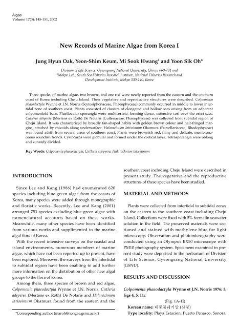
New Records of Marine Algae from Korea I
New Records of Marine Algae from Korea I
New Records of Marine Algae from Korea I

You also want an ePaper? Increase the reach of your titles
YUMPU automatically turns print PDFs into web optimized ePapers that Google loves.
<strong>Algae</strong><br />
Volume 17(3): 145-151, 2002<br />
INTRODUCTION<br />
Since Lee and Kang (1986) had enumerated 620<br />
species including blue-green algae <strong>from</strong> the coasts <strong>of</strong><br />
<strong>Korea</strong>, many species were added through monographic<br />
and floristic works. Recently, Lee and Kang (2001)<br />
arranged 753 species excluding blue-green algae with<br />
nomenclatural accounts based on these works.<br />
Meanwhile, many other species have been identified<br />
<strong>from</strong> various works and supplimented to the marine<br />
algal flora <strong>of</strong> <strong>Korea</strong>.<br />
With the recent intensive surveys on the coastal and<br />
island environments, numerous members <strong>of</strong> marine<br />
algae, which have not been reported up to present, have<br />
been explored. Moreover, the surveys <strong>from</strong> the intertidal<br />
to subtidal region have been enabling to add further<br />
more information on the distribution <strong>of</strong> other new algal<br />
groups to the flora <strong>of</strong> <strong>Korea</strong>.<br />
Among them, three species <strong>of</strong> brown and red algae,<br />
Colpomenia phaeodactyla Wynne et J.N. Norris, Cutleria<br />
adspersa (Mertens ex Roth) De Notaris and Halarachnion<br />
latissimum Okamura found <strong>from</strong> the eastern and the<br />
*Corresponding author (maroh@nongae.gsnu.ac.kr)<br />
<strong>New</strong> <strong>Records</strong> <strong>of</strong> <strong>Marine</strong> <strong>Algae</strong> <strong>from</strong> <strong>Korea</strong> I<br />
Jung Hyun Oak, Yeon-Shim Keum, Mi Sook Hwang 1 and Yoon Sik Oh*<br />
Division <strong>of</strong> Life Science, Gyeongsang National University, Chinju 660-701 and<br />
1 Mokpo Lab., South Sea Fisheries Research Institute, National Fisheries Research and<br />
Development Institute, Mokpo 530-140, <strong>Korea</strong><br />
Three species <strong>of</strong> marine algae, two browns and one red were newly reported <strong>from</strong> the eastern and the southern<br />
coast <strong>of</strong> <strong>Korea</strong> including Cheju Island. Their vegetative and reproductive structures were described. Colpomenia<br />
phaeodactyla Wynne et J.N. Norris (Scytosiphonaceae, Phaeophyceae) commonly occurred in middle to lower intertidal<br />
zone <strong>of</strong> southern coast. Plants consisted <strong>of</strong> clusters <strong>of</strong> elongated and hollow sacs arising <strong>from</strong> an adherent<br />
colpomenioid base. Plurilocular sporangia were multiseriate, forming dense, extensive sori over the erect sacs.<br />
Cutleria adspersa (Mertens ex Roth) De Notaris (Cutleriaceae, Phaeophyceae) was collected <strong>from</strong> subtidal region <strong>of</strong><br />
Cheju Island. It was characterized by broadly fan-shaped habits with golden brown colour and hair-fringed margins,<br />
attached by rhizoids along undersurface. Halarachnion latissimum Okamura (Furcellariaceae, Rhodophyceae)<br />
was found adrift <strong>from</strong> several areas <strong>of</strong> southern coast. Plants were brownish red, filmy and delicate, membranaceous<br />
roundish fronds. Cystocarps were globular and formed under the cortical layer. Tetrasporangia were oblong<br />
and zonately divided.<br />
Key Words: Colpomenia phaeodactyla, Cutleria adspersa, Halarachnion latissimum<br />
southern coast including Cheju Island were described in<br />
present study. The vegetative and the reproductive<br />
structures <strong>of</strong> these species have been studied.<br />
MATERIAL AND METHODS<br />
Plants were collected <strong>from</strong> intertidal to subtidal zones<br />
on the eastern to the southern coast including Cheju<br />
Island. Collections were fixed with 5% formalin seawater<br />
solution in the field. The preserved materials were sectioned<br />
and stained with methylene blue for light<br />
microscopy. Observation and photomicrography were<br />
conducted using an Olympus BX50 microscope with<br />
PM10 photography system. Specimens examined in present<br />
study were deposited in the herbarium <strong>of</strong> Division<br />
<strong>of</strong> Life Science, Gyeongsang National University<br />
(GSNU).<br />
RESULTS AND DISCUSSION<br />
Colpomenia phaeodactyla Wynne et J.N. Norris 1976: 5,<br />
figs 4, 5, 11c<br />
(Fig. 1A-1I)<br />
<strong>Korea</strong>n name: 대롱불레기말 (신칭)<br />
Type locality: Playa Estacion, Puerto Penasco, Sonora,
146 <strong>Algae</strong> Vol. 17(3), 2002<br />
Fig. 1. Colpomenia phaeodactyla Wynne et J.N. Norris.<br />
A. Plant <strong>from</strong> Sangjokam, Goseong. B. Erect sacs with adventitious branches (arrows). C. Colpomenioid base. D. Cross section <strong>of</strong><br />
base. E. Cortex <strong>of</strong> base. F. Tufts <strong>of</strong> hair cells. G. Cortical cells and ascocysts in surface view. H. Ascocysts (arrow) and cuticle<br />
(arrowhead) in cross section. I. Plurilocular sporangia (Scales A: 5 cm, B: 2 cm, C: 1 cm, D: 500 µm, E, H: 20 µm, F, I: 30 µm, G: 50<br />
µm).<br />
Gulf <strong>of</strong> Californica, Mexico<br />
Distribution: Northern Gulf <strong>of</strong> California, Pacific Baja<br />
California, Mexico, Costa Rica (Wynne and Norris 1976),<br />
Chile (Santelices 1989; Santelices and H<strong>of</strong>fmann 1997)<br />
and central Japan (Yoshida et al. 2000) and <strong>Korea</strong> (present<br />
study).<br />
Specimens examined: Sangjokam, Goseong,<br />
Gyeongnam, 14 II 2002 (J.H. Oak), Sachon, Namhaedo,<br />
Gyeongnam, 16 III 2001 (Y.S. Oh), Geumodo, Yeosu, 8 III<br />
2001 (Y.S. Oh), Jindo, Jeonnam, 18 II 2000; 25 II 2001 (Y.S.<br />
Oh and M.S. Hwang).<br />
Plants commonly occurred in middle to lower intertidal<br />
zone and tidal pools <strong>of</strong> southern coast. Thalli were<br />
dark brown, consisted <strong>of</strong> clusters <strong>of</strong> elongated and hollow<br />
sacs (Fig. 1A), 15-25 cm long and 0.5-1.5 cm broad,<br />
arising <strong>from</strong> an adherent colpomenioid base, 0.8-1.5 cm<br />
in diameter and 800 µm thick (Fig. 1C). Cortical layer <strong>of</strong><br />
the base was 1-2 cells thick and cortical cells were<br />
cuboidal, 10-13 µm long and 10-15 µm broad. Medullary<br />
cells were elongated, 75-125 µm long and 25-45 µm<br />
broad in longitudinal section (Fig. 1D-E). The sacs were<br />
5-15 <strong>from</strong> a single base, tapered toward the base and<br />
apex, 100-160 µm thick. They were occasionally undulated<br />
or contorted and produced adventitious branches<br />
(Fig. 1B). Cortical layer was 1-2 cortical cells thick with a<br />
cuticle outside. The cortical cells were cuboidal, 10-15<br />
µm in diameter (Fig. 1G). Medullae consisted <strong>of</strong> 3-4 layers<br />
<strong>of</strong> spherical to subspherical cells with 20-75 µm in<br />
cross section, elongated cells with 60-140 µm long and<br />
25-90 µm broad in longitudinal section. Ascocysts were<br />
globular in surface, ovoid to obovoid in cross section, 20-<br />
25 µm long and 10-15 µm broad (Fig. 1H). Hairs were<br />
tufts, colorless, consisting <strong>of</strong> elongated cells with 30-45<br />
µm long and 10 µm broad (Fig. 1F). Plurilocular sporangia<br />
were biseriate, 30-45 µm long, forming dense, extensive<br />
sori lacking a cuticle over the erect sacs (Fig. 1I).<br />
Unilocular sporangia were not found.<br />
Colpomenia phaeodactyla grew on rocks covered with<br />
muddy sands in middle to lower intertidal zone along<br />
the southern coast. In the field, however, the apparent<br />
cylindrical sacs <strong>of</strong> C. phaedactyla could be misidentified<br />
as enlarged forms <strong>of</strong> Scytosiphon lomentaria (Lyngbye)
Link or elongated forms <strong>of</strong> Colpomenia bullosa (Sauders)<br />
Yamada. C. phaeodactyla was distinguished <strong>from</strong> C. bullosa<br />
by having the delicate feature <strong>of</strong> the sacs and its<br />
clustered aspect <strong>from</strong> common basal system (Wynne and<br />
Norris 1976). The s<strong>of</strong>t and delicate texture was due to<br />
thinner sacs with the range <strong>of</strong> 100-160 µm, compared to<br />
C. bullosa with 120-230 µm in thickness.<br />
The general features <strong>of</strong> the plants were in good accordance<br />
with the original description given by Wynne and<br />
Norris (1976). However, the plants <strong>of</strong> our collections<br />
formed occasionally undulated or contorted sacs and<br />
produced adventitious branches in contrast to the morphological<br />
feature <strong>of</strong> the type with a smooth and entire<br />
habit.<br />
Colpomenia phaeodactyla was reported <strong>from</strong> the south-<br />
Oak et al.: <strong>New</strong> <strong>Records</strong> <strong>of</strong> <strong>Marine</strong> <strong>Algae</strong> <strong>from</strong> <strong>Korea</strong> 147<br />
Fig. 2. Cutleria adspersa (Mertens ex Roth) De Notaris.<br />
A. Plant growing on rocks mixed with corallines in subtial zone. B. Plant with fan-shaped habit. C. Hair-fringed margins. D.<br />
Trichothallic filaments with meristems (arrow) in cross section. E. Trichothallic meristems (arrow) in surface view. F.<br />
Rectangular cortical cells. G. Subspherical medullary cells in cross section. H. Elongated rectangular medullary cells in longitudinal<br />
section. I. Rhizoids <strong>from</strong> undersurface (Scales A: 2 cm, B: 3 cm, C: 500 µm, D, E: 50 µm, F: 20 µm, G-I: 50 µm).<br />
eastern Pacific, including the Gulf <strong>of</strong> California, Costa<br />
Rica and Chile, while C. bullosa usually occurred at the<br />
pacific coast <strong>of</strong> North America (Wynne and Norris 1976;<br />
Santelices 1989). In the northwestern Pacific, C. bullosa<br />
occurred in the northern part <strong>of</strong> Japan (Yoshida et al.<br />
2000) and C. phaeodactyla was mostly restricted in the<br />
southern to central part as currently observed in the distribution<br />
<strong>of</strong> the plants in <strong>Korea</strong>.<br />
Cutleria adspersa (Mertens ex Roth) De Notaris 1842:<br />
10<br />
(Fig. 2A-2I)<br />
<strong>Korea</strong>n name: 겹부채채찍말 (신칭)<br />
Type locality: Cadiz, Spain<br />
Distribution: Northeast Atlantic (Feldmann and
148 <strong>Algae</strong> Vol. 17(3), 2002<br />
Fig. 3. Halarachnion latissimum Okamura.<br />
A. Plant <strong>from</strong> Namhaedo (Tetrasporophyte). B. Plant <strong>from</strong> Namhaedo (Carposporophyte). C. Apex showing axial cells. D.<br />
Cortical cells. E. Rhizoids (arrow) <strong>from</strong> subcortical cells transversing thallus. F. Tetrasporangia (arrow) immersed in cortex. G.<br />
Globular tetrasporangia (arrow) in surface view. H. Elliptical tetrasporangia divided zonately (Scales A: 3 cm, B: 2 cm, C: 500<br />
µm, D, G: 30 µm, E, F: 50 µm, H: 20 µm).<br />
Magne 1964), Mediterranean (Ribera et al. 1992),<br />
Australia (Phillips 1997), Japan (Yoshida et al. 2000) and<br />
<strong>Korea</strong> (present study)<br />
Specimens examined: Seongsanpo, Cheju Island, 10<br />
III 2001; 25 III 2002 (Y.S. Oh).<br />
Plants grew on rocks in the subtidal zone <strong>of</strong> Cheju<br />
Island (Fig. 2A). Thalli were up to 4 cm high, golden<br />
brown coloured, fan-shaped upright habits with hairfringed<br />
margins (Fig. 2B, C). Rhizoids arose <strong>from</strong> epidermal<br />
cells along lower surface <strong>of</strong> prostrate portion (Fig.<br />
2I). Thalli showed marginal trichothallic growth <strong>from</strong><br />
the basal meristem <strong>of</strong> tufts <strong>of</strong> uniseriate apical filaments<br />
at the edges (Fig. 2D, E). The filaments were 20-25 mm in<br />
diameter. Fronds were polystichous, 100-120 µm thick.<br />
Cortices were phaeoplastic, 1-2 cells thick, and upper<br />
cortical cells were rectangular, 7-15 µm long and 5-10 µm<br />
broad (Fig. 2F). Medulla was 5-7 cells thick and<br />
medullary cells were colorless, subspherical and 15-40<br />
µm in cross section (Fig. 2G), elongated rectangular, 70-<br />
95 µm long and 18-25 µm broad in longitudinal section<br />
(Fig. 2H). Sporangia were not found.<br />
In <strong>Korea</strong>, only one species in the genus Cutleria, C.<br />
cylindrica was commonly recorded in the eastern and the<br />
southern coast, whereas three species, including C.<br />
adspersa and C. multifida, were widely distributed in<br />
Japan (Kitayama et al. 1992; Sasaki et al. 1987; Yoshida et<br />
al. 2000). In present study, C. adspersa was firstly found<br />
<strong>from</strong> the subtidal area <strong>of</strong> Seongsanpo, Cheju Island. It<br />
grew on rocks covered with coralline algae, such as<br />
Amphiroa spp. and Marginisporum spp. and shaded by<br />
Sargassum forest in 5-10 m deep bottom.<br />
The genus Cutleria showed a heteromorphic life history.<br />
An erect bladed gametophyte alternated with a small<br />
encrusting sporophyte, which was once called Aglaozonia<br />
by culture studies (Falkenberg 1879; Kuckuck 1899;<br />
Yamanouchi 1912). Sporophytes and fertile thalli were
not found in present study.<br />
Cutleria adspersa was similar to C. mollis and C. hancockii<br />
in having broadly fan-shaped blades developed<br />
<strong>from</strong> marginal zones <strong>of</strong> the thallus. Cutleria mollis known<br />
in Australia appeared more erect habit with s<strong>of</strong>t and<br />
thinner blade than C. adspersa (Allender and Kraft 1983).<br />
However, C. adspersa was fairly similar to C. mollis in size<br />
and thickness <strong>of</strong> the thalli; the former observed in present<br />
study was 4 cm long and 100-120 µm thick, while<br />
the latter 5 cm long and 95-125 µm thick (Allender and<br />
Kraft 1983).<br />
C. hancockii commonly occurred in the northern coast<br />
<strong>of</strong> the Gulf <strong>of</strong> California (Dawson 1944). It was distinguished<br />
<strong>from</strong> C. adspersa in having light coloured small<br />
and loosely anchored sporophyte as well as the middle<br />
to lower intertidal habitat (Dawson 1944). However,<br />
there were little differences in morphological features<br />
<strong>from</strong> the fan-shaped gametophytes <strong>of</strong> the two species<br />
(Dawson 1944). In addition, C. hancockii was occasionally<br />
collected <strong>from</strong> the subtidal area and showed much more<br />
morphological variation than the original description<br />
(Riosmena-Rodriguez et al. 2001). We could not compare<br />
Oak et al.: <strong>New</strong> <strong>Records</strong> <strong>of</strong> <strong>Marine</strong> <strong>Algae</strong> <strong>from</strong> <strong>Korea</strong> 149<br />
Fig. 4. Halarachnion latissimum Okamura.<br />
A. Carpogonial branch. B. Auxiliary cell. C. Connecting filaments (arrows) <strong>from</strong> carpogonial derivative cells. D. Gonimoblast<br />
initial and connecting filament (arrow). E, F. Gonimoblast development and connecting filaments (arrows). G. Globular cystocarps<br />
immersed under the cortical layer (Scales A-F: 10 µm, G: 50 µm).<br />
the features <strong>of</strong> sporophytes <strong>of</strong> the two species, because<br />
sporophytes <strong>of</strong> C. adspersa were not available in present<br />
study. Therefore these three species should be investigated<br />
to confirm their specific delimitations in detail.<br />
Halarachnion latissimum Okamura 1933: 9, pl. 306<br />
(Figs 3A-4G)<br />
<strong>Korea</strong>n name: 매끈곱단이 (신칭)<br />
Type locality: Enoshima, Japan<br />
Distribution: Japan (Okamura 1933; Yoshida et al.<br />
2000) and <strong>Korea</strong> (present study)<br />
Specimens examined: Pogyo, Goseong, Gyeongnam,<br />
7 II 2002 (J.H. Oak and Y.-S. Keum); Mibeob, Namhaedo,<br />
Gyeongnam, 14 III 2002 (Y.-S. Keum); Yeonampo,<br />
Pohangshi, 17 III 2002 (Y.S. Oh).<br />
Thalli were brownish red, delicately membranaceous,<br />
roundish, cuneate or subcordate with entire margin (Fig.<br />
3A-B), arising <strong>from</strong> a small basal disc, 0.7-2 mm in diameter.<br />
They were up to 20 cm long, 15 cm broad and 100-<br />
200 µm thick, and <strong>of</strong>tenly undulate to lobed in mature.<br />
Thalli were multiaxial, initiated by several apical cells,<br />
forming subterminal cells (Fig. 3C). The subterminal
150 <strong>Algae</strong> Vol. 17(3), 2002<br />
cells cut <strong>of</strong>f lateral initials towards the surface and<br />
branched several times to form cortex. Cortical layer was<br />
1-3 cells thick and cortical cells were subspherical to<br />
polygonal in surface, 8-12 µm in diameter (Fig. 3D).<br />
Subcortical cells were subspherical to stellate with elongated<br />
arms, 13-18 µm in diameter. Medulla has a few<br />
slender filaments (rhizoids), which were transeversely<br />
located. The rhizoids arose <strong>from</strong> the inner subcortical<br />
cells to grow transversely out <strong>of</strong> the medulla. They were<br />
usually unbranched and consisted <strong>of</strong> 3-5 elongated cells,<br />
55-150 µm long and 4-5 µm broad (Fig. 3E).<br />
Tetrasporophytes were 15-20 cm long and 13-15 cm<br />
broad (Fig. 3A). Tetrasporangia were scattered over the<br />
frond and globular in surface (Fig. 3G), elliptic in cross<br />
section (Fig. 3F). They were zonately divided, 28-34 µm<br />
long and 18-22 µm broad (Fig. 3H). Carposporophytes<br />
were 10-15 cm long and 10 cm broad (Fig. 3B).<br />
Carpogonial branches consisted <strong>of</strong> 3-4 cells were borne<br />
<strong>from</strong> subcortical cells (Fig. 4A). Intercalary cells <strong>of</strong> them<br />
were usually distended to one side. After fertilization,<br />
the carpogonium and the intercalary cells divided transversely<br />
into two derivative cells (Fig. 4C). The carpogonial<br />
derivative cells produced 1-2 connecting filaments.<br />
The second connecting filament was cut <strong>of</strong>f <strong>from</strong> each<br />
derivative cell opposite to the first (Fig. 4C). The connecting<br />
filaments were septate and branched. Auxiliary<br />
cells arisen <strong>from</strong> the subcortical cells produced 3-4 lateral<br />
branches with small cortical cells. They were round to<br />
broadly obovate, 20-28 µm long and 13-20 µm broad<br />
(Fig. 4B). After fusion between the connecting filament<br />
and the auxiliary cell, the auxiliary cell initiated a hemispherical<br />
lobe that developed gonimoblast initial (Fig.<br />
4D-E). The gonimoblast initials were divided and developed<br />
into gonimoblasts (Fig. 4F). The gonimoblasts grew<br />
inwards in the thallus. Cystocarps were globular, 200-<br />
350 µm in diameter and densely scattered over the whole<br />
frond and immersed under the cortical layer (Fig. 4G).<br />
Spermatangia were not found.<br />
Plants adrift were collected <strong>from</strong> the beaches <strong>of</strong> the<br />
eastern and the southern coasts. Most <strong>of</strong> them were<br />
found at the sheltered area with sandy and rocky substrata.<br />
Matured plants attached to pebbles <strong>of</strong>tenly<br />
occurred at the shallow tidal pools in lower intertidal<br />
zone.<br />
Halarachnion latissimum was characterized by filmy,<br />
delicate membranous habit and zonately divided<br />
tetrasporangia. These features were similar to<br />
Schizymenia dubyi (Chauvin) J. Agardh commonly<br />
occurred in <strong>Korea</strong>, but H. latissimum showed more mem-<br />
branaceous, rounded to subcordate thallus, and thinner<br />
cortical layers consisted <strong>of</strong> 1-2 cells thick than those <strong>of</strong> S.<br />
dubyi. Additionally, H. latissimum was distinguished<br />
<strong>from</strong> S. dubyi by carposporangia showing inward development.<br />
In this study, it was remarkable to recognise<br />
this species as the first member <strong>of</strong> the family<br />
Furcellariaceae in <strong>Korea</strong>.<br />
At present two more species were known in the genus<br />
Halarachnion: H. ligulatum (Woodward) Kützing and H.<br />
parvum Yamada. H. parvum endemic to Japan showed a<br />
smaller habit with ca. 1 cm in height (Yamada 1941).<br />
Among them H. ligulatum, a subtidal species in the<br />
Atlantic, was similar in simple blade morphology to H.<br />
latissimum. H. ligulatum was distinguished <strong>from</strong> H. latissimum<br />
by occasionally much-divided thallus with very<br />
narrow segments and <strong>of</strong>tenly proliferous branches at the<br />
margins and the surfaces (Knauss and Hommersand<br />
1989). In addition, H. ligulatum was monoecious with<br />
spermatangia and carposporangia on same thallus<br />
(Knauss and Hommersand 1989), whereas spermatangia<br />
were not found on carposporic plants <strong>of</strong> H. latissimum in<br />
present study.<br />
Halarachnion ligulatum showed a heteromorphic life<br />
history in which tetrasporophyte formed discoid crust<br />
corresponding to Cruoria rosea (Crouan frat.) Crouan frat.<br />
(Boillot 1965; Kornmann and Sahling 1977; Maggs 1983).<br />
However, we could collect cystocarpic and tetrasporic<br />
plants with erect and membranaceous habit <strong>of</strong> H. latissimum.<br />
It agreed well with the report that H. latissimum<br />
has an isomorphic life history (Okamura 1933). The discrepancies<br />
in the life history between H. ligulatum and H.<br />
latissimum led to question their relationship and suggested<br />
that two species could be separated at the generic<br />
level. As the genera in the family Furcellariaceae showed<br />
both heteromorphic and isomorphic life history, the further<br />
study needs to confirm their relationships.<br />
REFERENCES<br />
Allender B.M. and Kraft G.T. 1983. The marine algae <strong>of</strong> Lord<br />
Howe Island (<strong>New</strong> South Wales): the Dictyotales and<br />
Cutleriales (Phaeophyta). Brunonia 6: 73-130.<br />
Boillot A. 1965. Sur l’alternance de generations heteromorphes<br />
d’une Rhodophycée, Halarachnion ligulatum (Woodward)<br />
Kützing (Gigartinales, Furcellariacées). C.R. Acad. Sc. Paris<br />
261: 4191-4193.<br />
Dawson E.Y. 1944. The marine algae <strong>of</strong> the Gulf <strong>of</strong> California.<br />
Allan Hancock Pacific Expeditions 3: 189-453, 47 pls.<br />
Falkenberg P. 1879. Die Befruchtigen und die<br />
Generationswechsel von Cutleria. Mitt. Zool. Stat. Neapel 1:
420-447.<br />
Feldmann J. and Magne M.F. 1964. Additions a l’inventaire de<br />
la flore marine de Rosc<strong>of</strong>f algues, champignons, lichens.<br />
Trav. Stat. Biol. Rosc<strong>of</strong>f 15: 1-23 [+5].<br />
Kitayama T. Kawai H. and Yoshida T. 1992. Dominance <strong>of</strong><br />
female gametophytes in field populations <strong>of</strong> Cutleria cylindrica<br />
(Cutleriales, Phaeophyceae) in the Tsugaru Strait,<br />
Japan. Phycologia 31: 449-461.<br />
Knauss M.E. and Hommersand M.H. 1989. Vegetative and<br />
reproductive development <strong>of</strong> Halarachnion ligulatum<br />
(Gigartinales, Rhodophyta). Br. phycol. Jour. 24: 39-52.<br />
Kornmann P. and Sahling P.-H. 1977. Meeresalgen von<br />
Helgoland. Benthische Grun-, Braun und Rotalgen.<br />
Helgolander Wiss. Meeresuntersuch. 29: 1-289.<br />
Kuckuck P. 1899. Beitrage zur Kenntnis der Meeresalgen. 9.<br />
Uber den Generationswechsel von Cutleria multifida (engl.<br />
Bot.) Grev. Wiss. Meeresunters. Abt. Helgol., N.F. 3: 95-117.<br />
Lee I.K. and Kang J.W. 1986. A Check List <strong>of</strong> <strong>Marine</strong> <strong>Algae</strong> in<br />
<strong>Korea</strong>. <strong>Korea</strong>n J. Phycol. 1: 311-325.<br />
Lee Y. and Kang S. 2001. A Catalogue <strong>of</strong> the Seaweeds in <strong>Korea</strong>.<br />
Cheju National University Press.<br />
Maggs C.A. 1983. A phenological study <strong>of</strong> the epiflora <strong>of</strong> two<br />
maerl beds in Galway Bay. Galway University College,<br />
Galway, Ireland.<br />
Okamura K. 1933. Icones <strong>of</strong> Japanese algae. Vol. VII, Tokyo.<br />
Phillips J.A. 1997. <strong>Algae</strong>. In Queensland Plants: Names and<br />
Distribution. (Henderson R.J.F., editor), pp. 223-240.<br />
Queensland Herbarium, Department <strong>of</strong> Environment,<br />
Indooroopilly, Queensland.<br />
Oak et al.: <strong>New</strong> <strong>Records</strong> <strong>of</strong> <strong>Marine</strong> <strong>Algae</strong> <strong>from</strong> <strong>Korea</strong> 151<br />
Ribera M.A. Gomez-Garreta A., Gallardo T., Cormaci M.,<br />
Furnari G. and Giaccone G. 1992. Check-list <strong>of</strong><br />
Mediterranean Seaweeds. I. Fucophyceae (Warming 1884).<br />
Bot. Mar. 35: 109-130.<br />
Riosmena-Rodriguez R., Paul-Chavez L. and Hinojosa-Arango<br />
G. 2001. Range extension <strong>of</strong> Cutleria hancockii Dawson<br />
(Cutleriales, Phaeophyta) to the southwestern gulf <strong>of</strong><br />
California, Mexico. Bot. Mar. 44: 461-465.<br />
Santelices B. 1989. Algas marinas de Chile distribucion, ecologia,<br />
utilizacion, diversidad. Ediciones Universidad Catolica<br />
de Chile, Santiago.<br />
Santelices B. and H<strong>of</strong>fmann A. 1997. Flora Marina de Chile<br />
Central. <strong>Marine</strong> Flora <strong>of</strong> Central Chile. Ediciones<br />
Universidad Catolica de Chile, Santiago.<br />
Sasaki S., Kikuchi K. and Matsuyama K. 1987. Cutleria<br />
cylindrica; a new record <strong>from</strong> Hokkaido. Jpn. J. Phycol. 35:<br />
289-290.<br />
Wynne M.J. and Norris J.N. 1976. The genus Colpomenia Derbes<br />
et Solier (Phaeophyta) in the Gulf <strong>of</strong> California. Smithson.<br />
Contrib. Bot. 35: 1-18.<br />
Yamada Y. 1941. Notes on some Japanese algae IX. Sci. Paps.<br />
Inst. Algol. Res. Fac. Sci. Hokkaido Imp. Univ. 2: 195-215.<br />
Yamanouchi S. 1912. The life history <strong>of</strong> Cutleria. Bot. Gaz. 54:<br />
441-502.<br />
Yoshida T., Yoshinaga K. and Nakajima Y. 2000. Check list <strong>of</strong><br />
marine algae <strong>of</strong> Japan. Jpn. J. Phycol. 48: 113-166.<br />
Accepted 30 August 2002