New Records of Marine Algae from Korea I
New Records of Marine Algae from Korea I
New Records of Marine Algae from Korea I

Create successful ePaper yourself
Turn your PDF publications into a flip-book with our unique Google optimized e-Paper software.
146 <strong>Algae</strong> Vol. 17(3), 2002<br />
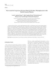
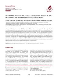
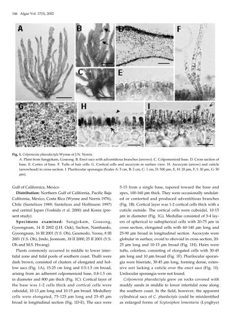
Fig. 1. Colpomenia phaeodactyla Wynne et J.N. Norris.<br />
A. Plant <strong>from</strong> Sangjokam, Goseong. B. Erect sacs with adventitious branches (arrows). C. Colpomenioid base. D. Cross section <strong>of</strong><br />
base. E. Cortex <strong>of</strong> base. F. Tufts <strong>of</strong> hair cells. G. Cortical cells and ascocysts in surface view. H. Ascocysts (arrow) and cuticle<br />
(arrowhead) in cross section. I. Plurilocular sporangia (Scales A: 5 cm, B: 2 cm, C: 1 cm, D: 500 µm, E, H: 20 µm, F, I: 30 µm, G: 50<br />
µm).<br />
Gulf <strong>of</strong> Californica, Mexico<br />
Distribution: Northern Gulf <strong>of</strong> California, Pacific Baja<br />
California, Mexico, Costa Rica (Wynne and Norris 1976),<br />
Chile (Santelices 1989; Santelices and H<strong>of</strong>fmann 1997)<br />
and central Japan (Yoshida et al. 2000) and <strong>Korea</strong> (present<br />
study).<br />
Specimens examined: Sangjokam, Goseong,<br />
Gyeongnam, 14 II 2002 (J.H. Oak), Sachon, Namhaedo,<br />
Gyeongnam, 16 III 2001 (Y.S. Oh), Geumodo, Yeosu, 8 III<br />
2001 (Y.S. Oh), Jindo, Jeonnam, 18 II 2000; 25 II 2001 (Y.S.<br />
Oh and M.S. Hwang).<br />
Plants commonly occurred in middle to lower intertidal<br />
zone and tidal pools <strong>of</strong> southern coast. Thalli were<br />
dark brown, consisted <strong>of</strong> clusters <strong>of</strong> elongated and hollow<br />
sacs (Fig. 1A), 15-25 cm long and 0.5-1.5 cm broad,<br />
arising <strong>from</strong> an adherent colpomenioid base, 0.8-1.5 cm<br />
in diameter and 800 µm thick (Fig. 1C). Cortical layer <strong>of</strong><br />
the base was 1-2 cells thick and cortical cells were<br />
cuboidal, 10-13 µm long and 10-15 µm broad. Medullary<br />
cells were elongated, 75-125 µm long and 25-45 µm<br />
broad in longitudinal section (Fig. 1D-E). The sacs were<br />
5-15 <strong>from</strong> a single base, tapered toward the base and<br />
apex, 100-160 µm thick. They were occasionally undulated<br />
or contorted and produced adventitious branches<br />
(Fig. 1B). Cortical layer was 1-2 cortical cells thick with a<br />
cuticle outside. The cortical cells were cuboidal, 10-15<br />
µm in diameter (Fig. 1G). Medullae consisted <strong>of</strong> 3-4 layers<br />
<strong>of</strong> spherical to subspherical cells with 20-75 µm in<br />
cross section, elongated cells with 60-140 µm long and<br />
25-90 µm broad in longitudinal section. Ascocysts were<br />
globular in surface, ovoid to obovoid in cross section, 20-<br />
25 µm long and 10-15 µm broad (Fig. 1H). Hairs were<br />
tufts, colorless, consisting <strong>of</strong> elongated cells with 30-45<br />
µm long and 10 µm broad (Fig. 1F). Plurilocular sporangia<br />
were biseriate, 30-45 µm long, forming dense, extensive<br />
sori lacking a cuticle over the erect sacs (Fig. 1I).<br />
Unilocular sporangia were not found.<br />
Colpomenia phaeodactyla grew on rocks covered with<br />
muddy sands in middle to lower intertidal zone along<br />
the southern coast. In the field, however, the apparent<br />
cylindrical sacs <strong>of</strong> C. phaedactyla could be misidentified<br />
as enlarged forms <strong>of</strong> Scytosiphon lomentaria (Lyngbye)