- Page 1 and 2: Third International Visual Field Sy
- Page 3 and 4: Third International visual i: ii~,d
- Page 5 and 6: CONTENTS E.L. Greve: Introduction .
- Page 7 and 8: Session IV. Methodology Chairman: H
- Page 9 and 10: of the R.N.L. may not be accompanie
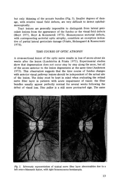
- Page 11 and 12: Docum. Ophthal. Proc. Series, Vol.
- Page 13 and 14: conduction disturbances. Their caus
- Page 15 and 16: Sofue 1975, Sommer, Miller, Pollack
- Page 17: areas but they can occur also in ot
- Page 21 and 22: metric strategies, it affords an ob
- Page 23 and 24: Docum. Ophthal. Proc. Series, Vol.
- Page 25 and 26: Table 1. Atrophic stages of maculop
- Page 27 and 28: VS=20/60 Fig. 3. Red-free fundus ph
- Page 29 and 30: I 20/20 l- 1.0 1.2- 20/25 ,- 20/30
- Page 31 and 32: It could be observed, in red-free f
- Page 33 and 34: VISUAL FIELD DEFECTS DUE TO TUMORS
- Page 35 and 36: 1. FIRST GROUP: THE SYMMETRIC TYPE
- Page 37 and 38: Table 4. Visual field defects due t
- Page 39 and 40: chiasm and above either of the opti
- Page 41 and 42: and 20/40 for the respective eyes t
- Page 43 and 44: advantages of applying other perime
- Page 45 and 46: Fig. 7. The pseud-macular sparing:
- Page 47 and 48: defect due to central integration o
- Page 49 and 50: Docum. Ophthal. Proc. Series, Vol.
- Page 51 and 52: disturbance of perception is visibl
- Page 53 and 54: Supraliminal stimuli still show a b
- Page 55 and 56: tion (Dannheim 1977, 1978). These f
- Page 57 and 58: Huber, A. Chiasmasyndrome - Klinik.
- Page 59 and 60: Table 1. Incidence of age, sex and
- Page 61 and 62: Table 3. Time interval between the
- Page 63 and 64: Table 5. Initial and final visual a
- Page 65 and 66: Table 7. State of the optic disc at
- Page 67 and 68: Visual field defects. Table 12 summ
- Page 69 and 70:
Effect of the time lag between the
- Page 71 and 72:
Table IS. Effect of systemic cortic
- Page 73 and 74:
In contrast to Group I, usually no
- Page 75 and 76:
series. The following case vividly
- Page 77 and 78:
Docum. Ophthal Proc. Series, Vol. 1
- Page 79 and 80:
well as cases due to Diabetes. As c
- Page 81 and 82:
a- b - ht:Mii,H Fig. 4. Typical def
- Page 83 and 84:
of both diseases, just as the infer
- Page 85 and 86:
of minor ONH therefore include a de
- Page 87 and 88:
Fig. 2. Visual field defect due to
- Page 89 and 90:
less complete lesions certainly are
- Page 91 and 92:
probably congenital hydrocephalus w
- Page 93 and 94:
area was involved as well. In 4 fie
- Page 95 and 96:
chiasma. Although transient papillo
- Page 97 and 98:
REFERENCES Bynke, H. & Heijl, A. Au
- Page 99 and 100:
*U. y: R-• 01.. 1Q69. V..T DY.ST
- Page 101 and 102:
98 y=’ -’ 40 cl.0 ,,' .4 ,' 3'
- Page 103 and 104:
REFERENCES Matsuda, H. & Nakabayash
- Page 105 and 106:
Perhaps we can bring this up in a c
- Page 107 and 108:
there have been no visual symptoms
- Page 109 and 110:
could understand supra-se&r meningi
- Page 111 and 112:
I found in pituitary adenoma and in
- Page 113 and 114:
Docum. Ophthal. Proc. Series, Vol.
- Page 115 and 116:
tided to compare the two eyes to se
- Page 117 and 118:
Fig. 4. (Lichter). Nucleus of a def
- Page 119 and 120:
Fig. 6. (Lichter). Nucleus of a def
- Page 121 and 122:
THE EARLY VISUAL FIELD DEFECT IN GL
- Page 123 and 124:
central tests were done with the be
- Page 125 and 126:
Fig, 4. Earliest field defect showi
- Page 127 and 128:
ences were normally distributed aro
- Page 129 and 130:
Docum. Ophthal. Proc. Series, Vol.
- Page 131 and 132:
was found. The first patients of th
- Page 133 and 134:
fibre bundle. This group has been f
- Page 135 and 136:
On the one hand the w.s.d. may be r
- Page 137 and 138:
REFERENCES Armaly, M.F. Selective p
- Page 139 and 140:
followed over a one-year period. So
- Page 141:
RESULTS Forty-nine out of 49 open a
- Page 144 and 145:
DISCUSSION AND CONCLUSIONS 1. 1. Al
- Page 146 and 147:
Table I. JR OD Month I.O.P. mm HG 1
- Page 148 and 149:
was almost always abnormal if the s
- Page 150 and 151:
perties. II. Dichoptic properties o
- Page 152 and 153:
until now had to watch for the appe
- Page 154 and 155:
In the following time pressure cont
- Page 156 and 157:
matous perimetric changes (Aulhorn
- Page 158 and 159:
Docum. Ophthal. Proc. Series, Vol.
- Page 160 and 161:
genital or closed angle glaucoma or
- Page 162 and 163:
60 50 40 30 20 10 0 MEDIANS bsx OHn
- Page 164 and 165:
those who developed glaucoma with t
- Page 166 and 167:
that did not yet show a break-throu
- Page 168 and 169:
CASE REPORTS Case 1.: a 52-year-old
- Page 170 and 171:
172 Goldmann-field Tiibinger nasal
- Page 172 and 173:
174 Friedman-fidd New Front Plate f
- Page 174 and 175:
Leblanc, R. Peripheral nasal field
- Page 176 and 177:
types of defects described, Goldman
- Page 178 and 179:
similar areas of the field. Additio
- Page 180 and 181:
Fig. 2. Visual field of the right e
- Page 182 and 183:
Table 1. The same type of defects s
- Page 184 and 185:
Docum. Ophthal. Proc. Series, Vol.
- Page 186 and 187:
tion tonometer) at time of initial
- Page 188 and 189:
treated eyes (5/61 or 8.2%) than in
- Page 190 and 191:
was not quite statistically signifi
- Page 192 and 193:
early, disappeared entirely. Howeve
- Page 194 and 195:
Docum. Ophthal. Proc. Series, Vol.
- Page 196 and 197:
The results of this investigation e
- Page 198 and 199:
30’ a) asb 32 10 32 100 320 1000
- Page 200 and 201:
OVER-ALL CONCLUSIONS Some degree of
- Page 202 and 203:
eye with a modified Goldmann-Weeker
- Page 204 and 205:
flow of axoplasm in the optic nerve
- Page 206 and 207:
Vanderburg, D. & Drance, S.M. Studi
- Page 208 and 209:
naga, Endo & Matsuo 1976). We initi
- Page 210 and 211:
up examinations. AU the eyes withou
- Page 212 and 213:
41 among 69 (59%), were located in
- Page 214 and 215:
oped relative field defects. In the
- Page 216 and 217:
utilizing the static method of peri
- Page 218 and 219:
Docum. Ophthal. Proc. Series, Vol.
- Page 220 and 221:
CASE PRESENTATIONS For the first tw
- Page 222 and 223:
DISCUSSION As a bundle of nerve fib
- Page 224 and 225:
REVERSIBILITY OF VISUAL FIELD DEFEC
- Page 226 and 227:
glaucoma with use of some agents bu
- Page 228 and 229:
Docum. Ophthal. Proc. Series, Vol.
- Page 230 and 231:
lyzer showed paracentral slight spo
- Page 232 and 233:
no disappearance. In 9 eyes progres
- Page 234 and 235:
closure glaucoma, in early stage id
- Page 236 and 237:
(Isayama & Tagami, 1977) in order t
- Page 238 and 239:
fiber bundle defects did not corres
- Page 240 and 241:
However, after the ocular pressure
- Page 242 and 243:
FllE3UENCY of FIWDIN6 EMly 6LAUCOYA
- Page 244 and 245:
gression. Each visual field was div
- Page 246 and 247:
DISCUSSIONS AND COMMENTS Aulhom dev
- Page 248 and 249:
Docum. Ophthal. Proc. Series, Vol.
- Page 250 and 251:
we 10 - 8- 0 eye 10 - 5- J-J o4 10
- Page 252 and 253:
REFERENCES Aulhom, E. & Harms, H. E
- Page 254 and 255:
Docum. Ophthal. Proc. Series, Vol.
- Page 256 and 257:
Docum. Ophthal. Proc. Series, Vol.
- Page 258 and 259:
no major effect of the water drinki
- Page 260 and 261:
Case b, right eye, 45" meridian +-+
- Page 262 and 263:
REFERENCES Aoyama, T. Pupillographi
- Page 264 and 265:
These authors of the second group d
- Page 266 and 267:
The behaviour of type 1) must be co
- Page 268 and 269:
isolated defect of the Bjerrum area
- Page 270 and 271:
choosing the direction of your prof
- Page 272 and 273:
How does one explain contraction on
- Page 274 and 275:
adequate way if we could understand
- Page 276 and 277:
analyse, you can see how well they
- Page 278 and 279:
(L. Ron&i and L. Barca. biological
- Page 280 and 281:
with a way to teach the clinician t
- Page 282 and 283:
Docum. Ophthal. Proc. Series, Vol.
- Page 284 and 285:
Docum. Ophthal. Proc. Series, Vol.
- Page 286 and 287:
The relevant quantities are defined
- Page 288 and 289:
The average number n of false scoto
- Page 290 and 291:
criteria will suffice for the separ
- Page 292 and 293:
Let us consider the two arrangement
- Page 294 and 295:
esults of an initial low-resolution
- Page 296 and 297:
practical applications, for patholo
- Page 298 and 299:
examination phase also provides inf
- Page 300 and 301:
Section: 1. Characters ‘READY’
- Page 302 and 303:
In order not to disturb the subject
- Page 304 and 305:
A B Fig. 5. Result with the semi-au
- Page 306 and 307:
Docum. Ophthal. Proc. Series, Vol.
- Page 308 and 309:
3 Fig. 2. Left visual field in a pa
- Page 311 and 312:
In the remaining 8 fields of 5 case
- Page 313 and 314:
Docum. OphthaI. Proc. Series, Vol.
- Page 315 and 316:
ROLE OF STANDARDISATION IN AUTOMATE
- Page 317 and 318:
REFERENCES Aulhorn, E Ueber die Aut
- Page 319 and 320:
Matsuo. Any other comment on Dr. Fa
- Page 321 and 322:
need an infinite number; I don’t
- Page 323 and 324:
not separate clearly between normal
- Page 325 and 326:
SUMMARY OF SESSION III: AUTOMATION
- Page 327 and 328:
RESULTS Visual fields were measured
- Page 329 and 330:
etinal edema was seen. The visual a
- Page 331 and 332:
DISCUSSION In the new fundus contro
- Page 333 and 334:
Fig. 8. Occlusion of arterial branc
- Page 335 and 336:
ACKNOWLEDGEMENTS The authors would
- Page 337 and 338:
NEW FUNOUS PHOTO PERIMITER-OPTICAL
- Page 339 and 340:
354 Fig. 4. Heredodegeneration of m
- Page 341 and 342:
_No. 9 Diag.NeuritiS optica retrobu
- Page 343 and 344:
Fig. 7 shows caecocentral scotoma p
- Page 345 and 346:
360 Fig. 1. (See text) Fig. 2. (See
- Page 347 and 348:
were obtained from several cases of
- Page 349 and 350:
significance of eye-rotation in per
- Page 351 and 352:
through an actual change in sensiti
- Page 353 and 354:
Docum. Ophthal. Proc Series, Vol 19
- Page 355 and 356:
angiography (FFA) and differential
- Page 357 and 358:
Fig. 2a. Results of FFA of case 2 (
- Page 359 and 360:
Case 4: A woman of 68 years was see
- Page 361 and 362:
-.. 378 Fig. 4.5. Results of FFA in
- Page 363 and 364:
3. indicating very sensitively the
- Page 365 and 366:
added distorting components are pre
- Page 367 and 368:
INVERSION Fig. 1. A tangent screen
- Page 369 and 370:
Case History VC is a 42-year-old Ca
- Page 371 and 372:
a=# 0 OTHERS * REDUCED SENSITIVITY
- Page 373 and 374:
gential errors has been made. In es
- Page 375 and 376:
that a functional anomaly is prefer
- Page 377 and 378:
Docum. Ophthal. Proc. Series, Vol.
- Page 379 and 380:
In the cases of this group, hemorra
- Page 381 and 382:
399
- Page 383 and 384:
are mainly represented by the PCS g
- Page 385 and 386:
oquine retinopathy, 2 eyes of centr
- Page 387 and 388:
Table 2. FVFA Profiles of PRD. 406
- Page 389 and 390:
REFERENCES Friedmann, AL The assesm
- Page 391 and 392:
Docum. Ophthal. Proc. Series, Vol.
- Page 393 and 394:
stimulus used by Verriest and Israe
- Page 395 and 396:
In Figure 3, threshold gradients ar
- Page 397 and 398:
Docum. Ophthal. Proc. Series, Vol.
- Page 399 and 400:
Matsuo: I would like to proceed to
- Page 401 and 402:
SUMMARY OF SESSION IV: METHODOLOGY
- Page 403 and 404:
puts in work all the processus inhe
- Page 405 and 406:
to the luminous conditions of backg
- Page 407 and 408:
MEASURES OF HOW WELL A PROCEDURE DI
- Page 409 and 410:
A general idea or model about the c
- Page 411 and 412:
Docum. Ophthal. Proc. Series, Vol.
- Page 413 and 414:
enough to understand the test. Fig.
- Page 415 and 416:
visual function is normal. Therefor
- Page 417 and 418:
ackground field up to 50’ of visu
- Page 419 and 420:
clearly isolated with this perimete
- Page 421 and 422:
15’ nasal IO 5’ 0’ 5” tempo
- Page 423 and 424:
Docum. Ophthal. Proc. Series, Vol.
- Page 425 and 426:
Fig 2, A new attachment (left) and
- Page 427 and 428:
and 6000 asb. in rabbit No. 2. Fig.
- Page 429 and 430:
Docum. Ophthal. Proc. Series, Vol.
- Page 431 and 432:
Case 2. Cihoretinal occlusion. Male
- Page 433 and 434:
detected by kinetic perimetry were
- Page 435 and 436:
Fig. 6. The thresholds of two prese
- Page 437 and 438:
Docum. Ophthal. PIOC. Series, Vol.
- Page 439 and 440:
peripheral parts of the retina, the
- Page 441 and 442:
2. Characteristics of Vertex Potent
- Page 443 and 444:
Regarding the characteristics of th
- Page 445 and 446:
Docum. Ophthal. Proc. Series, Vol.
- Page 447 and 448:
changes could be detected without P
- Page 449 and 450:
An isolated nasal PVF defect in the
- Page 451 and 452:
Docum. Ophthal. PIOC. Series, Vol.
- Page 453 and 454:
determination of the achromatic inc
- Page 455 and 456:
field ‘and also other methods, fo
- Page 457 and 458:
REPORT OF THE IPS RESEARCH GROUP ON
- Page 459 and 460:
white or colour perimetry. Lakowski
- Page 461 and 462:
heard. I am not a neophyte in Japan
- Page 463:
T. Aoyama, 265 M.F. Armaly, 177 E.