Chapter 3 Photosynthetic Reaction Centers - Biophysical Society
Chapter 3 Photosynthetic Reaction Centers - Biophysical Society
Chapter 3 Photosynthetic Reaction Centers - Biophysical Society

Create successful ePaper yourself
Turn your PDF publications into a flip-book with our unique Google optimized e-Paper software.
<strong>Chapter</strong> 3<br />
<strong>Photosynthetic</strong> <strong>Reaction</strong><br />
<strong>Centers</strong>:<br />
So little time, so much to do<br />
John H. Golbeck<br />
Department of Biochemistry<br />
and<br />
Molecular Biology<br />
The Pennsylvania State University<br />
University Park, PA 16802<br />
USA<br />
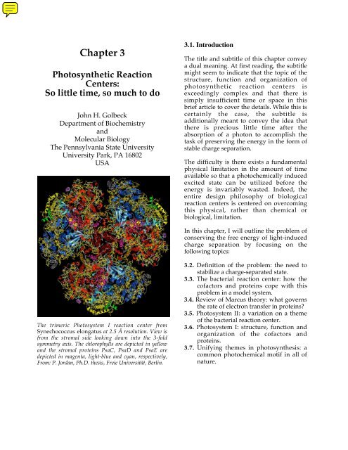
The trimeric Photosystem I reaction center from<br />
Synechococcus elongatus at 2.5 Å resolution. View is<br />
from the stromal side looking down into the 3-fold<br />
symmetry axis. The chlorophylls are depicted in yellow<br />
and the stromal proteins PsaC, PsaD and PsaE are<br />
depicted in magenta, light-blue and cyan, respectively,<br />
From: P. Jordan, Ph.D. thesis, Freie Universität, Berlin.<br />
3.1. Introduction<br />
The title and subtitle of this chapter convey<br />
a dual meaning. At first reading, the subtitle<br />
might seem to indicate that the topic of the<br />
structure, function and organization of<br />
photosynthetic reaction centers is<br />
exceedingly complex and that there is<br />
simply insufficient time or space in this<br />
brief article to cover the details. While this is<br />
certainly the case, the subtitle is<br />
additionally meant to convey the idea that<br />
there is precious little time after the<br />
absorption of a photon to accomplish the<br />
task of preserving the energy in the form of<br />
stable charge separation.<br />
The difficulty is there exists a fundamental<br />
physical limitation in the amount of time<br />
available so that a photochemically induced<br />
excited state can be utilized before the<br />
energy is invariably wasted. Indeed, the<br />
entire design philosophy of biological<br />
reaction centers is centered on overcoming<br />
this physical, rather than chemical or<br />
biological, limitation.<br />
In this chapter, I will outline the problem of<br />
conserving the free energy of light-induced<br />
charge separation by focusing on the<br />
following topics:<br />
3.2. Definition of the problem: the need to<br />
stabilize a charge-separated state.<br />
3.3. The bacterial reaction center: how the<br />
cofactors and proteins cope with this<br />
problem in a model system.<br />
3.4. Review of Marcus theory: what governs<br />
the rate of electron transfer in proteins?<br />
3.5. Photosystem II: a variation on a theme<br />
of the bacterial reaction center.<br />
3.6. Photosystem I: structure, function and<br />
organization of the cofactors and<br />
proteins.<br />
3.7. Unifying themes in photosynthesis: a<br />
common photochemical motif in all of<br />
nature.
E<br />
3.2. Definition of the Problem: The Need<br />
to Stabilize a Charge-Separated State.<br />
In this section, I will outline the problems<br />
associated with extracting free energy from<br />
a photochemically generated chargeseparated<br />
state.<br />
3.2.1. Definition of the Problem: The<br />
Absorption of Light<br />
The absorption of light by a chromophore<br />
occurs in ca. 10 -15 sec from the ground<br />
electronic state of a molecule (Figure 1). If,<br />
for the purpose of this discussion, we<br />
suppose that the second singlet excited state<br />
is populated, internal conversion will result<br />
in the loss of vibrational and rotational<br />
energy and the transfer of electronic energy<br />
to the first excited singlet state in ca. 10 -12<br />
sec. The residence time in the ground<br />
vibrational and rotational level of the first<br />
excited state is, in comparison, relatively<br />
long, up to 10 -8 sec, for chlorophylls.<br />
1 st<br />
Excited<br />
Triplet<br />
Ground<br />
State<br />
Delay prior to<br />
Phosphorescence<br />
10-4 - 102 s<br />
(Emission ˜ 10 -15 s)<br />
Intersystem<br />
Crossing<br />
10-8 s<br />
Internal<br />
Conversion<br />
Non-radiative<br />
decay<br />
102 - 10-11 s<br />
Absorption ˜ 10 -15 s<br />
Nonradiative decay ˜10 -8 s<br />
Delay prior to Fluorescence 10 -8 s<br />
2 nd<br />
Excited<br />
Singlet<br />
1 st<br />
Excited<br />
Singlet<br />
Figure 1. Lifetimes of absorption, fluorescence<br />
and phosphorescence at the equilibrium<br />
internuclear distance of the ground state. The<br />
energy levels are not depicted to scale.<br />
˜ 10-12 s<br />
(Emission ˜ 10 -15 s)<br />
2<br />
At this point, three processes compete for<br />
de-excitation: non-radiative decay (thermal<br />
processes), intersystem crossing (formation<br />
of triplet states), and fluorescence (reemission<br />
of a photon). In any given<br />
molecule, the process with the shortest<br />
lifetime will prevail. Hence, in the absence<br />
of a competing process, fluorescence<br />
represents the upper limit of an excited<br />
state lifetime. A corollary to this statement<br />
is that every molecule that absorbs light<br />
would fluoresce were it not for the existence<br />
of faster de-excitation processes.<br />
Take home lesson #1: In photosynthesis, the<br />
energy of the photon can be productively utilized<br />
only between the limits of absorption (10 -15 sec)<br />
and fluorescence (10 -8 sec) of the chlorophyll<br />
molecule.<br />
There exists a fourth possible fate for the<br />
singlet excited state: photochemistry, which<br />
is electron transfer to a separate molecule,<br />
producing a charge-separated (cationanion)<br />
pair. For this to happen, the excited<br />
state must be followed by a rapid electron<br />
transfer step, one that exceeds the rates of<br />
any de-excitation process.<br />
For a quantum yield ≥ 0.99 (definition: the<br />
ratio of the number of charge separated<br />
states divided by the number of photons<br />
used), this step must be several orders-ofmagnitude<br />
faster than 10 -8 sec if<br />
fluorescence is the primary de-excitation<br />
mode. Thus, charge separation must occur<br />
within ca. 10 -10 sec (100 ps) following the<br />
absorption of the photon. Clearly, any<br />
design of a photochemical reaction center<br />
that depends on simple diffusion chemistry<br />
between two molecules in solution will not<br />
succeed.<br />
Chlorophyll a is the major light absorbing<br />
and the sensitizing (S) chromophore in<br />
oxygenic photosynthetic systems, including<br />
Photosystem I and Photosystem II in<br />
cyanobacteria, algae and higher plants<br />
(Figure 2). In this role it has several major<br />
advantages; it absorbs light in the red and<br />
blue regions of the visible spectrum, it has a<br />
large molar extinction coefficient, and it has<br />
a high fluorescence yield.
Figure 2. Structure of chlorophyll a, the antenna<br />
and sensitizer molecule of Photosystem I. Note that<br />
pheophytin a lacks the central Mg 2+ atom;<br />
chlorophyll b has a –CHO group in place of the CH 3<br />
group in ring II; bacteriochlorophyll, a has a single<br />
bond in place of the double bond in ring II; bacteriopheophytin,<br />
a has a single bond in place of the<br />
double bond in ring II and lacks the central Mg 2+<br />
atom; and bacteriochlorophyll b has a –CHO group<br />
in place of the methyl group and a single bond in<br />
place of the double bond in ring II.<br />
Why should this matter? It does because the<br />
absorption of a photon by a molecule of<br />
chlorophyll a leads within 10 -12 sec to the<br />
lowest vibrational and rotational level of the<br />
first excited singlet state. The absence of<br />
non-radiative decay routes lengthens the<br />
excited state lifetime to the maximum<br />
allowed for the extraction of energy.<br />
This excited state is stable for up to 10 -8 sec,<br />
after which the molecule returns to the<br />
ground state with the emission of a photon.<br />
This is the maximum interval allowed for<br />
the extraction of work via photochemistry.<br />
In contrast, a non-fluorescent molecule<br />
would have a much shorter lifetime in the<br />
singlet excited state, making it even more<br />
difficult to extract the energy of the excited<br />
state in the time available.<br />
3<br />
Take home lesson #2: Chlorophyll a is an<br />
extremely good chromophore for photosynthetic<br />
systems because it is both an efficient absorber of<br />
light and because the excited state lifetime is<br />
long.<br />
Problem 1. If the energy in a photon, E = hn<br />
= hc/l where h is Planck's constant, and n,<br />
l and c are the frequency, wavelength, and<br />
speed of light, what is the energy, in Joules<br />
and electron-volts (eV), of the photon<br />
absorbed by (i) the bacterial<br />
photosynthetic reaction center special pair<br />
at its peak at 865 nm in the near-infrared<br />
region, and (ii) the Photosystem II reaction<br />
center at 680 nm in the visible region, and<br />
(iii) the Photosystem I reaction center at<br />
700 nm at the edge of the visible region?<br />
3.2.2. Definition of the Problem: Creation<br />
of Charge Separation<br />
In principle, an immobilized pair of donoracceptor<br />
molecules could be used for rapid<br />
electron transfer. In the following example,<br />
we will assume that the sensitizer also<br />
serves the role of electron donor:<br />
S – A + hu -> S* – A -> S + – A -<br />
The electron in the excited singlet state of<br />
the sensitizer S finds an electron deficient<br />
molecule within van der Waals distance,<br />
and electron transfer results, creating a<br />
reductant A - . Meanwhile, the hole left in the<br />
sensitizer results in the creation of an<br />
oxidant, S + . Thus, a charge-separated state<br />
S + - A - is produced.<br />
S + meanwhile could become reduced by a<br />
reductant in solution, A - could become<br />
oxidized by an oxidant in solution, and the<br />
chemical free energy of the photon could<br />
thereby be extracted to carry out useful<br />
work. The problem with this scheme is that<br />
S + and A - are in very close proximity,<br />
making A - the closest reductant to S + and<br />
the S + the closest oxidant to A - . Hence, the
charge-separated state would be shortlived.<br />
In practice, the charge-separated state has a<br />
several possible fates, including:<br />
S + – A - -> (S* – A) -> S – A + hu<br />
or<br />
S + – A - -> (S* – A) -> S – A + ∆<br />
where hn is a fluorescent photon and ∆ is<br />
heat released as vibrational and rotational<br />
energy.<br />
In biological reaction centers, the first<br />
charge-separated state between the<br />
chlorophyll donor and the chlorophyll<br />
acceptor has a lifetime of ca. 10 ns. Because<br />
diffusion chemistry is so much slower, a<br />
simple donor-acceptor pair is not very<br />
useful in a biochemical environment.<br />
3.2.3. Definition of the Problem:<br />
Stabilization of Charge Separation<br />
What can be done to solve the problem of<br />
the short lifetime of the charge-separated<br />
state? One solution is to extend the distance<br />
between S + and A - by adding a second<br />
closely spaced acceptor molecule, A 2:<br />
S – A 1 – A 2 + hu -> S* – A 1 – A 2 -><br />
S + – A 1 - – A2 -> S + – A 1 – A2 -<br />
What incentive would the electron have to<br />
move from A 1 - to A 2? This could happen by<br />
introducing a drop in Gibbs free energy<br />
between A 1 and A 2, thereby altering the<br />
equilibrium constant. While this results in a<br />
loss of thermodynamic efficiency (i.e. the<br />
free energy conserved as D + A - divided by<br />
the energy of the photon), this is the price<br />
that must be paid for an increased lifetime<br />
of the charged-separated state. A similar<br />
strategy might be to add a second closely<br />
spaced donor molecule, D:<br />
D – S – A1 + hu -> D – S* – A1 -><br />
-<br />
D – S + - +<br />
– A1 -> D – S – A1<br />
4<br />
In this example, the hole would be<br />
transferred from S + to D (which is<br />
equivalent to an electron transfer from D to<br />
S + ) if the equilibrium constant were<br />
similarly altered by introducing a drop in<br />
Gibbs free energy between D and S + .<br />
Take home lesson #3: an increased lifetime of the<br />
charge-separated state can be purchased by a loss<br />
of Gibbs free energy in a system that contains<br />
additional electron acceptors or donors.<br />
In summary, the working strategy of a<br />
photosynthetic reaction center involves:<br />
• Efficient absorption by a<br />
chromophore/sensitizer,<br />
• Rapid and efficient charge separation<br />
between the sensitizer and an acceptor,<br />
• Rapid charge delocalization with an<br />
second donor or acceptor to increase the<br />
lifetime of the charge-separated state.<br />
Therefore, the key to understanding the<br />
efficiency of photosynthetic electron<br />
transfer is the relationship between the rate<br />
of electron transfer and the parameters of<br />
distance, Gibbs free energy, and<br />
reorganization energy.<br />
3.3. The bacterial reaction center: how the<br />
cofactors and proteins cope with this<br />
problem in a model system.<br />
However, before we begin to examine this<br />
relationship in depth, we need to look at<br />
how a naturally occurring photosystem is<br />
constructed and how it operates. We shall<br />
use the bacterial reaction center (Rhodobacter<br />
sphaeroides) for this purpose because it is the<br />
simplest and best-understood model<br />
system.<br />
3.3.1. Global View of the Bacterial<br />
<strong>Reaction</strong> Center: Overall Function<br />
If charge separation is the goal of<br />
photochemical reaction centers, just what<br />
are the products of this reaction?
hn<br />
BPh<br />
Q A<br />
Cyt c<br />
P865<br />
ATP<br />
ADP + P i<br />
Q B<br />
H +<br />
Q<br />
QH 2<br />
H +<br />
H +<br />
Cyt c<br />
Cyt bc 1<br />
complex<br />
ATP Synthase<br />
Figure 3 Overall operation of the bacterial reaction<br />
center. The design philosophy is to promote a series<br />
of embedded cycles, one involving electrons and<br />
another involving protons. The reaction center<br />
reduces quinone to quinol, and the cytochrome bc 1<br />
complex oxidizes quinol to quinone. During this<br />
process, four protons are translocated across the<br />
membrane, producing an electrochemical gradient.<br />
The gradient represents the stored energy of the<br />
photon, which is used by ATP synthase to produce<br />
the final product of bacterial photosynthesis, ATP.<br />
The short answer is that the bacterial<br />
reaction center reduces ubiquinone to<br />
dihydroubiquinol in a reaction that<br />
consumes two protons on the inside of a<br />
closed biological membrane (Figure 3). The<br />
dihydroubiquinol is oxidized by the<br />
cytochrome bc 1 complex (in a<br />
mechanistically complex reaction termed<br />
the protonmotive Q-cycle) in a reaction that<br />
translocates up to four protons to the<br />
outside of the membrane. The resulting<br />
electrochemical proton gradient (∆m H+)<br />
represents stored free energy derived from<br />
the photon. [See <strong>Chapter</strong> 1 in this textbook,<br />
section 1-8,4]. The energy in the<br />
electrochemical proton gradient is<br />
converted to chemical bond energy in the<br />
synthesis of adenosine triphosphate (ATP)<br />
by the membrane-bound ATP-synthase<br />
enzyme.<br />
H +<br />
5<br />
3.3.2. Global View of the Bacterial<br />
<strong>Reaction</strong> Center: Internal Operation<br />
The bacterial reaction center uses a strategy<br />
of transferring the electron from the<br />
primary electron acceptor, bacteriopheophytin<br />
a, to two additional electron<br />
acceptor molecules, termed Q A and Q B, to<br />
stabilize charge separation in a suitable time<br />
domain. Both are quinones (Figure 4).<br />
A soluble reduced cytochrome c 2+ reduces<br />
the oxidized bacteriochlorophyll sensitizer<br />
with a peak absorbance at 865 nm, termed<br />
P865, thereby preventing the charge<br />
+<br />
recombination between oxidized P865 and<br />
-<br />
the electron QB . The net effect is that<br />
-<br />
reduced QB has no oxidized donor in close<br />
proximity, thus allowing for a long-lived<br />
-<br />
product P865 QA QB . The oxidized<br />
cytochrome c 3+ molecule diffuses from the<br />
membrane-bound reaction center and<br />
accepts an electron from the cytochrome bc 1<br />
complex.<br />
Figure 4 Structures of oxidized phylloquinone, the<br />
intermediate electron acceptor in Photosystem I,<br />
and oxidized plastoquinone, the intermediate<br />
electron acceptor in Photosystem II. Note the<br />
naphthoquinone and benzoquinone head groups and<br />
the extended phytyl-like tail.<br />
3.3.3. Global View of the Bacterial<br />
3.3.3. Global View of the <strong>Reaction</strong> Center:<br />
Operation of the Cycle<br />
The first photon absorbed leads to the<br />
charge-separated state (Figure 5):<br />
-<br />
P865 QA QB The second photon absorbed leads to the<br />
unstable charge-separated state:<br />
- -<br />
P865 QA QB
One proton is taken from the medium as the<br />
electron moves from Q A to Q B, resulting in:<br />
P 865 Q A (Q BH) -<br />
A second proton is subsequently taken up<br />
from the medium, resulting in:<br />
P 865 Q A (Q BH 2)<br />
The doubly reduced and protonated<br />
ubiquinol QH 2 has a low affinity for the Q B<br />
binding site and diffuses in the membrane<br />
to the cytochrome bc 1 complex. The Q B<br />
binding site has a high affinity for oxidized<br />
ubiquinone, which diffuses in the<br />
membrane from the cytochrome bc 1<br />
complex.<br />
The net result is that the Q B site is<br />
‘recharged’ with fresh quinone, thereby<br />
allowing another round of light-induced<br />
reduction of ubiquinone to ubiquinol.<br />
H +<br />
QH 2<br />
P685 Q A (Q BH 2 )<br />
P685 Q A (Q BH) -<br />
H +<br />
Q<br />
P685 Q A Q B<br />
hn<br />
P685 Q A - QB -<br />
Cyt c 2+<br />
Cyt c 3+<br />
P685 Q A - QB<br />
P685 Q A Q B -<br />
Cyt c 3+<br />
Cyt c 2+<br />
Figure 5. Cyclic operation of the bacterial reaction<br />
center. Note that Q A is only capable of a single<br />
electron reduction to the semiquinone radical<br />
state, whereas Q B is capable of double reduction<br />
followed by double protonation to the fullyreduced<br />
hydroquinone state. The reduced<br />
hydroquinone is loosely bound to its site and is<br />
displaced by an oxidized quinone for another<br />
round of light-induced turnover.<br />
6<br />
3.3.4. Global View of the Bacterial<br />
<strong>Reaction</strong> Center: Cofactor Arrangement<br />
The atomic resolution x-ray crystal structure<br />
of the purple bacterial reaction center from<br />
Rhodobacter sphaeroides (PDB entry 1AIJ)<br />
shows that the L and M subunits form a<br />
heterodimeric core of membrane-spanning<br />
a-helices arranged along a pseudo-C 2<br />
symmetry axis (Figure 6). The most striking<br />
feature of the structure is that that there are<br />
no charged amino acids within the<br />
membrane-spanning a-helices. The electron<br />
transfer components are embedded entirely<br />
within the hydrophobic core.<br />
Figure 6 Depiction of the Rhodobacter sphaeroides<br />
reaction center. The a-helices are depicted in<br />
red/yellow; b-sheets in cyan; the bacteriochlorophyll<br />
a and bacteriopheophytin a molecules in green; and<br />
the non-heme iron in blue. The menbrane thickness<br />
can be estimated by the length of the membranespanning<br />
a-helices. From the Jena Image Library<br />
for Biological Molecules; http://www.imb-<br />
The most striking feature of the structure is<br />
jena.de/IMAGE.html<br />
As shown in Figure 7, two molecules of<br />
bacteriochlorophyll a function in a dimer<br />
configuration as the primary electron<br />
donor. A bridging bacteriochlorophyll a<br />
molecule serves as a transient intermediate<br />
electron acceptor, which is difficult to<br />
observe spectroscopically because of the<br />
short lifetime, and a bacteriopheophytin a<br />
serves as the primary electron acceptor. A
tightly bound ubiquinone, Q A, is the<br />
secondary electron acceptor, and a mobile<br />
ubiquinone, Q B, is the terminal electron<br />
acceptor.<br />
Spectroscopic and structural studies show:<br />
• The electron transfer cofactors are very<br />
closely spaced.<br />
• Only one branch of cofactors functions<br />
in electron transfer from P 865 to Q B.<br />
• The non-heme iron can be replaced with<br />
Zn 2+ without loss of function.<br />
As shown in Figure 7, the approximate<br />
Figure 7. Cofactors of the bacterial reaction<br />
center. The edge-to-edge distances between the<br />
cofactors are depicted along with the electron<br />
transfer rates. The distances are only<br />
approximate and refer to the closest approach of<br />
the aromatic rings (sufficiently accurate for the<br />
purpose of this discussion). Note the pseudo C 2<br />
axis of symmetry that spans the<br />
bacteriochlorophyll a dimer and the non-heme<br />
iron.<br />
As shown in Figure 7, the approximate<br />
edge-to-edge distance between the<br />
bacteriochlorophyll a dimer, P 865, and the<br />
bridging bacteriochlorophyll a molecule is<br />
ca. 6 Å, and the distance between the<br />
bridging bacteriochlorophyll a and the<br />
bacteriopheophytin a acceptor is ca. 5 Å.<br />
7<br />
The electron is transferred between P865* and the bacteriopheophytin a acceptor, a<br />
distance of ca. 10 Å, in 2.8 ps. The<br />
bacteriopheophytin a acceptor is separated<br />
from QA by ca. 10 Å, and the electron is<br />
transferred in 200 ps. Finally, the electron is<br />
-<br />
transferred from QA to QB in 100 µs over a<br />
distance of 15 Å.<br />
Clearly, there is a relationship between<br />
distance and rate of electron transfer. One<br />
can raise the following two questions: what<br />
factors govern the rate of each electron<br />
transfer step, and what is the role of the<br />
protein?<br />
Problem 2. The fastest chemical reaction<br />
known in biology is the first electron<br />
transfer in bacterial photosynthesis, in<br />
which an initial distance of ca. 6 Å is<br />
covered in a time period of 2.8 ps. The next<br />
fastest biological reaction is the electron<br />
transfer step from reduced BPh - to Q A, in<br />
which a distance of ca. 10 Å is covered in a<br />
time period of 200 ps. Calculate the<br />
average speed of these electron transfer<br />
steps in km/h. How fast is this relative to<br />
the speed of light (10 8 m sec -1 ), the cruise<br />
speed of a Boeing 747 (1000 km h -1 ) or a<br />
fast walk (6 km h -1 )?<br />
Why does speed matter in photosynthetic<br />
systems? The answer has to do with our<br />
previous finding that the initial electron<br />
transfer process must occur faster than the<br />
fluorescence lifetime of the singlet excited<br />
state of the chlorophyll sensitizer molecule.<br />
In practice, this means that the electron<br />
transfer cofactors must be exceedingly close<br />
together in photosynthetic systems<br />
3.4. Review of Marcus Theory: What<br />
Governs the Rate of Electron Transfer in<br />
Proteins?<br />
We will now briefly review the relationship<br />
between rate, distance, and driving force in<br />
biological electron transfer reactions.
3.4.1. Review of Marcus Theory: Overview<br />
of Proteins<br />
With metal ions in solution, electron<br />
transfer occurs when the two are in van der<br />
Waals contact, but with metal cofactors in<br />
proteins, electron transfer must occur over<br />
distances up to 15 Å or greater. Rates of<br />
chemical reactions are described along a<br />
potential energy surface in traditional<br />
transition state theory. As reactants gain<br />
energy from thermal collisions, they<br />
overcome an activation energy barrier to<br />
achieve the transition state, after which they<br />
spontaneously decay to product.<br />
In traditional theory, the formation of the<br />
transition state invariably leads to product.<br />
This is a so-called ‘adiabatic’ reaction. In<br />
thermodynamics, this term is used to<br />
indicate that no heat flows into or out of a<br />
system, but in electron transfer theory, it<br />
means that the system makes no transition<br />
to other states, remaining on the lowerorder<br />
electronic surface.<br />
In traditional theory, electron transfer<br />
occurs at the intersection of the two<br />
potential energy surfaces, a condition<br />
attained by thermal fluctuations of the two<br />
nuclei. The horizontal displacement<br />
represents the adjustment of the nuclear<br />
coordinates to the different electronic<br />
configurations for the reactant (D - A) and<br />
product (D A - ).<br />
In proteins, due to the fact that the electron<br />
transfer must occur between metals<br />
cofactors that are separated by large<br />
distances, an additional electronic term is<br />
required for electron transfer, the ‘electronic<br />
matrix coupling element’. This term<br />
effectively removes the degeneracy of the<br />
reactant and product states at the<br />
intersection.<br />
3.4.2. Review of Marcus Theory: Fermi’s<br />
Golden Rule<br />
Fermi’s Golden Rule applies to long-range<br />
electron transfer processes in proteins, in<br />
which the electron donors and acceptors are<br />
8<br />
only weakly coupled due to their longdistance<br />
separation. These are so-called<br />
‘non-adiabatic reactions’:<br />
k et µ 2p/h * |H AB| 2 * (FC) (1)<br />
where k et is the electron transfer rate. h is<br />
Planck’s constant |H AB| is the electronic<br />
matrix coupling element, and FC is the<br />
Franck-Condon factor.<br />
The rule has two terms. The first term,<br />
|H AB| 2 , states that the electron transfer rate<br />
is proportional to the square of the weak<br />
coupling of the reactant and product<br />
electronic states. The donor and acceptor<br />
wave functions must span the space<br />
separating the donor and acceptor<br />
molecules; hence, this parameter concerns<br />
the variable of distance. The overlap of<br />
wavefunctions falls off exponentially with<br />
edge-to-edge distance between cofactors,<br />
modified by a term that is different for free<br />
space, proteins, and covalently bound<br />
molecules. We will examine this factor more<br />
closely later.<br />
The second term, the Franck Condon factor,<br />
relates to the nuclear position of the reactant<br />
molecules and their environment, and is<br />
similar to traditional transition state theory.<br />
This term relates to the overlap of the<br />
reactant and product harmonic oscillators,<br />
and includes such factors as the effect of<br />
temperature, the reorganization energy, and<br />
the Gibbs free energy difference between<br />
the products and reactants. We shall<br />
examine this term first.<br />
3.4.3. Review of Marcus Theory: The<br />
Franck-Condon Term<br />
Marcus used harmonic oscillators as<br />
descriptions of electron donors and<br />
acceptors. However, instead of using two<br />
separate potential energy surfaces for the<br />
reactants, he used a single surface to<br />
describe the potential energy of the<br />
precursor complex as a function of its<br />
nuclear configuration (Figure 8).
Figure 8. Intersecting parabolic potential<br />
energy surfaces used to represent classical<br />
reactant and product harmonic oscillators for<br />
three electron transfer reactions with different<br />
free energies. Note that in the top panel, -∆G 0<br />
< l, in the middle panel -∆G 0 = l and in the<br />
lower panel -∆G 0 > l. The quantum<br />
mechanical harmonic oscillator wavefunctions<br />
are superimposed on these parabolas; the fastest<br />
electron transfer occurs at maximum overlap.<br />
The optimum rate of electron transfer therefore<br />
occurs when -∆G 0 = l. Reproduced with<br />
permission from Moser et al. 1992;<br />
http://www.nature.com/<br />
The latter includes its rotational,<br />
vibrational, and translational motion<br />
including those of the molecules in the<br />
surrounding solution. In keeping with this<br />
idea, a single surface was used for the<br />
product complex. Marcus recognized that<br />
the free energy of activation, ∆G ‡<br />
, has a<br />
9<br />
quadratic dependence on the reorganization<br />
energy (l) and on the standard Gibbs free<br />
energy difference between the products and<br />
reactants (∆G 0 ) according to the expression<br />
∆G ‡<br />
µ [-(∆G 0 + l) 2 /4l]. The rate of electron<br />
transfer, ket, is related to these terms<br />
according to the following equation:<br />
k et µ exp [-(∆G 0 + l) 2 /4lk BT] (2)<br />
where k B is Boltzmann’s constant, and T is<br />
the absolute temperature. [For derivation of<br />
the Franck-Condon term, see <strong>Chapter</strong> I,<br />
section I-13 in this textbook].<br />
The expression for ∆G ‡<br />
defines a parabola.<br />
As -∆G 0 increases, the rate of electron<br />
transfer increases, but only until -∆G 0 = l, at<br />
which point the rate is maximum. As -∆G 0<br />
increases further, the rate of electron<br />
transfer decreases. There are hence three<br />
regions to the parabola. In the ‘normal’<br />
region, -∆G 0 < l, and ket increases if -∆G 0<br />
increases or if l decreases. At the optimum, -<br />
∆G 0 = l, ket is maximum. In the ‘inverted’<br />
region, -∆G 0 > l , and ket decreases if -∆G 0<br />
increases or if l decreases.<br />
In Figure 9, the rate of electron transfer is<br />
shown as a function of ∆G 0 for two<br />
photosynthetic reactions; the<br />
physiologically-useful electron transfer over<br />
ca. 10 Å from BPh - to Q A (top), and the<br />
physiologically-unproductive backreaction<br />
over 22 Å from Q A - to P + .<br />
An analysis over a range of temperatures<br />
suggests a constant value of l of 0.7 eV in<br />
proteins, and a common hw of 70 meV<br />
(basically, the width of the parabola), which<br />
is over twice the 25 meV Boltzmann energy<br />
(kT) available at room temperature.<br />
Although I will not discuss this parameter<br />
further, the high frequency protein<br />
vibrational modes are important because<br />
they are likely to be coupled to electron<br />
transfer.<br />
The most striking feature of this plot is the<br />
relative insensitivity of the electron transfer<br />
rate to the thermodynamic driving force,<br />
especially at values near the optimum rate.
Take home lesson #4: Nature does not appear to<br />
rely primarily on altering the Gibbs free energy<br />
between product and reactant to modulate the<br />
rate of electron transfer in proteins.<br />
Figure 9. Free energy dependence on electron<br />
transfer rate in the bacterial reaction center.<br />
The Gibbs free energy was changed by<br />
replacing the native Q A with compounds<br />
with different redox potentials. The fits are<br />
with l = 0.7 eV and with hw = 150 meV<br />
(dotted), 70 meV (solid, 30 meV (large,<br />
dashed) and 10 meV (small, dashed).<br />
Reproduced with permission from Moser et<br />
al., 1992; http://www.nature.com/<br />
Problem 3. Calculate the drop in<br />
Gibbs free energy that would be<br />
needed to accelerate the electron<br />
transfer rate between two cofactors<br />
by a factor of 10, given l = 0.7 eV in<br />
proteins and no change in distance or<br />
reorganization energy.<br />
10<br />
3.4.4. Review of Marcus Theory: The<br />
Electronic Coupling Term<br />
The electronic coupling term depends on<br />
the distance between the electron donor and<br />
electron acceptor, and the nature of the<br />
intervening medium. The equation for the<br />
electronic coupling term is:<br />
| H AB| 2 µ exp (-bR) (3)<br />
where H AB is the coupling probability at<br />
close contact, R is the edge-to-edge distance<br />
between the electron donor and the electron<br />
acceptor, and depends on the nature of the<br />
intervening medium (Figure 10). b is 0.7 Å -1<br />
in covalently bonded molecules, 1.4 Å -1 in<br />
proteins and 2.8 Å -1 in a vacuum.<br />
Figure 10. Free energy optimized rate vs edge-toedge<br />
distance relationships for electron transfer<br />
in (top) the photosynthetic system and (bottom)<br />
in the Ru-cyt c and Ru-Mb system. The vertical<br />
line represents van der Waals contact.<br />
Reproduced with permission from Moser et al.,<br />
1992; http://www.nature.com/
In proteins, this corresponds to a 10-fold<br />
change in the rate of electron transfer for<br />
every 1.7 Å of distance. Therefore, unlike<br />
the weak contribution by the Franck-<br />
Condon term, the rate of electron transfer in<br />
proteins is strongly sensitive to the<br />
electronic coupling term.<br />
Take home lesson #5: Biological systems take<br />
good advantage of altering the distance between<br />
product and reactant to modulate the rate of<br />
electron transfer in proteins.<br />
3.4.5. Review of Marcus Theory: The<br />
Moser-Dutton Simplification<br />
Given the finding of a constant hw = 70 meV<br />
and l = 0.7 eV in proteins, Moser and<br />
Dutton (Moser et al. 1992) simplified the<br />
equation to:<br />
k et = 10 ^ [15 - 0.6R – 3.1(∆G 0 + l) 2 /l] (4)<br />
or<br />
log 10 k et = 15 - 0.6R – 3.1(∆G 0 + l) 2 /l (5)<br />
This simplified equation states that when<br />
electron transfer cofactors are at van der<br />
Waals distance, electron transfer rates are<br />
10 15 sec -1 .<br />
There are three correction terms. One<br />
involves distance: as the spacing between<br />
electron transfer cofactors increases, the<br />
logarithm of the rate decreases by 0.6*R,<br />
where R is the edge-to-edge distance. The<br />
second involves the difference in the<br />
standard Gibbs free energy between<br />
reactants and products, and the final term<br />
involves the reorganization energy, l. Note<br />
that when -∆G 0 = l, the rate is optimum;<br />
any increase or decrease in Gibbs free<br />
energy relative to the reorganization energy<br />
leads to a lower (suboptimal) rate of<br />
electron transfer.<br />
Thus, one can calculate the optimum rate of<br />
electron transfer (to within an order-ofmagnitude)<br />
by simply knowing the edge-toedge<br />
distance between donor and acceptor<br />
pairs (Figure 11).<br />
11<br />
10 10<br />
10 7<br />
10 4<br />
10 1<br />
10 -2<br />
Time, s<br />
10 -5<br />
10 -8<br />
10 -11<br />
10 -14<br />
5<br />
1 century<br />
1 yr<br />
1 h<br />
1 s<br />
1 ms<br />
1 µs<br />
1 ns<br />
10<br />
Note that when the edge-to-edge distance<br />
between cofactors is 10 Å, the electron<br />
transfer rate is ca. 1 ns. Doubling this<br />
distance to 20 Å results in an electron<br />
transfer rate of ca. 1 ms, and a distance of 30<br />
Å results in a rate of almost an hour.<br />
This relationship may explain, in part, why<br />
an energy-transducing biological membrane<br />
is >30 Å in thickness: charge recombination<br />
between an oxidized cofactor on one side<br />
and a reduced cofactor on the other side of<br />
the membrane must be prevented on<br />
timescales that these cofactors carry out<br />
their respective biochemical roles in<br />
metabolism.<br />
To summarize:<br />
15<br />
20 25<br />
Distance, Å<br />
Figure 11. The Moser-Dutton simplification of<br />
the Marcus relationship: electron transfer rate<br />
in proteins as a function of distance, assuming<br />
optimal conditions, i.e. –∆G 0 = l.<br />
• b is usually considered constant in<br />
proteins, although a more sophisticated<br />
treatment is available that takes into<br />
account variations of b in proteins (see<br />
below).<br />
• ∆G 0 can modulate the rate up to 10 5 fold<br />
over the available range of 0 to 1.3 eV in<br />
biochemical systems. In reality, however,<br />
∆G 0 is usually constrained by the need to<br />
conserve free energy,<br />
30<br />
35<br />
40<br />
45
• l is difficult to measure experimentally<br />
but is typically assumed to be 0.7 eV in a<br />
protein mileu.<br />
• R, however, is a free variable that is<br />
effective over a 10 12 fold range with few<br />
restrictions.<br />
A more complete treatment has been<br />
developed (Page et al., 1999) that takes into<br />
account variations in polypeptide structure.<br />
This equation includes a packing density (r)<br />
of protein atoms in the volume between<br />
redox centers to account for variations in b:<br />
Log 10k et=13–(1.2–0.8r)(R–3.6)–3.1(∆G 0 +l) 2 /l<br />
where r is the fraction of the volume<br />
between redox cofactors that is within the<br />
united van der Waals radius of intervening<br />
atoms.<br />
Thus, although the fundamental concepts of<br />
electron transfer have been refined and<br />
expanded in recent years, equation (4) will<br />
be sufficient to make important points<br />
regarding the placement of cofactors in<br />
biological systems.<br />
Take home lesson #6: In bioenergetic complexes,<br />
metal centers and organic cofactors exist in part<br />
to cope with the problem of long-distance<br />
electron transfer across membranes.<br />
3.4.6. Review of Marcus Theory:<br />
Implications for <strong>Photosynthetic</strong> Systems<br />
If a 40 Å biological membrane needs to be<br />
spanned, such as in the chloroplast or<br />
mitochondria, meaningful rates are possible<br />
only if several intervening cofactors are<br />
involved. This is why all transmembrane<br />
redox complexes use multiple cofactors.<br />
As we have seen, photosynthetic systems<br />
are highly constrained. Electron transfer<br />
rates must be achieved that exceed the 10 -8 s<br />
limit imposed on the excited state of the<br />
sensitizer by fluorescence. Given a 99%<br />
quantum yield, electron transfer rates must<br />
exceed 10 -01 s. This can only be<br />
accomplished by placing the primary<br />
12<br />
electron acceptor a very short distance from<br />
the excited sensitizer in the initial stage of<br />
charge separation.<br />
If we look at the placement of cofactors in<br />
the bacterial reaction center, we see that this<br />
condition is met. Looking back to Figure 7,<br />
we see that the distance between the<br />
bacteriochlorophyll special pair, P 865, and<br />
the bridging bacteriochlorophyll is ca. 6 Å,<br />
and the distance between the bridging<br />
bacteriochlorophyll and the primary<br />
acceptor bacteriopheophytin a, BPh, is ca. 5<br />
Å. The rate of electron transfer between P 865<br />
and BPh has been measured to be 2.8 ps,<br />
which corresponds roughly to the sum of<br />
the optimal rates of electron between these<br />
three components. The distance between<br />
BPh and Q A is 10 Å and the electron transfer<br />
rate is 200 ps, which again is roughly in line<br />
with the 1 ns time predicted in Figure 11.<br />
Thus, the reason for the need for the earliest<br />
steps of electron transfer to be faster than<br />
10 -8 s is accomplished by placing the<br />
electron transfer cofactors in exceedingly<br />
close contact. No other multicofactor<br />
enzyme, to my knowledge, places cofactors<br />
at these close distances.<br />
Problem 5. Using the relationship between<br />
optimal rates and edge-to-edge distance<br />
for of long-distance electron transfer in<br />
proteins, calculate how many electron<br />
transfer cofactors would be needed to span<br />
a 40 Å thick biological membrane in a time<br />
period of 10 µs. Assume for the purpose of<br />
this argument each cofactor is a transition<br />
metal with a radius of 1.5 Å.<br />
The question arises whether there is a role<br />
for the Franck-Condon factor in<br />
photosynthetic electron transfer. It is true<br />
that each step is accomplished with a drop<br />
in Gibbs free energy, but this may simply be<br />
to ensure that the equilibrium is biased<br />
toward product formation. However, each<br />
forward electron transfer step also competes<br />
with an inherent charge recombination step.
+ -<br />
For example, P865 recombines with QA with<br />
a half-time of about 100 ms. The -∆G o is<br />
very large for this reaction, and this may<br />
have the otherwise-paradoxical effect of<br />
pushing the reaction into the ‘inverted’<br />
Marcus region. This would tend to slow<br />
down the charge recombination rate,<br />
thereby further guaranteeing that forward<br />
electron transfer remains highly efficient.<br />
Whether this actually happens in any<br />
known photochemical reaction center,<br />
however, is still the topic of discussion.<br />
3.5. Photosystem II: a variation on a theme<br />
of the bacterial reaction center.<br />
We will now examine the components and<br />
electron transfer process in Photosystem II.<br />
3.5.1. Details of Quinone-Type <strong>Reaction</strong><br />
<strong>Centers</strong>: Photosystem II<br />
The purple bacterial reaction center shares a<br />
striking similarity to Photosystem II of<br />
plants and cyanobacteria. Electron transfer<br />
on the acceptor side is virtually identical in<br />
that plastoquinone (see Figure 4) is reduced<br />
to dihydroplastoquinol at the Q B site (Figure<br />
12), which subsequently leaves the binding<br />
site to interact with the cytochrome b 6f<br />
complex. An oxidized plastoquinone that<br />
has returned from the cytochrome b 6f<br />
reoccupies the Q B site for another round of<br />
light-induced reduction. Because the<br />
tertiary acceptor is a mobile quinone, both<br />
the bacterial reaction center and<br />
Photosystem II are termed ‘Type II’ reaction<br />
centers.<br />
The location of polypeptides in the 3.8 Å Xray<br />
crystal structure of Photosystem II (PDB<br />
entry 1FE1) is depicted in Figure 12. Note<br />
the presence of the five core transmembrane<br />
a-helices A through E on the D1 and D2<br />
proteins that together comprise the<br />
heterodimer, and the presence of six ahelices<br />
on CP43 and CP47 that comprise the<br />
antenna proteins. It is interesting that the<br />
D1/D2 heterodimeric proteins of<br />
Photosystem II overlay almost perfectly the<br />
13<br />
core heterodimeric proteins of the bacterial<br />
reaction center.<br />
Nonetheless, electron transfer on the<br />
oxidizing side is different from the bacterial<br />
reaction center in that the primary electron<br />
donor, P680, has a redox potential in excess<br />
of +1V, and a<br />
Figure 12. The components of the Photosystem II<br />
reaction center depicted with redox potential on the<br />
y-axis and rate of electron transfer on the x-axis.<br />
Note the similarity in the identity of the cofactors<br />
and the electron transfer rates with the bacterial<br />
reaction center.<br />
Nonetheless, electron transfer on the<br />
oxidizing side is different from the bacterial<br />
reaction center in that the primary electron<br />
donor, P680, has a redox potential in excess<br />
of +1V, and a Mn 4 cluster exists that<br />
oxidizes two water molecules to dioxygen<br />
in a reaction that requires four photons. A<br />
redox-active tyrosine (Y Z) links P680 + to the<br />
Mn 4 cluster and, in cooperation with a<br />
nearby histidine residue, participates in<br />
moving the released protons to the aqueous<br />
phase. The details of the Mn 4 cluster and the<br />
chemistry of water oxidation are not well<br />
understood. A model of the Mn 4 wateroxidizing<br />
cluster is depicted in Figure 14<br />
superimposed on the 3.5 Å electron density<br />
map of Photosystem II (Zouni et al. 2001).<br />
Because of the oxidation of water and the<br />
release of dioxygen, the Photosystem II<br />
reaction
Figure 13. Model for Photosystem II from<br />
Synechococcus elongatus based on the X-ray crystal<br />
structure at 3.8 Å. a) Top view from the lumenal side<br />
showing polypeptides A through E on D1 and D2, the<br />
reaction center core proteins. Also depicted are the<br />
peripheral antenna proteins CP43 and CP47, each of<br />
which consists of six membrane spanning a-helices. b)<br />
Side view showing the membrane-spanning subunits<br />
as well as the Mn cluster, the PsbO protein and the<br />
bound cytochrome. Reproduced with permission from<br />
Zouni et al., 2001; http://www.nature.com/<br />
/<br />
14<br />
center does not operate in a cycle as does<br />
the bacterial reaction center. Rather, the<br />
released protons from water oxidation are<br />
deposited on one side of the thylakoid<br />
membrane to help build the electrochemical<br />
gradient.<br />
The electrons derived from water pass<br />
through the cytochrome b 6f complex and<br />
find their way to Photosystem I via a small,<br />
copper-containing protein named<br />
plastocyanin (under copper-limiting<br />
conditions, cytochrome c 6 can substitute for<br />
plastocyanin in certain cyanobacteria and<br />
algae). Photosystem I ultimately promotes<br />
these electrons to a reduction level sufficient<br />
to reduce NADP + . The electron transfer<br />
pathway in plants and cyanobacteria is<br />
hence linear, with Photosystem II and<br />
Photosystem I operating in series.<br />
Figure 14. Model for the Mn 4 cluster in<br />
Photosystem II from Synechoccus elongatus<br />
based on the 3.8 Å x-ray crystal structure and<br />
on EXAFS data. Reproduced with permission<br />
from Zouni et al., 2001;<br />
http://www.nature.com/<br />
Take home message #7: In higher plant, algal<br />
and cyanobacterial photosynthesis, Photosystem<br />
II functions as a non-cyclic photochemical<br />
reaction center, removing electrons from water<br />
and transferring them through the cytochrome<br />
b 6f complex to Photosystem I, which ultimately<br />
reduces NADP + .
3.6. Photosystem I: Structure, Function and<br />
Organization of Cofactors and Proteins.<br />
With the description of Marcus theory and<br />
Type II reaction centers as background, we<br />
now move to an analysis of Photosystem I.<br />
3.6.1. Details of Photosystem I: General<br />
Overview<br />
Photosystem I shares many common<br />
features with Type II reaction centers. In<br />
particular, a protein heterodimer consisting<br />
of PsaA and PsaB comprises the heart of the<br />
reaction center. The electron transfer<br />
cofactors include a pair of chlorophyll a<br />
molecules as the primary electron donor, a<br />
chlorophyll a monomer as the primary<br />
electron acceptor, and a phylloquinone (2methyl-3-phytyl-1,4-naphthoquinone)<br />
as a<br />
secondary electron acceptor (see Figure 4).<br />
Two molecules of phylloquinone exist per<br />
reaction center, but whether one or both are<br />
active in transferring electrons is still under<br />
investigation.<br />
The differences with Type II reaction<br />
centers exist primarily on the electron<br />
acceptor side. Photosystem I utilizes a [4Fe-<br />
4S] cluster that, unlike the non-heme iron in<br />
the bacterial reaction center, functions in<br />
electron transfer. This interpolypeptide<br />
[4Fe-4S] cluster is a rather uncommon motif,<br />
found in the nitrogenase Fe protein and in<br />
only a few other proteins. Its purpose is to<br />
intercept the electron from the singly<br />
reduced phylloquinone and vector it<br />
toward the stromal (cytoplasmic) phase.<br />
Two additional [4Fe-4S] clusters, termed F A<br />
and F B, participate in this process by<br />
providing a pathway for electrons to leave<br />
the reaction center. These clusters are<br />
bound to a stromal-bound polypeptide<br />
labeled PsaC. The electrons are picked up<br />
by the soluble [2Fe-2S] protein, ferredoxin, a<br />
one-electron carrier protein, which can in<br />
turn form a complex with ferredoxin<br />
:NADP + oxidoreductase to reduce NADP +<br />
to NADPH. The latter enzyme can be<br />
thought of as a 1-electron, 2-electron<br />
accumulator. Photosystem I is termed a<br />
15<br />
‘Type I’ reaction center due to the presence<br />
of iron-sulfur clusters as electron acceptors.<br />
3.6.2. Details of Photosystem I: the Pre-<br />
Crystal Model<br />
Cyanobacterial Photosystem I was known<br />
from biochemical and genetic studies<br />
(Golbeck, 1994) to consist of 11 subunits (an<br />
additional subunit was discovered in the<br />
high-resolution x-ray crystal structure.) A<br />
cartoon of the cyanobacterial reaction<br />
center, which was assembled prior to the<br />
crystal structure from biochemical,<br />
spectroscopic and genetic evidence<br />
(Golbeck 1992), is depicted in Figure 15.<br />
Figure 15. Pre-crystal cartoon of Photosystem I<br />
based on spectroscopic, biochemical and genetic<br />
evidence. P700 was shown to be a chlorophyll a<br />
special pair by EPR; F X was found to be an<br />
interpolypeptide cluster by EPR and biochemical<br />
studies; two phylloquinones were known from<br />
chemical extraction; and PsaC, PsaD and PsaE<br />
were deduced to be stromal proteins from<br />
biochemical studies. With permission, from the<br />
Annual Review of Plant Physiology and Plant<br />
Molecular Biology, Volume 43 © 1992 by<br />
Annual Reviews; www.annualreviews.org<br />
The primary electron transfer cofactors<br />
were proposed to be located on the PsaA<br />
and PsaB polypeptides, which form a<br />
heterodimer, and on the peripheral PsaC
protein, which is similar to a small, dicluster<br />
bacterial ferredoxins. Two other peripheral<br />
proteins, PsaD and PsaE were also found to<br />
be located on the stromal ridge along with<br />
PsaC. The function of PsaD and PsaE is to<br />
assist in docking ferredoxin, and an<br />
additional function of PsaE may be to<br />
regulate cyclic electron transfer around<br />
Photosystem I. The function of PsaF is<br />
related to plastocyanin docking, at least in<br />
algal and higher plant Photosystem I.<br />
The assembly of the stromal Photosystem I<br />
subunits is known from resolution and<br />
reconstitution studies (Golbeck, 1995): the<br />
F X iron-sulfur cluster and the F A/F B clusters<br />
must be present for PsaC to bind, and PsaC<br />
must be present for PsaD and PsaE to bind.<br />
The assembly of F X, in turn, requires the<br />
participation of a membrane-bound<br />
rubredoxin (Shen et al. 2002). This orderly<br />
assembly probably ensures that the three<br />
stromal subunits do not bind to PsaA/PsaB<br />
heterodimers that lack F X.<br />
One interesting detail is that Photosystem I<br />
is isolated from cyanobacterial membranes<br />
as a trimer, whereas it is isolated from<br />
higher plant membranes as a monomer. The<br />
deletion of PsaL in cyanobacteria results in<br />
the isolation of monomers, but functions for<br />
the other low-molecular mass subunits<br />
could not be definitively be assigned due to<br />
a lack of a phenotype in other mutants.<br />
3.6.3. Details of Photosystem I: High-<br />
Resolution EM and Crystallization Efforts<br />
High-resolution transmission electron<br />
microscopic images provided the first visual<br />
depiction of the Photosystem I reaction<br />
center. In this technique, thousands of highresolution<br />
electron micrographs are selected<br />
and averaged, thereby allowing the general<br />
contours of membrane proteins to be<br />
visualized (Boekema et al. 1987).<br />
The images showed clearly the trimeric<br />
nature of cyanobacterial Photosystem I<br />
(Figure 16). The trimers are shaped as a disk<br />
with a radius of 105 Å and a thickness of 65<br />
Å (not including the ‘stromal ridge’, which<br />
16<br />
would add another ca. 30 Å), and the<br />
monomers within the trimer are<br />
asymmetrical. The images showed three<br />
prominent ‘bumps’ on the stromal surface<br />
of each monomer that, from biochemical<br />
resolution and reconstitution studies, were<br />
thought to represent the three stromal<br />
proteins. Images constructed from deletion<br />
mutants, allowed the unambiguous<br />
identification of PsaC, PsaD and PsaE. The<br />
remaining 9 polypeptides, including PsaA<br />
and PsaB, are integral membrane-spanning<br />
subunits of Photosystem I.<br />
Figure 16. High-resolution electron microscopic<br />
images of Photosystem I. The figure represents 1970<br />
top views and 457 side views that were selected<br />
from electron micrographs and subsequently<br />
aligned to different references. The classification<br />
shows that the Photosystem I trimeric complex<br />
consists of three very similar, if not identical, units.<br />
Reprinted from Biochim. Biophys. Acta, 974,<br />
Boekema EJ, Dekker JP, Rögner M, Witt I, Witt<br />
HT, Van Heel M. ‘Refined analysis of the trimeric<br />
structure of the isolated photosystem I complex from<br />
the thermophilic cyanobacterium Synechococcus<br />
sp’, pp. 81-87. Copyright (1989) with permission<br />
from Elsevier.
At about the same period in time,<br />
Photosystem I was reported crystallized<br />
from the oxygenic cyanobacterium<br />
Synechococcus elongatus (Witt et al. 1987).<br />
During the 12 years that elapsed from this<br />
first report of crystals to the atomic model at<br />
2.5 Å resolution, intermediate models were<br />
constructed from electron density maps at<br />
resolutions of 6 Å and 4 Å.<br />
As we will see in the next several sections, a<br />
wealth of information could be obtained<br />
from these intermediate models. The<br />
crystals were photochemically active, and<br />
the high water content allowed the<br />
diffusion of reductants and oxidants, a<br />
necessary precondition for magnetic<br />
resonance and optical spectroscopic studies.<br />
3.6.4. Details of Photosystem I: X-Ray<br />
Crystallography at 6 Å Resolution<br />
The unit cell in the Photosystem I crystals<br />
(Krauß et al. 1993) contains two trimers as<br />
shown in Figure 17. The crystals are ca. 80%<br />
water, and the crystal contacts involve only<br />
a very few amino acids, occurring primarily<br />
between PsaE on the stromal side of one<br />
Photosystem I trimer and PsaF on the<br />
lumenal side of another Photosystem I<br />
trimer.<br />
The key to the interpretation of the 6 Å<br />
electron density map lies in the presence of<br />
the three iron-sulfur clusters. Due to their<br />
extremely high electron density, F X, F B and<br />
F A were readily located, and nearby were<br />
six regions of electron density that<br />
resembled the six chlorophyll a molecules<br />
involved in electron transfer in the bacterial<br />
reaction center.<br />
This was the first indication that bridging<br />
chlorophylls were present between the<br />
primary donor (P700) and the primary<br />
acceptor (A 0) chlorophylls. Thus, the basic<br />
photochemical motif of six chlorophylls was<br />
found to be conserved between the bacterial<br />
reaction center and Photosystem I. A total<br />
of 28 a -helices and 45 chlorophyll a<br />
molecules were located in the 6 Å map, with<br />
17<br />
the PsaA/PsaB heterodimer represented by<br />
nine a-helices.<br />
The interpolypeptide location of F X that was<br />
proposed from biochemical and EPR<br />
studies was confirmed, and a pseudo C 2<br />
axis of symmetry was found to run through<br />
P700 and the F X cluster. The big surprise,<br />
however, was that the vector that connects<br />
F A and F B is tilted at an angle of ca. 62 o from<br />
the membrane normal. Thus, the stromal<br />
PsaC protein is tilted away from the<br />
membrane plane, a geometry that breaks<br />
the C 2 axis of symmetry of the reaction<br />
center as a whole. One important<br />
implication from this finding is that because<br />
of the distances involved, electron transfer<br />
must be must occur serially through the<br />
three iron-sulfur clusters.<br />
Figure 17. Schematic view of the unit cell and<br />
the packing scheme of Photosystem I trimers.<br />
Reprinted from J Mol Biol 272, Schubert WD,<br />
Klukas O, Krauß N, Saenger W, Fromme P, Witt<br />
HT. ‘Photosystem I of Synechococcus elongatus<br />
at 4 Å resolution: comprehensive structure<br />
analysis’, pp. 741-769. Copyright (1997), with<br />
permission from Elsevier.
Problem 6. The center-to-center distance<br />
between F X and the closest iron-sulfur<br />
cluster in PsaC (F A) is 14.9 Å, between F X<br />
and the most distant iron-sulfur cluster in<br />
PsaC is 22 Å between F A the F B is 12.3 Å<br />
(see Figure 15). Show why electron<br />
transfer must occur serially through the<br />
three iron-sulfur clusters.<br />
3.6.5. Details of Photosystem I: X-Ray<br />
Crystallography at 4 Å<br />
In the 4 Å electron density map of<br />
Photosystem I (PDB entry 1C51), nearly all<br />
of the transmembrane a-helices could be<br />
identified and the positions of ca. 80 of the<br />
96 total chlorophyll a antenna molecules<br />
could be located (Schubert et al. 1997).<br />
Because the side chains of the amino acids<br />
could not be resolved at this resolution<br />
(Figure 18), the membrane-spanning<br />
subunits were assigned indirectly.<br />
For example, the identification of PsaF, PsaI,<br />
PsaJ, PsaK, PsaL and PsaM was aided by<br />
prior biochemical studies, including<br />
chemical cross-linking to identify nearest<br />
neighbors, proteolysis and biotinylation<br />
studies to identify surface-exposed<br />
subunits, and mutagenesis studies to<br />
determine susceptibility of the subunits to<br />
detergents and chaotropic agents (Chitnis<br />
2001). Through these techniques, the<br />
following polypeptides were found to be<br />
neighbors: PsaE/PsaF/PsaJ; and PsaL/PsaI.<br />
Due to its size and hydrophobicity, PsaK<br />
was predicted to span the membrane twice,<br />
and a PsaL deletion mutant formed only<br />
monomers, allowing it to be localized near<br />
the C 3 symmetry axis.<br />
PsaD and PsaE were long known to be<br />
stromal subunits, and PsaC contains the<br />
two iron-sulfur clusters, F A and F B. Only the<br />
PsaM subunit was assigned by default. One<br />
interesting finding was that surface-located<br />
a-helices present on PsaA and on PsaB are<br />
comparable to the surface a-helices present<br />
in the bacterial reaction center. The<br />
18<br />
significance of these a-helices will become<br />
apparent shortly. The phylloquinones were<br />
not identified on this electron density map,<br />
which was not unexpected because at this<br />
resolution they also resemble the side<br />
chains of bulky amino acids.<br />
Figure 18. Cartoon of Photosystem I at 4 Å resolution.<br />
a) top view from the stromal side, b) side view. The blue<br />
and green colors represent the heterodimer PsaA/PsaB<br />
(not identified at this resolution). The surface a-helices<br />
(n and n’) are visible as are the core polypeptides i(i’),<br />
j(j’), k(k’), l(l’) and m(m’). The a-helical part of PsaD is<br />
depicted, as are the two small a-helical regions of PsaC.<br />
Reproduced with permission from Krauß et al., 1996;<br />
http://www.nature.com/
Electron paramagnetic resonance (EPR)<br />
spectroscopy (see appendix in Ohnishi et al.<br />
1998 for a short explanation of EPR) proved<br />
especially valuable in providing structural<br />
information that supplemented the 4 Å<br />
model of Photosystem I. In particular, the<br />
distance between P700 + and the<br />
-<br />
phylloquinone anion radical, A1 , was found<br />
to be 25.4 Å by measuring the dipolar<br />
coupling between the two spin systems, and<br />
the orientation of the dipolar coupling axis<br />
with respect to the crystallographic axes (i.e.<br />
the tilt angle of the P700-A1 axis relative to<br />
the membrane normal) was determined by<br />
measuring the angular dependence of the<br />
dipolar coupling in single crystals of<br />
Photosystem I (Bittl and Zech 2001).<br />
Figure 19. Electron paramagnetic resonance<br />
spectroscopy was the first structural technique to<br />
provide information on the location of the<br />
phylloquinones in Photosystem I. The position of<br />
the quinones (grey circles) is superimposed on the 4<br />
Å model of the Photosystem I, showing the<br />
transmembrane m (m’) helix which provides the<br />
ligands for P700, and the surface-located n (n’)<br />
helix, which was thought to bind the quinone. The<br />
experimental parameters obtained of distance (r)<br />
and angle (f) define a circle, which when<br />
superimposed on the low-resolution model of PS I,<br />
provided a highly plausible location for the<br />
phylloquinones. The location was later verified in<br />
the high-resolution 2.5 Å model of Photosystem I.<br />
Reprinted from Biochim Biophys Acta 1507, Bittl<br />
R, Zech SG. ‘Pulsed EPR spectroscopy on shortlived<br />
intermediates in Photosystem I’, pp. 194-211.<br />
Copyright (2001), with permission from Elsevier.<br />
19<br />
The distance and angle measurements<br />
define a circle, and when this circle is<br />
superimposed on the 4 Å model of<br />
Photosystem I, the quinones can be located<br />
at the intersection of the membranespanning<br />
m/m’ a-helices and the surface<br />
n/n’ a-helices mentioned above (Figure<br />
19). It is interesting that Q A and Q B exist in<br />
roughly the same relative locations in the<br />
bacterial reaction center, at the intersection<br />
of a membrane-spanning a-helix and a<br />
surface a-helix.<br />
Independently, the three-dimensional<br />
structures of unbound PsaC, PsaD and PsaE<br />
were probed by NMR spectroscopy (Figure<br />
20). PsaC (PDB entry 1K0T) was found to be<br />
folded in a manner similar to that of low<br />
molecular weight dicluster ferredoxins<br />
(Antonkine et al. 2002). Both iron-sulfur<br />
cluster binding motifs and clusters exhibit a<br />
local pseudo-C 2-symmetry, which also<br />
applies but to a lesser extent to other<br />
regions of the protein. PsaC differs from<br />
dicluster ferredoxins, however, in three<br />
regions: a minor 2-residue extension of the<br />
N-terminus, a sequence insertion of 8<br />
residues in the middle of the loop<br />
connecting the two consensus iron-sulfur<br />
binding motifs, and a C-terminal extension<br />
of 15 residues.<br />
PsaD exists as a dimer in solution and has at<br />
least two domains, one structured and the<br />
remainder unstructured (Xia et al. 1998).<br />
The structured domain contains a small<br />
amount of b-sheet. It is likely that PsaD falls<br />
into the category of a ‘natively<br />
unstructured’ protein because it assumes it<br />
final 3-dimensional shape only when bound<br />
to Photosystem I.<br />
PsaE (PDB entries 1PSF, 1QP2) was found<br />
to consist of a five-stranded b-sheet with<br />
(+1, +1, +1, -4x) topology and a single turn<br />
of a 3(10) helix between the b D and b E<br />
strands (Falzone et al. 1994). Interestingly, a<br />
portion of the structure is similar to the Src<br />
homology 3 (SH3) domain, a motif found in<br />
membrane-associated proteins involved in<br />
signal transduction in eukaryotic<br />
organisms.
Figure 20. NMR solution structure of unbound<br />
PsaE (PDB entry 1QP2) and PsaC (PDB entry<br />
1K0T). (Top) Ribbon diagram of the lowest energy<br />
structure of PsaE from Nostoc sp. PCC 8009. The<br />
protein is a 5-stranded 3 10 b-sheet sheet with an ahelical<br />
turn between the pentultimate and last<br />
strands. (Bottom) Sausage diagram of PsaC<br />
constructed from 30 energy-minimized structures.<br />
The iron atoms are shown in red and the labile<br />
sulfides are shown in yellow; the N- and C-termini<br />
are truncated for clarity. PsaE courtesy of Juliette<br />
Lecomte. PsaC from J Biol Inorg Chem ‘Solution<br />
structure of the unbound, oxidized Photosystem I<br />
subunit PsaC, containing [4Fe-4S] clusters F A and<br />
F B: a conformational change occurs upon binding to<br />
Photosystem I’, Antonkine ML, Liu G, Bentrop D,<br />
Bryant DA, Bertini I, Luchinat C, Golbeck JH,<br />
Stehlik D., vol. 7, pp. 461-472, Figure 7, 2002; ©<br />
Springer.<br />
20<br />
With the publication of the 4 Å model of<br />
Photosystem I, a new symmetry-related<br />
issue arose: the orientation of PsaC relative<br />
to the axis that connects the two iron-sulfur<br />
clusters. The consequence of the local<br />
pseudo-C 2-symmetry in PsaC mentioned<br />
above is that its orientation on the<br />
Photosystem I core is ambiguous on a lowresolution<br />
electron density map. The<br />
orientation of PsaC could, in principle, be<br />
determined on a high-resolution electron<br />
density map of Photosystem I due to the<br />
non-symmetry related elements in PsaC,<br />
such as the internal loop and the extended<br />
C-terminus.<br />
However, the resolution of the 4 Å map was<br />
insufficient to reveal these details, and the<br />
orientation of PsaC was ultimately solved<br />
using biochemical and biophysical<br />
techniques (Golbeck 2001). In the end, it<br />
was shown that the cluster nearest to F X is<br />
F A and the cluster nearest to the stromal<br />
surface is F B. Since the location of F A and F B<br />
relative to the protein axis was known from<br />
earlier mutagenesis and electron<br />
paramagnetic resonances studies (Zhao et<br />
al. 1992), the overall orientation of PsaC<br />
could be determined without ambiguity.<br />
Given the midpoint potentials of F A (-520<br />
mV) and F B (-580 mV), this orientation<br />
implies that a small thermodynamically<br />
uphill electron transfer step is associated<br />
with the terminal electron acceptors.<br />
3.6.6. Details of Photosystem I: Cofactor<br />
Arrangement at Atomic Resolution<br />
The 2.5 Å atomic resolution X-ray crystal<br />
structure of Photosystem I (PDB entry 1JB0)<br />
(Jordan et al. 2001) finally revealed the<br />
location of 12 polypeptides, 96 chlorophylls,<br />
2 phylloquinones, three [4Fe-4S] clusters, 22<br />
carotenoids, 4 lipids and 1 Ca 2+ molecule<br />
(Figure 21). The presence of the 4 lipids<br />
came as a surprise; they appear to be an<br />
integral part of the structure and not just a<br />
contaminant from isolation. Their function<br />
is at this time unknown. At this resolution,<br />
the non-symmetry related elements are<br />
visible in PsaC, confirming its orientation<br />
such that F A is proximal to F X and F B is<br />
proximal to the ferredoxin binding site.
Figure 21. a) Top view and b) end view of the<br />
Photosystem I reaction center (PDB entry 1JB0)<br />
showing the C 3 axis of symmetry (arrow). The reaction<br />
center polypeptides are largely membrane-intrinsic ahelices<br />
consisting of PsaA and PsaB (red and blue), PsaF<br />
(yellow), PsaL (grey), PsaM (pink) and three stromal<br />
proteins, PsaC (magenta), PsaD (blue) and PsaE (cyan).<br />
The thickness of the biological membrane can be<br />
estimated from the height of the membrane-spanning<br />
helices. The electron transfer cofactors from P700 to F X<br />
are embedded within the membrane phase and thereby<br />
shielded from the solvent. Photosystem I exists in the<br />
membrane of cyanobacteria as a trimer. Reproduced with<br />
permission from Jordan et al., 2001;<br />
http://www.nature.com/<br />
21<br />
There exist significant differences in<br />
structure with the unbound form of PsaC,<br />
primarily in the arrangement of the N- and<br />
C-termini with respect to the [4Fe-4S] core<br />
domain (Antonkine et al. 2003). These<br />
changes are thought to be important in the<br />
process of binding to the Photosystem I core<br />
(Figure 22). The N-terminal, b-sheet that<br />
represents the core portion of PsaD is firmly<br />
bound to PsaA and PsaB, but a long,<br />
extended arm of the a ‘C-clamp’ reaches<br />
over the surface of PsaC and attaches itself<br />
via the C- terminus, to PsaB (Figure 21). It is<br />
a remarkable feature; its presence may lock<br />
the correct PsaC orientation with respect to<br />
the PsaA and PsaB heterodimer; it also<br />
induces conformational changes in PsaC,<br />
thereby establishing the magnetic<br />
properties of F A and F B observed in the<br />
wild-type complex, allowing for the<br />
efficient reduction of ferredoxin. In contrast,<br />
PsaE has nearly the same conformation in<br />
the unbound state as in the bound state, and<br />
appears to have little effect on the<br />
properties of the iron-sulfur clusters in<br />
PsaC.<br />
The atomic resolution structure showed that<br />
P700 is a heterodimer, consisting of a<br />
chlorophyll a’ (the 13’ epimer of chlorophyll<br />
a) ligated to the PsaA subunit via an axial<br />
histidine nitrogen to the central Mg 2+ atom,<br />
and a chlorophyll a ligated to the PsaB<br />
subunit via an axial histidine nitrogen to the<br />
central Mg 2+ atom (Figure 23). The existence<br />
of a chlorophyll a’ as a component of P700<br />
had been predicted from earlier chemical<br />
extraction studies (Watanabe et al. 1985).<br />
There are three H-bonds to the chlorophyll<br />
a’ molecule, which are thought to aid in its<br />
selective insertion into the PsaA<br />
polypeptide, but which also break the<br />
otherwise nearly perfect symmetry of the<br />
electron transfer cofactor chain.<br />
On both branches, a water molecule serves<br />
as the axial ligand to the bridging<br />
chlorophyll a molecules and surprisingly, a<br />
methionine sulfur is the axial ligand to the<br />
primary acceptor chlorophyll a molecules.<br />
This is the first reported instance of a sulfur<br />
ether as a ligand to a chlorophyll molecule.
Figure 22. Comparison of 30 superimposed NMR<br />
solution structures of unbound PsaC (A) with the<br />
X-ray crystal structure of bound PsaC (B). The<br />
regions where the two structures show significant<br />
differences are identified in red. The inset in (A)<br />
represents a superposition based on residues 68 to<br />
80, and indicate a helical secondary structure of the<br />
C-terminus. J Mol Biol 327, Antonkine ML, Jordan<br />
P, Fromme P, Krauß N, Golbeck J, Stehlik, D.<br />
‘Assembly of protein subunits within the stromal<br />
ridge of Photosystem I. Structural changes between<br />
unbound and sequentially-bound PS I-bound<br />
polypeptides and correlated changes of the magnetic<br />
properties of the terminal clusters’, pp. 671-697.<br />
Copyright (2003), with permission from Elsevier.<br />
The phylloquinones were found where they<br />
had been predicted to be from the magnetic<br />
resonances studies. On both branches, the<br />
phylloquinones are bound to the protein via<br />
an H-bond from the peptide backbone of a<br />
leucine to the oxygen ortho to the phytyl tail<br />
on the quinone, and there is a tryptophan<br />
residue in p contact with the phylloquinone.<br />
The presence of a bifurcating electron<br />
transfer chain on the PsaA-side and PsaBside<br />
(Figure 23) begs the question whether<br />
electron transfer in Photosystem I is<br />
22<br />
bidirectional or unidirectional (as in the<br />
bacterial reaction center). There is no<br />
inherent requirement for unidirectional<br />
electron transfer, because unlike Type II<br />
reaction centers, both quinones in Type I<br />
reaction centers remain bound and either or<br />
both can, in principle, function exclusively<br />
as a one-electron redox cofactor. However,<br />
there exist subtle differences in the<br />
distances and geometries of the electron<br />
transfer cofactors on the PsaA-branch and<br />
the PsaB-branch, and this makes it<br />
exceedingly unlikely that electron transfer is<br />
precisely equivalent along both sides.<br />
Figure 23. The arrangement of electron transfer<br />
cofactors and the center-to-center distances. Note<br />
the bifurcating electron transfer pathway that<br />
diverges at P700 (eC-A1 and eC-B1) and converges<br />
at the interpolypeptide F X iron-sulfur cluster. In<br />
cyanobacteria, the electron transfer pathway is<br />
known to proceed up the PsaA-bound cofactors (eC-<br />
A3 and Q K-A) but whether the PsaB-side cofactors<br />
(eC-B3 and Q K-B) support substantial electron<br />
transfer is still under investigation. The bridging<br />
chlorophyll on the PsaA chain is ligated through a<br />
water by a PsaB-side residue, and the the bridging<br />
chlorophyll on the PsaB chain is ligated through a<br />
water by a PsaA-side residue. The electron is<br />
handed transferred from F X through F A and F B to<br />
soluble ferredoxin. Note the high degree of<br />
similarity in P700 through Q K-A and Q K-B in<br />
Photosystem I with P865 through Q A and Q B in the<br />
bacterial reaction center. Reproduced with<br />
permission from Jordan et al., 2001;<br />
http://www.nature.com/
Problem 7. Download the file 1JBO from<br />
the protein database. Locate the main<br />
structural features of the Photosystem I<br />
reaction center that are responsible (i) for<br />
its ability to transfer electrons across the<br />
membrane and to reduce ferredoxin, (ii)<br />
for the stability of the PsaA/PsaB dimer,<br />
and (iii) for the binding of the stromal<br />
polypeptides PsaC, PsaD and PsaE. (ii)<br />
Identify the role of key amino acids.<br />
3.6.7. Details of Photosystem I: Electron<br />
Transfer Rates<br />
The forward and backward electron transfer<br />
rates for Photosystem I are depicted in<br />
Figure 24. Electron transfer from A 0 to A 1<br />
occurs with a lifetime of ca. 20 ps, (Hastings<br />
et al. 1994), which is similar to the lifetime<br />
of the primary acceptor in the bacterial<br />
reaction center. This lifetime is also<br />
consistent with the distances between the<br />
primary electron donor and acceptor.<br />
Electron transfer from A 1 to F X is biphasic,<br />
with lifetimes of ca. 20 ns and 200 ns (Sétif<br />
and Brettel 1993). The reason for the<br />
biphasic electron transfer kinetics is unclear.<br />
One possibility is that one kinetic phase<br />
represents electron transfer up the PsaAside<br />
cofactors while the other kinetic phase<br />
represents electron transfer up the PsaBside<br />
cofactors (Joliot and Joliot 1999).<br />
Another possibility is that both kinetic<br />
phases represent unidirectional electron<br />
transfer among phylloquinones in two<br />
slightly different environments The only<br />
certainty at this time is the PsaA-side<br />
phylloquinone is active in electron transfer<br />
(Boudreaux et al. 2001; Yang et al. 1998).<br />
The electron transfer rates between F X, F B<br />
and F A are not known with certainty;<br />
however, the electron reaches ferredoxin in<br />
500 ns (Sétif and Bottin 1994). Because of the<br />
distances involved, it is also likely that the<br />
electron transfer step between F A and F B is<br />
faster than the step between F X and F A. The<br />
remaining reactions involve solution<br />
23<br />
biochemistry; the electron is transferred<br />
from F B to soluble ferredoxin, which in turn<br />
interacts with ferredoxin:NADP + oxidoreductase<br />
to reduce NADP + to NADPH.<br />
Meanwhile, plastocyanin or cytochrome c 6<br />
reduces P700 + using an electron taken from<br />
the cytochrome b 6f complex, thereby<br />
returning Photosystem I to its initial state<br />
and poising it for another round of lightinduced<br />
turnover. Thus, Photosystem I is an<br />
enzyme that can be classified as a lightdriven,<br />
plastocyanin:ferredoxin oxidoreductase.<br />
Figure 24. Plot of the midpoint potential vs x-ray<br />
distance in the membrane normal for the electron<br />
transfer cofactors in Photosystem I. The forward<br />
electron transfer rates are shown along with the<br />
charge recombination rates. Note that the forward<br />
rates are usually three orders of magnitude faster<br />
than the backward rates, thereby ensuring a high<br />
quantum yield in Photosystem I. The equilibrium<br />
constants are also depicted here. Reproduced from<br />
Biochim Biophys Acta 1507, Vassiliev IR,<br />
Antonkine ML, Golbeck JH. ‘Iron-sulfur clusters in<br />
type I reaction centers’, pp. 139-160. Copyright<br />
(2001), with permission from Elsevier.
Problem 8. How much energy, in eV and kJ,<br />
is stored at pH 7 in reducing equivalents<br />
when two electrons are transferred from<br />
the special pair of Photosystem I (E m =<br />
+430 mV) to the primary quinone, A 1,<br />
whose E m 7 is -800 mV?<br />
3.6.8. Details of Photosystem I:<br />
Arrangement of the Antenna Chlorophylls<br />
In addition to the six chlorophyll a<br />
molecules that are proposed to serve an<br />
electron transfer function, there exist 90<br />
additional chlorophyll a molecules that<br />
serve as antenna. Their function is to<br />
increase the optical cross-section by feeding<br />
excitons to the trapping center, P700. The<br />
reason for the existence of antenna<br />
chlorophylls is that the solar flux is<br />
insufficient to excite the trapping<br />
chlorophyll, P700, at rates comparable with<br />
the maximum throughput ca. 1000 electrons<br />
second in Photosystem I. Were the antenna<br />
to be absent, and only the six photoactive<br />
chlorophylls to function as antenna, then<br />
the solar flux would still be limited to<br />
promoting a very sub-marginal electron<br />
transfer rate (See Problem 9).<br />
In principle, the plant or cyanobacterium<br />
could compensate by increasing the number<br />
of reaction centers per unit area. However,<br />
this would be undesirable if only because<br />
iron, in particular, is difficult to assimilate<br />
(it is present in the environment as the<br />
highly-insoluble oxide) and the synthesis of<br />
protein is metabolically expensive.<br />
It makes more biophysical and biochemical<br />
sense simply to increase the number of<br />
metabolically inexpensive chromophores<br />
and place them close together in a protein<br />
environment in which they can rapidly<br />
transfer the exciton among themselves, and<br />
ultimately to the trapping center, P700. This<br />
is the same principle used by parabolic dish<br />
antennas for the reception of microwaves;<br />
the dish focuses the electromagnetic<br />
radiation on a single active transistor that<br />
24<br />
converts the concentrated radio waves to an<br />
electric current. This is a lot cheaper than<br />
ganging hundreds or thousands of<br />
transistors together in an array to detect the<br />
weak radiation field.<br />
As in most reaction centers, the<br />
Photosystem I chlorophylls are not all in the<br />
same environment. This leads in a<br />
distribution of spectral maxima and to a<br />
correspondingly broader absorption range<br />
for visible quanta. Most of the chlorophylls<br />
are bound to the PsaA/PsaB heterodimer,<br />
but each of the small membrane-spanning<br />
polypeptides (PsaF, PsaI, PsaJ, PsaK, PsaL,<br />
PsaM and PsaX) binds from one to four<br />
antenna chlorophyll molecules. Thus, the<br />
small, membrane-spanning polypeptides<br />
are antenna chlorophyll proteins, a function<br />
that would not have been uncovered easily<br />
through deletion mutagenesis studies.<br />
The six N-terminal a-helices in Photosystem<br />
I can be thought of as an accessory<br />
chlorophyll protein that is covalently bound<br />
to the reaction center protein. When<br />
observed from the side, the antenna<br />
chlorophylls are found to be distributed in<br />
two rings (Figure 25), with one ring near the<br />
stromal side and the other ring near the<br />
lumenal side of the membrane. Two of the<br />
antenna chlorophylls are positioned<br />
between the antenna ring and the<br />
photoactive chlorophylls. It is thought that<br />
they provide a pathway for exciton transfer<br />
between the antenna bed and the reaction<br />
center core pigments.<br />
There are also 22 b-carotene molecules that<br />
probably serve protective as well as antenna<br />
functions in the reaction center. A triplet<br />
state on a chlorophyll a can be transferred to<br />
a b-carotene and then converted harmlessly<br />
to vibrational motion (heat) through<br />
internal conversion. Here it is perhaps<br />
significant that b-carotene molecules do not<br />
fluoresce strongly; thus, the energy is<br />
dumped to heat very quickly. It is also<br />
significant that every photochemical<br />
reaction center and nearly every antenna<br />
chlorophyll protein has b-carotene<br />
embedded throughout their structures.
Figure 25. The arrangement of antenna chlorophylls<br />
and b-carotenes relative to the electron transfer chain<br />
in the PS I reaction center. (a) Top view from the<br />
stromal side showing the three iron-sulfur clusters<br />
(yellow), the six photoactive chlorophylls (purple),<br />
the antenna chlorophylls on PsaA/PsaB (yellow) on<br />
peripheral subunits (other colors) and the bcarotenes<br />
(black). The latter are clustered in the six<br />
regions shown in dotted lines. The four lipids<br />
associated with PS I are depicted with Roman<br />
numberals. (b) Side view showing the<br />
inhomogeneous distribution of chlorophyll molecules<br />
near the stromal edge and near the luminal edge of<br />
the Photosystem I complex. Reproduceed with<br />
permission from Jordan et al., 2001;<br />
http://www.nature.com/<br />
3.6.9. Details of Photosystem I: Algae and<br />
Higher Plants<br />
Photosystem I in algae and higher plants is<br />
accompanied by a light-harvesting complex<br />
I which is composed of four different<br />
subunits. Each of these subunits is a dimer,<br />
consisting of either Lhca1/Lhca4,<br />
Lhca2/Lhca2 or Lhca3/Lhca3. Two copies<br />
of Lhca1/Lhca4 and one copy each of<br />
Lhca2/Lhca2 and Lhca3/Lhca3 are thought<br />
to bind to one side of the plant Photosystem<br />
I complex as revealed by high-resolution<br />
electron microscopy (Figure 26).<br />
25<br />
PsaM and probably PsaX are missing in<br />
algal and plant Photosystem I complexes,<br />
but up to new proteins are present, PsaG,<br />
PsaH, PsaN and PsaO. It is reasonable to<br />
assume that these low molecular mass<br />
polypeptides play a role in binding or<br />
energy transfer from the light-harvesting<br />
complex I proteins. One should note that<br />
PsaL is present in plant Photosystem I; the<br />
latter exists as a monomer in the<br />
memberane, and hence PsaL must have a<br />
role in addition to trimerization of the<br />
reaction center.<br />
Figure 26. Comparison of the Photosystem I trimer<br />
from cyanobacteria (C) with a Photosystem I dimer<br />
from higher plants (D). The core region of PS I is<br />
superimposed on the higher plant dimer on the right.<br />
The remaining density indicates the position of the<br />
four LHCI light-harvesting chlorphyll-proteins.<br />
Reprinted with permission from Boekema et al., 2001;<br />
Copyright (2001), American Chemical <strong>Society</strong>.<br />
Problem 9. Assuming a solar irradiance of<br />
1000 µEinsteins m- 2 sec -1 calculate how<br />
photons are absorbed by a chlorophyll<br />
molecule each second. (One Einstein is a<br />
mole of photons.) Assume the chlorin ring of<br />
a chlorophyll a molecule is circular with a<br />
radius of 8 Å. If there were fast and efficient<br />
energy transfer between individual<br />
chlorophyll molecules, how many would<br />
need to cooperate to sustain a rate of<br />
electron transfer of 300 e - reaction center -1<br />
sec -1 ? Compare this with the number of<br />
chlorophylls in the Photosystem I core and<br />
peripheral antenna proteins that serve one<br />
P700. Is photosynthesis optimized for high<br />
light or low light conditions?
3.7. Unifying Themes in Photosynthesis: a<br />
Common Photochemical Motif in all of<br />
Nature<br />
In this last section, we will see that all<br />
known photosynthetic reaction centers<br />
share a common photochemical motif.<br />
3.7.1. Unifying Themes in Photosynthesis:<br />
The <strong>Reaction</strong> <strong>Centers</strong> in Green Sulfur<br />
Bacteria<br />
Just as Photosystem II has a bacterial<br />
counterpart in the purple bacterial reaction<br />
center, Photosystem I has a bacterial<br />
counterpart in green sulfur bacteria (i.e.<br />
Chlorobium tepidum) and heliobacteria. The<br />
reaction centers in these strict anaerobes<br />
have not been extensively studied, but they<br />
are known to contain a dimeric<br />
bacteriochlorophyll a (P895) as the special<br />
pair, a monomeric chlorophyll a as the<br />
acceptor, and iron-sulfur clusters as<br />
terminal electron acceptors. They are<br />
therefore classified as Type I reaction<br />
centers.<br />
Nevertheless, there are several differences<br />
with Photosystem I. One is that, in spite of<br />
considerable effort, quinones have not been<br />
proven to participate in forward electron<br />
transfer. Another is that the reaction center<br />
is a homodimer rather than a heterodimer.<br />
Finally, there are fewer polypeptides in<br />
these reaction centers. In Chlorobium<br />
tepidum, the reaction center only consists of<br />
5 polypeptides: a homodimeric core, a<br />
bound cytochrome c 551, a PsaC-like 2[4Fe-4S]<br />
protein, and a low molecular mass protein<br />
of unknown function. The antenna structure<br />
is also different; in the green sulfur bacteria,<br />
about 150,000 to 200,000 chlorophyll<br />
molecules self-assemble without protein<br />
within a specialized ‘sack-like’ structure<br />
called the chlorosome. This is probably a<br />
specialized adaptation to life in a very low<br />
light environment.<br />
An unusual, water-soluble antenna protein,<br />
termed the Fenna-Matthews-Olson (FMO)<br />
protein, is also found between the<br />
chlorosome and the reaction center. The<br />
crystal structure of this protein was solved<br />
26<br />
decades ago; what makes it special is the<br />
absence of carotenoids. It may serve as a<br />
conduit for excitation energy from the<br />
chlorosome antenna chlorophylls to the<br />
reaction center. Little structural information<br />
exists on the green sulfur bacterial reaction<br />
center apart from some scanning<br />
transmission electron micrographs of the<br />
latter that show a dimer with two centers of<br />
mass on each side of a cavity, as expected<br />
for a homodimer.<br />
3.7.2. Unifying Themes in Photosynthesis:<br />
Type I and Type II <strong>Reaction</strong> <strong>Centers</strong><br />
It is now clear that all biological reaction<br />
centers share a common motif in terms in<br />
the early stages of photochemical charge<br />
separation. The primary differences<br />
between the Type I and Type II reaction<br />
centers exist on the electron acceptor side.<br />
In Type II reaction centers, the electron<br />
moves from a fixed quinone to a mobile<br />
quinone in a two-electron and two-proton<br />
charge accumulation process. The released<br />
hydroquinol remains within the membrane<br />
phase.<br />
In Type I reaction centers, a single electron<br />
is vectored out of the membrane phase into<br />
the stromal/cytoplasmic phase from a fixed<br />
quinone through a series of three ironsulfur<br />
clusters, to soluble ferredoxin. The<br />
similarity of the bacterial reaction center<br />
with Photosystem II, and the similarity of<br />
the green sulfur bacterial and heliobacterial<br />
reaction centers with Photosystem I has<br />
been evident for a number of years, but the<br />
relationship between the Type I and Type II<br />
reaction centers as a whole has only recently<br />
become evident on both the structural and<br />
functional level.<br />
The relationship between the reaction center<br />
and antenna polypeptides in Photosystem I,<br />
Photosystem II and the purple bacterial<br />
reaction center is depicted in Figure 27. The<br />
six N-terminal a-helices of Photosystem I<br />
are analogous to the six a-helical proteins<br />
CP43 and CP47 in Photosystem II. The five<br />
C-terminal a-helices of PS I are analogous to
the five a-helices in the D1 and D2 of<br />
Photosystem II and to the helices of the L<br />
and M subunits of the bacterial reaction<br />
center.<br />
Figure 27. Schematic depiction of the polypeptides<br />
that comprise the antenna and reaction center<br />
portion of Photosystem I (PS I), Photosystem II (PS<br />
II) and the purple bacterial reaction center (PbRC).<br />
Note that the antenna chlorophyll proteins<br />
comprise the six N-terminal a-helices, and that the<br />
reaction center core comprises the five C-terminal ahelices<br />
of PsaA and PsaB. Their counterparts in<br />
Photosystem II are CP43 and CP47, which<br />
represent the antenna chlorophyll proteins, and D1<br />
and D2, which represent the reaction center core<br />
proteins. D1 and D2, in turn, are analogous to the<br />
L and M polypeptides of the purple bacterial<br />
reaction center. The latter contain a separate set of<br />
antenna chlorophyll proteins that are unrelated to<br />
those in Photosystem I or Photosystem II. J Mol Biol<br />
280, ‘A common ancestor for oxygenic and<br />
anoxygenic photosynthetic systems: A comparison<br />
based on the structural model of photosystem I’,<br />
Schubert WD, Klukas O, Saenger W, Witt HT,<br />
Fromme P, Krauß N., pp. 297-314. Copyright<br />
(1998), with permission from Elsevier.<br />
27<br />
Thus, Photosystem I is a bipartite protein<br />
that contains two distinct domains<br />
associated with light harvesting and chargeseparation.<br />
The Photosystem I and<br />
Photosystem II polypeptides share only 14%<br />
sequence similarity in cyanobacteria. The<br />
similarity with the bacterial reaction center<br />
is barely over that predicted by chance.<br />
Figure 28. The relationship between the central<br />
core region of PS I with the purple bacterial<br />
reaction center. (Top) Both reaction centers<br />
consist of a heterodimer, each of which is<br />
composed of five transmembrane helices and two<br />
surface helices. Each subunit of PS I additionally<br />
contains six additional a-helices on the Ctermini,<br />
which binding chlorophyll and serve as<br />
to increase the optical cross-section. (Bottom) A<br />
superposition of the transmembrane a-helices in<br />
the bacterial reaction center (red) and the central<br />
transmembrane a-helices in PS I. The primary<br />
electron donors are superimposed exactly in this<br />
depiction. Reproduced with permission from<br />
Schubert et al., 1998. J Mol Biol 280, ‘A common<br />
ancestor for oxygenic and anoxygenic<br />
photosynthetic systems: A comparison based on<br />
the structural model of photosystem I’, Schubert<br />
WD, Klukas O, Saenger W, Witt HT, Fromme P,<br />
Krauß N., pp. 297-314. Copyright (1998), with<br />
permission from Elsevier
Yet, the three dimensional arrangement of<br />
the five C-terminal polypeptides in<br />
Photosystem I closely matches the three<br />
dimensional arrangement of the D1/D2<br />
polypeptides in Photosystem II. Indeed,<br />
they even resemble the three dimensional<br />
arrangement of polypeptides in the L/M<br />
subunits of the bacterial reaction center<br />
(Figure 28).<br />
Similarly, the three dimensional<br />
arrangement of polypeptides in the six core<br />
antenna helices of Photosystem I closely<br />
matches the three-dimensional arrangement<br />
of polypeptides in the antenna proteins,<br />
CP43 and CP47.<br />
Type II: Quinone Type<br />
Photosystem II P680 680 B I QA (Chl Chl a) 1 or 2 Chl a Ph<br />
870/960 B<br />
Purple Bacteria P870/960 B I QA Type I: Iron-Sulfur Type<br />
(BChl BChl) 2 BChl<br />
BPh<br />
Photosystem I P700 700 B A0 A 1<br />
(Chl Chl a) 2<br />
Green Bacteria P840 840 ?<br />
(BChl BChl a) ?<br />
Chl a Chl a Phylloquinone<br />
? A 0 ?<br />
Fe Q B<br />
Plastoquinone Plastoquinone<br />
Fe Q B<br />
Ubi Ubi/Menaquinone Menaquinone Ubiquinone<br />
BChl BChl663 663 Menaquinone<br />
Menaquinone?<br />
[F X F A F B ]<br />
3 [4Fe-4S]<br />
[F X F A F B ]<br />
3 [4Fe-4S]<br />
Figure 29. The relationship between Type II<br />
(Photosystem II and the purple bacterial reaction<br />
center) and Type I (Photosystem I and the green<br />
sulfur bacterial reaction center). A bridging<br />
chlorophyll and a quinone have not been identified<br />
in the green bacterial reaction center, and there is<br />
some indication that the latter may not exist. This<br />
would be the first anomaly among an otherwise<br />
unified structural and functional motif for the early<br />
electron transfer cofactors in photosynthetic<br />
systems. Note that the motif breaks down at the<br />
terminal electron acceptors; hence, the alternate<br />
name ‘quinone’ type and ‘iron-sulfur’ type reaction<br />
centers for Type II and Type I reaction centers,<br />
respectively.<br />
It therefore appears that but one<br />
overwhelmingly successful design strategy<br />
has evolved in nature to cope with the<br />
problem of generating and stabilizing lightinduced<br />
charge separation so as to produce<br />
a high quantum yield (Figure 29). The<br />
differences between the reaction centers are<br />
one of details; important details to be<br />
28<br />
certain, such as the need to attain different<br />
redox potential spans in Photosystem I and<br />
Photosystem II so as to reduce NADP + and<br />
oxidize water, respectively (Figure 30).<br />
Figure 30 A comparison of the redox potentials of<br />
Photosystem I with Photosystem II. Note that the<br />
primary electron donors and the quinones are offset<br />
from one another by about 700 mV. The cofactors<br />
are highly similar in both instances, indicating that<br />
the protein environment is what modulates the<br />
midpoint potential of a given electron transfer<br />
component.<br />
What is fascinating is that the same<br />
photochemical motif is capable of poising<br />
the primary donor of Photosystem II, P680,<br />
at ca.1 V and the primary donor of<br />
Photosystem I, P700, at +400 mV, and of<br />
poising plastoquinone in the Q A site of<br />
Photosystem II at ca. 0 V and the<br />
phylloquinone in the A 1 site of Photosystem<br />
I at ca. –800 mV.<br />
The challenge for the future will be to<br />
discover how the protein environment<br />
modulates the redox potentials of the<br />
electron transfer cofactors so that the<br />
diverse activities such as NADP +<br />
photoreduction and water splitting can be<br />
carried out within the environment of a<br />
biological membrane [See <strong>Chapter</strong> I, section<br />
I-10 in this textbook].
Problem 10. The release of one dioxygen<br />
molecule requires the transfer of four<br />
electrons from two molecules of water to<br />
two molecules of NADP + . Given the E m,7 of<br />
the 2H 2O 2H + + O 2 couple as +0.82 V and<br />
the E m,7 of the NADP + + 2 e - + H + <br />
NADPH couple as –0.32 V, and given the<br />
energies of the photons absorbed by<br />
Photosystem II and Photosystem I from<br />
Problem 1, calculate the net thermodynamic<br />
efficiency of the electron transfer steps in<br />
photosynthesis. Note that this is a minimum<br />
value, due to additional energy stored in a<br />
protonmotive gradient that leads to ATP<br />
production.<br />
Acknowledgments<br />
I thank William Cramer and Donald Bryant<br />
for a critical reading of this chapter and for<br />
many helpful suggestions. This material is<br />
based upon work supported by the<br />
National Science Foundation under Grant<br />
MCB-0117079 and by the Department of<br />
Energy under Grant DE-FG02-98ER20314.<br />
References<br />
Antonkine ML, Jordan P, Fromme P, Krauß<br />
N, Golbeck J, Stehlik D. 2003. Assembly<br />
of protein subunits within the stromal<br />
ridge of Photosystem I. Structural<br />
changes between unbound and<br />
sequentially-bound PS I-bound<br />
polypeptides and correlated changes of<br />
the magnetic properties of the terminal<br />
iron-sulfur clusters. J. Mol. Biol. 327:<br />
671-697.<br />
Antonkine ML, Liu G, Bentrop D, Bryant<br />
DA, Bertini I, Luchinat C, Golbeck JH,<br />
Stehlik D. 2002. Solution structure of the<br />
unbound, oxidized Photosystem I<br />
subunit PsaC, containing [4Fe-4S]<br />
clusters F A and F B: a conformational<br />
change occurs upon binding to<br />
Photosystem I. J Biol Inorg Chem 7:461-<br />
472.<br />
29<br />
Bittl R, Zech SG. 2001. Pulsed EPR<br />
spectroscopy on short-lived<br />
intermediates in Photosystem I. Biochim<br />
Biophys Acta 1507:194-211.<br />
Boekema EJ, Dekker JP, Van Heel MG,<br />
Rögner M, Saenger W, Witt I, Witt HT.<br />
1987. Evidence for a trimeric<br />
organization of the photosystem I<br />
complex from the thermophilic<br />
cyanobacterium Synechococcus sp. FEBS<br />
Lett. 217:283-286.<br />
Boekema EJ, Dekker JP, Rögner M, Witt I,<br />
Witt HT, Van Heel M. 1989. Refined<br />
analysis of the trimeric structure of the<br />
isolated photosystem I complex from<br />
the thermophilic cyanobacterium<br />
Synechococcus sp. Biochim. Biophys.<br />
Acta 974:81-7.<br />
Boekema EJ, Jensen PE, Schlodder E, van<br />
Breemen JF, van Roon H, Scheller HV,<br />
Dekker JP. 2001. Green plant<br />
photosystem I binds light-harvesting<br />
complex I on one side of the complex.<br />
Biochemistry 40:1029-36.<br />
Boudreaux B, MacMillan F, Teutloff C,<br />
Agalarov R, Gu F, Grimaldi S, Bittl R,<br />
Brettel K, Redding K. 2001. Mutations in<br />
both sides of the Photosystem I reaction<br />
center identify the phylloquinone<br />
observed by electron paramagnetic<br />
resonance spectroscopy. J Biol Chem<br />
276:37299-37306.<br />
Chitnis PR. 2001. Photosystem I: Function<br />
and Physiology. Annu Rev Plant Physiol<br />
Plant Mol Biol 52:593-626.<br />
Falzone CJ, Kao YH, Zhao JD, Bryant DA,<br />
Lecomte JTJ. 1994. Three-dimensional<br />
solution structure of PsaE from the<br />
cyanobacterium Synechococcus sp. strain<br />
PCC 7002, a photosystem I protein that<br />
shows structural homology with SH3<br />
domains. Biochemistry 33:6052-6062.<br />
Golbeck J. 1992. Structure and function of<br />
Photosystem-I. Annual Review of Plant<br />
Physiology and Plant Molecular Biology<br />
43:293-324.<br />
Golbeck JH. 1994. Photosystem I in<br />
Cyanobacteria. In: Bryant DA, editor.<br />
The Molecular Biology of<br />
Cyanobacteria. The Netherlands:<br />
Kluwer Academic Publishers. p 179-220.<br />
Golbeck JH. 1995. Resolution and<br />
Reconstitution of Photosystem I. In:
Song P. S. and Horspeels, W. M.,<br />
editorw. CRC Handbook of Organic<br />
Photochemistry and Photobiology.<br />
Bocca Raton, FL: CRC Press. p 1407-<br />
1419.<br />
Golbeck JH. 1999. A comparative analysis of<br />
the spin state distribution of in vivo and<br />
in vitro mutants of PsaC. A biochemical<br />
argument for the sequence of electron<br />
transfer in Photosystem I as F X -> F A -><br />
F B -> ferredoxin/flavodoxin. Photosyn.<br />
Res. 61:107-149.<br />
Golbeck JH. 2001. Iron-sulfur clusters in<br />
Type I reaction centers. Biochim.<br />
Biophys. Acta. 1507: 139-160.<br />
Hastings G, Kleinherenbrink FAM, Lin S,<br />
Mchugh TJ, Blankenship RE. 1994.<br />
Observation of the teduction and<br />
teoxidation of the primary electron<br />
acceptor in Photosystem I. Biochemistry<br />
33:3193-3200.<br />
Joliot P, Joliot A. 1999. In vivo analysis of the<br />
electron transfer within Photosystem I:<br />
are the two phylloquinones involved?<br />
Biochemistry 38:11130-11136.<br />
Jordan P, Fromme P, Witt HT, Klukas O,<br />
Saenger W, Krauß N. 2001. Three<br />
dimensional structure of Photosystem I<br />
at 2.5 Å resolution. Nature 411:909-917.<br />
Krauß N, Hinrichs W, Witt I, Fromme P,<br />
Pritzkow W, Dauter Z, Betzel C, Wilson<br />
KS, Witt HT, Saenger W. 1993. 3-<br />
Dimensional structure of System-I of<br />
photosynthesis at 6 Å resolution. Nature<br />
361:326-331.<br />
Krauß N, Schubert WD, Klukas O, Fromme<br />
P, Witt HT, Saenger W. 1996.<br />
Photosystem I at 4 Å resolution<br />
represents the first structural model of a<br />
joint photosynthetic reaction centre and<br />
core antenna system. Nature Struct<br />
Biology 3:965-973.<br />
Moser CC, Keske JM, Warncke K, Farid RS,<br />
Dutton PL. 1992. Nature of biological<br />
electron transfer. Nature 355:796-802.<br />
Ohnishi T, Sled VD, Yano T, Yagi T,<br />
Burbaev DS, Vinogradov AD. 1998.<br />
Structure-function studies of iron-sulfur<br />
clusters and semiquinones in the<br />
NADH-Q oxidoreductase segment of<br />
the respiratory chain. Biochim. Biophys.<br />
Acta 1365:301-308.<br />
30<br />
Page CC, Moser CC, Chen X, Dutton PL.<br />
1999. Natural engineering principles of<br />
electron tunnelling in biological<br />
oxidation-reduction. Nature 402:47-52<br />
Schubert WD, Klukas O, Krauß N, Saenger<br />
W, Fromme P, Witt HT. 1997.<br />
Photosystem I of Synechococcus elongatus<br />
at 4 Å resolution: comprehensive<br />
structure analysis. J Mol Biol 272:741-<br />
769.<br />
Schubert WD, Klukas O, Saenger W, Witt<br />
HT, Fromme P, Krauß N. 1998. A<br />
common ancestor for oxygenic and<br />
anoxygenic photosynthetic systems: A<br />
comparison based on the structural<br />
model of photosystem I. J Mol Biol<br />
280:297-314.<br />
Sétif P, Brettel K. 1993. Forward electron<br />
transfer from phylloquinone-A(1) to<br />
iron sulfur centers in spinach<br />
Photosystem-I. Biochemistry 32:7846-<br />
7854.<br />
Sétif P, Bottin H. 1994. Laser flash<br />
absorption spectroscopy study of<br />
ferredoxin reduction by Photosystem I<br />
in Synechocystis sp. PCC 6803: Evidence<br />
for submicrosecond and microsecond<br />
kinetics. Biochemistry 33:8495-8504.<br />
Shen G, Zhao J, Reimer SK, Antonkine ML,<br />
Cai Q, Weiland SM, Golbeck JH, Bryant<br />
DA. 2002. Assembly of photosystem I. I.<br />
Inactivation of the rubA gene encoding a<br />
membrane-associated rubredoxin in the<br />
cyanobacterium Synechococcus sp. PCC<br />
7002 causes a loss of photosystem I<br />
activity. J Biol Chem 277:20343-20354.<br />
Shen G, Antonkine ML, van der Est A,<br />
Vassiliev IR, Brettel K, Bittl R, Zech SG,<br />
Zhao J, Stehlik D, Bryant DA, Golbeck<br />
JH. 2002. Assembly of photosystem I. II.<br />
Rubredoxin is required for the in vivo<br />
assembly of F X in Synechococcus sp. PCC<br />
7002 as shown by optical and EPR<br />
spectroscopy. J Biol Chem 277:20355-<br />
20366.<br />
Vassiliev IR, Antonkine ML, Golbeck JH.<br />
2001. Iron-sulfur clusters in type I<br />
reaction centers. Biochim Biophys Acta<br />
1507:139-60.<br />
Watanabe T, Kobayashi M, Hongu A,<br />
Nakazato M, Hiyama T, Murata N. 1985.<br />
Evidence that a chlorophyll a' dimer<br />
constitutes the photochemical reaction
center 1 (P700) in photosynthetic<br />
apparatus. FEBS Lett. 191:252-256.<br />
Witt I, Witt HT, Gerken S, Saenger W,<br />
Dekker JP, Roegner M. 1987.<br />
Crystallization of reaction center I of<br />
photosynthesis. Low-concentration<br />
crystallization of photoactive protein<br />
complexes from the cyanobacterium<br />
Synechococcus sp. FEBS Lett. 221:260-264.<br />
Xia Z, Broadhurst RW, Laue ED, Bryant DA,<br />
Golbeck JH, Bendall DS. 1998. Structure<br />
and properties in solution of PsaD, an<br />
extrinsic polypeptide of Photosystem I.<br />
Eur J Biochem 255:309-316.<br />
Yang F, Shen G, Schluchter WM, Zybailov<br />
B, Ganago AO, Vassiliev IR, Bryant DA,<br />
Golbeck JH. 1998. Deletion of the PsaF<br />
polypeptide modifies the environment<br />
of the redox-active phylloquinone.<br />
Evidence for unidirectionality of<br />
electron transfer in Photosytem I. J.<br />
Phys. Chem. 102:8288-8299.<br />
Zhao J, Li N, Warren P, Golbeck J, Bryant D.<br />
1992. Site-directed conversion of a<br />
cysteine to aspartate leads to the<br />
assembly of a [3Fe-4S] cluster in PsaC of<br />
Photosystem- I - The photoreduction of<br />
F A is independent of F B. Biochemistry<br />
31:5093-5099.<br />
Zouni A, Witt HT, Kern J, Fromme P, Krauß<br />
N, Saenger W, Orth P. 2001. Crystal<br />
structure of Photosystem II from<br />
Synechococcus elongatus at 3.8 A<br />
resolution. Nature 409:739-743.<br />
31