The Molecular Design of Visual Transduction
The Molecular Design of Visual Transduction
The Molecular Design of Visual Transduction

You also want an ePaper? Increase the reach of your titles
YUMPU automatically turns print PDFs into web optimized ePapers that Google loves.
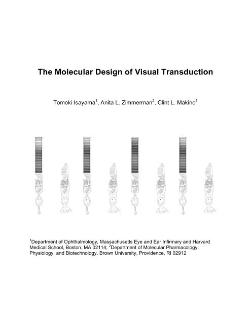
<strong>The</strong> <strong>Molecular</strong> <strong>Design</strong> <strong>of</strong> <strong>Visual</strong> <strong>Transduction</strong><br />
Tomoki Isayama 1 , Anita L. Zimmerman 2 , Clint L. Makino 1<br />
1 Department <strong>of</strong> Ophthalmology, Massachusetts Eye and Ear Infirmary and Harvard<br />
Medical School, Boston, MA 02114; 2 Department <strong>of</strong> <strong>Molecular</strong> Pharmacology,<br />
Physiology, and Biotechnology, Brown University, Providence, RI 02912
Our daily lives are so resplendent with activities that include or require vision,<br />
that it is easy to overlook the complexity <strong>of</strong> the underlying processes involved. Here we<br />
describe the first step in vision…detection <strong>of</strong> light by our photoreceptors. This input<br />
stage sets limits on what we can and cannot see. <strong>The</strong> goal <strong>of</strong> this resource page is to<br />
impart a better understanding <strong>of</strong> the molecular design behind visual transduction.<br />
Structure and Function <strong>of</strong> Rods and Cones<br />
<strong>The</strong> photoreceptors are located in the deepest layer <strong>of</strong> the retina (Fig.1), farthest<br />
from the incoming light. <strong>The</strong>re are two kinds, rods and cones, so named for their overall<br />
shapes. Rods operate in very low light, such as at night. Cones operate under brighter<br />
conditions and provide the basic units for color vision. <strong>The</strong> recently discovered<br />
intrinsically photoreceptive ganglion cells will be excluded from this review because<br />
while they respond to light, they have not been shown to contribute to image-forming<br />
vision.<br />
Rods and cones are specialized unipolar neurons. All vertebrate visual receptors<br />
follow a simple blueprint. <strong>The</strong>y can be divided into two portions, termed inner and outer<br />
segments, according to their radial position within the retina (Fig.1B). <strong>The</strong> inner<br />
segment consists <strong>of</strong> the cell body and contains the cellular organelles found in other<br />
neurons, including a synaptic terminal. <strong>The</strong> outer segment is an elaborate, modified<br />
cilium that contains the biochemical machinery needed for visual transduction. <strong>The</strong><br />
components <strong>of</strong> the phototransduction enzyme cascade are packed into stacks <strong>of</strong><br />
flattened, membranous vesicles (“disks”) that in rods are enclosed by the plasma<br />
membrane <strong>of</strong> the outer segment (Fig.1C). Cones have a similar structure, but the disk<br />
membranes are an extension <strong>of</strong> the plasma membrane, arranged into a series <strong>of</strong><br />
infoldings (Fig.1B).<br />
Most neurons maintain a resting membrane potential <strong>of</strong> about -60 to -70 mV and<br />
when excited, they open cation channels and allow Na + to flow in. <strong>The</strong> resulting<br />
depolarization opens voltage-gated Ca 2+ channels at the synapse. Ca 2+ flows in and<br />
promotes fusion <strong>of</strong> synaptic vesicles which release neurotransmitter. Rods and cones<br />
work “backwards”. At rest, that is in darkness, rods and cones are depolarized to -35 to<br />
-45 mV because channels in the outer segment membrane that pass Na + are already<br />
open. An efflux <strong>of</strong> K + through voltage-gated channels at the inner segment balances the<br />
influx <strong>of</strong> cations at the outer segment, completing an electrical circuit, referred to as the<br />
“dark” or circulating current (Fig.1D). Since photoreceptors are already depolarized in<br />
darkness, Ca 2+ channels are open and the influx <strong>of</strong> Ca 2+ ions supports a steady release<br />
<strong>of</strong> the neurotransmitter glutamate from rod and cone terminals onto second order<br />
neurons (Fig.1D, left). When rods and cones receive light, the channels in their outer<br />
segments close and the membrane hyperpolarizes towards the equilibrium potential for<br />
K + . At the synapse, voltage-sensitive Ca 2+ channels close and neurotransmitter release<br />
subsides (Fig.1D, right).<br />
<strong>The</strong> Phototransduction Cascade<br />
How does photon capture lead to closure <strong>of</strong> ion channels in the outer segment?<br />
<strong>The</strong> following outline will focus on rods since their phototransduction pathway is better<br />
1
understood, but cones operate in a qualitatively similar way.<br />
Figure 1. Cellular plan <strong>of</strong> vertebrate photoreceptors. A. Encapsulation <strong>of</strong> a thin layer <strong>of</strong><br />
brain tissue--the neural retina (tan)--within the vertebrate eye (left). <strong>The</strong> retina (right) is<br />
laminated with neurons that detect light, process the information, and convey it to higher<br />
visual centers in the brain. Light must pass through all layers <strong>of</strong> the retina to reach the<br />
photoreceptors. B. Morphology <strong>of</strong> rods and cones. C. Outer segment disk membranes<br />
containing the biochemical components <strong>of</strong> the light-detecting pathway. D. <strong>The</strong> “dark”<br />
current. Na + and a lesser amount <strong>of</strong> Ca 2+ enter through cyclic nucleotide-gated<br />
channels in the outer segment membrane while K + departs through voltage-gated<br />
channels in the inner segment. In darkness (left), the rod is depolarized and releases<br />
the neurotransmitter glutamate continuously. In response to light (right), channels in the<br />
outer segment membrane close, the rod hyperpolarizes, and glutamate release<br />
decreases. PL, photoreceptor layer; ONL, outer nuclear layer; OPL, outer plexiform<br />
layer; INL, inner nuclear layer; IPL, inner plexiform layer; GCL, ganglion cell layer; NFL,<br />
nerve fiber layer.<br />
In order for light to be seen, it must first be absorbed by the visual pigment<br />
rhodopsin. Rhodopsin consists <strong>of</strong> two parts. One part is a protein called opsin, and the<br />
other is 11-cis retinal. Opsin was sequenced independently by Ovchinnikov in the<br />
former Soviet Union and by Hargrave in the United States. Opsin is a single polypeptide<br />
2
chain with 7 helical segments that span the membrane (Fig.2A). <strong>The</strong> cloning <strong>of</strong> opsin by<br />
Nathans, then a grad student, paved the way for the discovery <strong>of</strong> other hepta-helical<br />
receptors that turned out to be the largest class <strong>of</strong> receptors in the human genome.<br />
Earlier in this century, opsin became the first hepta-helical receptor to have its X-ray<br />
crystal structure solved, thanks to Okada and Palczewski (Fig.2B).<br />
<strong>The</strong> other part <strong>of</strong> rhodopsin, 11-cis retinal (Fig.2C), binds to residue number 296,<br />
a lysine, on the seventh helix <strong>of</strong> opsin (Fig.2A). A Nobel prize was awarded to George<br />
Wald for identifying 11-cis retinal as the chromophore and for working out the<br />
photochemistry. 11-cis Retinal is a derivative <strong>of</strong> vitamin A, which cannot be made de<br />
novo by the body and must be taken in constantly in the diet. Hence vitamin A<br />
deficiency causes night blindness; depletion <strong>of</strong> 11-cis retinal leads to fewer functional<br />
rhodopsins and lower sensitivity <strong>of</strong> the rods -- the cells that do the heavy lifting during<br />
low light situations.<br />
<strong>The</strong> structure <strong>of</strong> 11-cis retinal makes it well suited to serve as the light-absorbing<br />
molecule in rod and cone pigments. Whenever a string <strong>of</strong> carbons are connected with<br />
alternating double and single bonds (Fig.2C), the bonds take on a special character, not<br />
exactly a single bond but not exactly a double bond either. Furthermore, the bonding<br />
electrons do not stay with a particular carbon pair, rather they de-localize over the entire<br />
conjugated chain. It does not take much energy to raise the electrons to the excited<br />
state. By itself, 11-cis retinal would absorb near UV light. But opsin perturbs the<br />
distribution <strong>of</strong> the electrons so that the excited state can be achieved with less energy,<br />
i.e., with longer wavelength light. Cone opsins have similar hepta-helical structures, but<br />
with different amino acid residues surrounding the bound 11-cis retinal; thus they tune<br />
the chromophore’s absorption to different wavelengths.<br />
Photon absorption isomerizes the retinal from the bent 11-cis form to the all-trans<br />
conformation (Fig.2C), and it does so with blazing speed--it only takes a few hundred<br />
femtoseconds (10 -13 sec). This discovery by Mathies, Shank and colleagues caused<br />
quite a stir because no one knew that nuclear rearrangements could occur on this time<br />
scale. Slower rearrangements in the surrounding opsin reposition its cytoplasmic loops,<br />
converting the protein to its active R* state. All subsequent steps occur in darkness.<br />
Thus, the chromophore converts the energy <strong>of</strong> a photon into a conformational change in<br />
protein structure. This structural change is but the first step in the phototransduction<br />
cascade, because most <strong>of</strong> the rhodopsin resides in disk membranes within the rod,<br />
physically separated from the plasma membrane where the ion channels are located.<br />
How does rhodopsin communicate with channels in the plasma membrane?<br />
Fung, Hurley and Stryer showed that rhodopsin is a G protein-coupled receptor<br />
and in rods and cones, named the corresponding G protein transducin (Fig.3A). Its Xray<br />
crystal structure was solved by Sigler and Hamm and co-workers. G proteins are<br />
heterotrimeric, consisting <strong>of</strong> , , and subunits. In its inactive state, transducin’s <br />
subunit has a GDP bound to it, but R* binds transducin and allows the GDP to<br />
dissociate (Fig.3B). GDP is the glue that holds the transducin heterotrimer together, so<br />
without GDP, the subunits come <strong>of</strong>f. R* and the subunit remain conjoined until a<br />
passing GTP binds the subunit, allowing it to detach from R*. R* repeats this action<br />
over and over until its activity is shut <strong>of</strong>f.<br />
3
Figure 2. Structure <strong>of</strong> rhodopsin. A. Amino acid sequence <strong>of</strong> bovine opsin. <strong>The</strong><br />
glycosylated N-terminus (violet residues) is located within the outer segment disk<br />
(intradiskal/extracellular) while the C-terminus (red residues) is exposed to the<br />
cytoplasm, with the seven helices (H1-H7) spanning the disk membrane. An eighth<br />
helix (H8) sits on the membrane surface. <strong>The</strong>re are seven phosphorylation sites (*) and<br />
two palmitoylated cysteines (gray squares) on the C-terminus. Two conserved cysteines<br />
(gray circles) on the first and second extracellular loops form a disulfide linkage. <strong>The</strong> 11cis<br />
retinal chromophore binds as a protonated Schiff base to Lysine 296 (black diamond).<br />
B. X-ray crystal structure <strong>of</strong> bovine rhodopsin. <strong>The</strong> color scheme for the helices (H1-H8)<br />
correspond to that in A. <strong>The</strong> blue spheres represent water molecules, and the arrows<br />
represent beta strand structure. Courtesy <strong>of</strong> T. Okada, Gakushuin University. C. <strong>The</strong><br />
light-absorbing chromophore <strong>of</strong> rhodopsin. Photoisomerization <strong>of</strong> the 11-cis retinal in<br />
opsin’s chromophore-binding pocket to the all-trans conformation (right) transforms<br />
rhodopsin to the activated state (R*).<br />
4
Transducin serves as an intermediary between the activated receptor and an<br />
effector. In this case, the effector is a phosphodiesterase (PDE) that enzymatically<br />
cleaves the phosphodiester bond <strong>of</strong> cGMP to form 5’-GMP (Fig.4). PDE is a<br />
heterotetramer that consists <strong>of</strong> a dimer <strong>of</strong> two catalytic subunits, and , each with an<br />
active site inhibited by a PDE subunit. <strong>The</strong> activated transducin subunit-GTP binds to<br />
PDE and relieves the inhibition on a catalytic subunit.<br />
Figure 3. Structure <strong>of</strong> transducin. A. Heterotrimeric composition <strong>of</strong> transducin (PDB ID:<br />
1GOT). Courtesy <strong>of</strong> K. Martemyanov, University <strong>of</strong> Minnesota. B. Activation <strong>of</strong><br />
transducin. R* binds the transducin heterotrimer and catalyzes the exchange <strong>of</strong> a bound<br />
GDP for a GTP, leading to the release <strong>of</strong> the -GTP subunit from transducin.<br />
So far, all <strong>of</strong> the steps have taken place on the disk membrane (Fig.4, left),<br />
separate from the ion channels in the plasma membrane <strong>of</strong> the outer segment. A small,<br />
soluble molecule, cGMP, links the two compartments. When activated, PDE hydrolyzes<br />
cGMP to 5’-GMP, the cGMP concentration inside the rod decreases, and cyclic<br />
nucleotide-gated ion channels respond by closing (Fig.4, right). Na + entry into the outer<br />
segment is blocked, and as a result the rod hyperpolarizes. <strong>The</strong> discovery by Fesenko<br />
and colleagues that these channels were gated directly by cGMP was a revelation in<br />
biology, because until that time people thought that cyclic nucleotides elicited their<br />
effects through the phosphorylation <strong>of</strong> proteins only. Cyclic nucleotide-gated channels<br />
are now widely studied in other neurons, as well as in kidney, heart and other organs.<br />
Seeing Single Photons<br />
Rods do more than just respond to light; Baylor, Lamb and Yau demonstrated by<br />
single cell recording that they possess quantal sensitivity. In order to signal individual<br />
photons, three important factors need to be addressed. First, background noise needs<br />
to be minimal. Second, the quantal response needs to be large. Third, the response<br />
must be reproducible.<br />
5
Figure 4. Bridging the gap between photon absorption at the disk membrane and<br />
channel closure at the plasma membrane. On the disk membrane (left), absorption <strong>of</strong> a<br />
photon by rhodopsin (R) leads to photoisomerization and formation <strong>of</strong> the active state <strong>of</strong><br />
the pigment (R*). R* catalyzes the transfer <strong>of</strong> a bound GDP for a GTP on transducin,<br />
causing the subunits to come <strong>of</strong>f, and leaving the transducin -GTP free to activate<br />
phosphodiesterase (PDE). A transducin -GTP can bind one PDE subunit and remove<br />
the inhibition from one <strong>of</strong> the PDE catalytic subunits ( or ). Activated PDE hydrolyzes<br />
cGMP, the intracellular messenger that is required for cGMP-gated channels in the<br />
outer segment plasma membrane to remain open (right). Reduction in the intracellular<br />
levels <strong>of</strong> cGMP closes channels, blocking entry <strong>of</strong> Na + and Ca 2+ and thereby decreases<br />
the circulating current.<br />
First: the quiet baseline. By analogy, let’s equate the detection <strong>of</strong> a single photon<br />
by a rod with a person trying to hear the sound <strong>of</strong> a single pin dropped onto the floor. It<br />
is far easier to hear the sound <strong>of</strong> the pin hitting the floor when it is quiet, and impossible<br />
when there is a lot <strong>of</strong> background noise. Consider the sources <strong>of</strong> noise in the visual<br />
transduction cascade. For any temperature above absolute zero, molecules are in<br />
constant motion, fidgeting and banging into each other. Sometimes enough energy<br />
accumulates within a rhodopsin molecule to set it <strong>of</strong>f, just as if it had absorbed a photon.<br />
At body temperature, the rhodopsin in your eye has a half-life for thermal isomerization<br />
exceeding 400 years (Fig.5A). That is 23 log units slower than light activation since<br />
photoisomerization takes a few hundred femtoseconds. Of course, for many rhodopsins<br />
thermal isomerization occurs much sooner than 400 years. Moreover, rods contain<br />
about a hundred million copies <strong>of</strong> rhodopsin, so there will be a thermal isomerization<br />
about once every minute.<br />
6
How does this affect the rod’s response? Figure 5B is a recording <strong>of</strong> a single<br />
rod’s response to dim flashes. Poisson statistics are obeyed; sometimes the rod does<br />
not respond, while at other times it detects a photon, and sometimes it detects two.<br />
Once in a while, due to thermal activation, the rod gives a “response” even though no<br />
photon was absorbed (Fig.5B, asterisk). Spontaneous events do not interfere with<br />
vision as long as their rate <strong>of</strong> occurrence is overwhelmed by the rate <strong>of</strong> responses<br />
arising from photon absorptions. But as the light becomes exceedingly dim, our<br />
absolute visual sensitivity is limited by this rate <strong>of</strong> thermal activation. So while rhodopsin<br />
is incredibly stable, it has not been grossly over-engineered.<br />
<strong>The</strong> next component in the phototransduction cascade, transducin, is the most<br />
stable G protein known. Random activation, that is spontaneous exchange <strong>of</strong> GDP for<br />
GTP, takes hours. Transducin is not nearly as stable as rhodopsin, but there are tenfold<br />
fewer <strong>of</strong> them in the outer segment than there are rhodopsins. Also as we shall see,<br />
the effect generated by a single transducin is a hundred times smaller than that <strong>of</strong> a<br />
single R*.<br />
PDE is even noisier than transducin, but there are ten-fold fewer PDE than<br />
transducins. Nevertheless, at any given instant in time, there are hundreds <strong>of</strong> active<br />
PDE in the absence <strong>of</strong> light. Most <strong>of</strong> the time, PDE shuts <strong>of</strong>f quickly without hydrolyzing<br />
too much cGMP. But once in a while, the PDE stays on long enough to produce a single<br />
photon response-like event. Luckily that only happens once every 10 or 20 minutes,<br />
which is less than the rate <strong>of</strong> thermal activation <strong>of</strong> rhodopsin. <strong>The</strong> activity <strong>of</strong> PDE<br />
actually serves an important function. In darkness, cGMP is constantly synthesized, so<br />
without dark PDE activity, cGMP would accumulate and open all <strong>of</strong> the cGMP-gated<br />
channels. For reasons that will be addressed later, the rod has a lot <strong>of</strong> channels but only<br />
a small percentage are normally open in darkness. If the rod were to contain too much<br />
cGMP, the floodgates would open and the cell would fill up with Na + and Ca 2+ since<br />
both go through the channel. This scenario is thought to occur in some forms <strong>of</strong> retinal<br />
disease, resulting in photoreceptor death and blindness.<br />
<strong>The</strong> last component <strong>of</strong> the cascade, the channel, is very, very noisy because it<br />
opens and closes very rapidly. Figure 5C shows a single channel that keeps flickering<br />
open and closed. Why use such a noisy channel? <strong>The</strong> channel has a greater tendency<br />
to open when cGMP is bound, and cGMP tends to stay bound to the open channel. For<br />
the channel to respond to changes in cGMP concentration, it must revert to the closed<br />
state to give the cGMP a chance to come <strong>of</strong>f. Once the channel is closed, if the<br />
concentration <strong>of</strong> cGMP is high, another cGMP will bind and re-open the channel. If the<br />
concentration <strong>of</strong> cGMP is low, the channel will stay closed since there will be fewer<br />
nucleotides around. Thus channel chatter ensures that the response to a decrease in<br />
cGMP levels will be quick.<br />
A high <strong>of</strong>f-rate constant <strong>of</strong> the closed channel for cGMP is also helpful, but then a<br />
lot <strong>of</strong> channels are needed in order to get enough <strong>of</strong> them to open at reasonable levels<br />
<strong>of</strong> cGMP. That’s one reason the rods have lots <strong>of</strong> channels, but as noted above the<br />
channel is a great source <strong>of</strong> noise. To keep the noise from being problematic, the rod<br />
does something clever. <strong>The</strong> channel is rather non-selective for cations…Na + , Ca 2+ , Mg 2+<br />
and even K + will permeate. Most <strong>of</strong> the current is carried by Na + since there is so much<br />
7
Figure 5. Noise in the system. A. Exponential decay <strong>of</strong> rhodopsin in darkness due to<br />
thermal isomerization. At 37°C, rhodopsin has a half-life <strong>of</strong> about 400 years. B. Dim<br />
flash responses <strong>of</strong> a mouse rod. <strong>The</strong> stimulus is so dim that not all the flashes elicit a<br />
response, but in some instances there is a “response” when no stimulus is presented<br />
(*). <strong>The</strong> line beneath the response shows the timing <strong>of</strong> flashes. T. Isayama,<br />
Massachusetts Eye & Ear Infirmary and Harvard Medical School. C. Current fluctuations<br />
<strong>of</strong> a single rod cyclic nucleotide-gated channel in saturating cGMP. <strong>The</strong> “fuzziness” in<br />
the open state results from unresolved transitions back to the closed state. Courtesy <strong>of</strong><br />
G. Matthews, State University <strong>of</strong> New York at Stony Brook.<br />
<strong>of</strong> it around, and the negative membrane voltage favors its entry. Na + is well behaved<br />
and passes through promptly. On the other hand, divalents, such as Ca 2+ and Mg 2+ ,<br />
take their time in going through, sometimes even stopping. When they stop, nothing<br />
else can get by because the channel is a single-file pore. This “divalent block” reduces<br />
the single channel conductance. Consequently, to get the proper amount <strong>of</strong> current the<br />
rod uses a large number <strong>of</strong> channels and averages out the noise.<br />
<strong>The</strong> general trend as we go further and further along the transduction pathway, is<br />
for molecules to become less and less stable. <strong>The</strong> effects <strong>of</strong> the noise generated by<br />
these elements are minimized and even put to good use as in the case <strong>of</strong> PDE and the<br />
channel.<br />
Having dealt with the background noise in our pin drop analogy, we next see that<br />
if the pin should fall on a s<strong>of</strong>t surface, it would barely make any sound at all. It would be<br />
far better if the pin were to fall onto a microphone connected to an amplifier and loud<br />
speakers. In biology, a strategy for achieving amplification is through the use <strong>of</strong> a<br />
biochemical cascade. A stimulus activates an enzyme, which in turn activates another<br />
enzyme, and so on. In rods, the first amplifying step is when R* activates transducin.<br />
After turning on transducin, it turns on another, and another (Fig.6A). All told, R* turns<br />
8
Figure 6. Amplification in the phototransduction cascade. A. Rhodopsin to transducin. A<br />
single R* can activate close to a hundred transducin molecules. B. Phosphodiesterase.<br />
Each transducin -GTP can bind to one PDE subunit, relieving inhibition on one <strong>of</strong> two<br />
PDE catalytic subunits ( or ) that hydrolyzes cGMP to 5’-GMP at the diffusion limit. A<br />
second transducin -GTP is required to activate the other PDE catalytic subunit. C.<br />
Protein interaction on the disk membrane. <strong>Transduction</strong> is improved by anchoring R*<br />
(orange), transducin (blue), and PDE (gray) to the phospholipid bilayer to ensure timely<br />
collisions (top). <strong>The</strong> high unsaturated fatty acid content <strong>of</strong> the disk membrane makes<br />
them very fluid because kinky double bonds disrupt the tight, orderly packing <strong>of</strong> the fatty<br />
acids (bottom). D. Cyclic GMP-gated channels. <strong>The</strong> dose-response relation for<br />
channel opening increases as the third power <strong>of</strong> cGMP concentration for [cGMP] < K0.5<br />
in response to a change in cGMP concentration, and the effect is enhanced as<br />
cooperativity rises. For comparison, solid and dashed red lines depict relations with Hill<br />
coefficients <strong>of</strong> n=1 and n=2, respectively, where more channels are open at a lower<br />
[cGMP], but increases in cGMP levels produce less dramatic effects. A. Zimmerman,<br />
Brown University.<br />
9
on close to a hundred transducins. Transducin activates PDE stochiometrically. But<br />
each PDE catalytic subunit enzymatically hydrolyzes cGMP as fast as cGMP can bind<br />
to it (Fig.6B).<br />
With a multi-step cascade, amplification increases over time. In this regard, it is<br />
significant that rhodopsin, transducin and PDE are all attached to the disk membrane<br />
(Fig.6C). Conversion <strong>of</strong> a 3-dimensional reaction volume to a 2-dimensional surface not<br />
only improves the overall probability for collisions between reactants, it also improves<br />
the odds for an effective collision. Imagine that R* and transducin have to be oriented in<br />
a specific manner in order to bind. In solution, right <strong>of</strong>f the bat, many R* and transducin<br />
pairs will be in exactly the wrong orientation when they collide. But in the membrane, R*<br />
and transducin can be fixed to increase the odds that they are facing the right direction.<br />
Membranes, however, can be more viscous than aqueous solutions, hindering<br />
the diffusion <strong>of</strong> membrane proteins. To minimize these effects, disk membranes are<br />
very special; they are low in cholesterol and have a high content <strong>of</strong> unsaturated fatty<br />
acids, properties that make the membrane very fluid (Fig.6C). As a result, the disk<br />
membrane is one <strong>of</strong> the most fluid in the body with a consistency like that <strong>of</strong> olive oil.<br />
<strong>The</strong> lateral diffusion rate for rhodopsin ranks among the fastest for any integral<br />
membrane protein. This feature is important because the rate <strong>of</strong> transducin activation is<br />
limited by how fast R* collides with transducin, which turns out to be about once every<br />
few milliseconds. <strong>The</strong>se two factors -- the restriction <strong>of</strong> the transduction components to<br />
the membrane and the membrane’s fluidity -- both optimize signal amplification.<br />
Additional amplification comes from the way in which the channel responds to<br />
changes in cGMP concentration. <strong>The</strong> dose response curve for the channel (current<br />
versus concentration <strong>of</strong> cGMP) (Fig.6D) follows the Hill equation:<br />
I/Imax = cGMP n /(cGMP n + K n ),<br />
where K is the concentration at which half the channels are open and n is the Hill<br />
coefficient. When n>1, there is cooperativity, that is the binding <strong>of</strong> one cGMP enhances<br />
additional binding and channel opening. For the native channel, n is about 3 (Fig.6D,<br />
gray line). <strong>The</strong> minimal amount <strong>of</strong> cGMP that must be present to open the channel is<br />
increased, but channel opening is very sensitive to changes in cGMP. Put another way,<br />
since multiple cGMP molecules are required to open the channel, it will close when only<br />
one or two cGMP molecules leave the channel, making it easily shut down by<br />
absorption <strong>of</strong> light. <strong>The</strong>refore the signal transduced by the disk membrane-bound<br />
components is further magnified at the plasma membrane by the cooperative nature <strong>of</strong><br />
cGMP binding to the channel.<br />
Ultimately, a single photon closes about 200 channels and thereby prevents the<br />
entry <strong>of</strong> about a million Na + ions into the rod.<br />
That brings us to the next problem. In the pin drop analogy, a loud sound can<br />
result from one pin dropped from a tall height, but it can also result from multiple pins<br />
hitting the same surface simultaneously from a lower height. How can we differentiate<br />
between the two? By making the signal from each hit the same.<br />
10
<strong>The</strong> size <strong>of</strong> the single photon response is determined by the shut<strong>of</strong>f <strong>of</strong> the<br />
phototransduction cascade. Rhodopsin shuts <strong>of</strong>f after binding to arrestin (Fig.7A).<br />
Arrestin is a very large protein, whose sheer bulk prevents any transducins from getting<br />
close enough to bind. A one step shut<strong>of</strong>f would be stochastic, that is sometimes it would<br />
happen quickly, sometimes slowly. <strong>The</strong> rod improves on this by phosphorylating R*<br />
(Fig.7A) at serine and threonine residues on the C terminus <strong>of</strong> the rhodopsin molecule<br />
that are targeted by the enzyme rhodopsin kinase (Fig.2A). <strong>The</strong>re are multiple<br />
phosphorylation sites, and each added phosphate simultaneously reduces R*’s activity<br />
and increases R*’s affinity for arrestin. Rieke and colleagues determined that a multi<br />
step shut<strong>of</strong>f averages out the variability in the timing and confers response<br />
reproducibility.<br />
Figure 7. Inactivation <strong>of</strong> the cascade. A. Shut<strong>of</strong>f <strong>of</strong> rhodopsin. Rhodopsin kinase<br />
(GRK1) phosphorylates serine and threonine residues at the C-terminus <strong>of</strong> R*.<br />
Phosphorylation <strong>of</strong> R* alone decreases transducin activation, but binding to arrestin is<br />
necessary to completely quench its activity. All trans-retinal comes <strong>of</strong>f and a new 11-cis<br />
retinal binds the opsin to regenerate rhodopsin. B. Shut<strong>of</strong>f <strong>of</strong> transducin and PDE.<br />
Transducin is inactivated by hydrolysis <strong>of</strong> the bound GTP to GDP. <strong>The</strong> slow, intrinsic<br />
GTPase activity <strong>of</strong> transducin is accelerated by the GAP (GTPase Activating Protein)<br />
complex, consisting <strong>of</strong> RGS9 (regulator <strong>of</strong> G protein signaling 9), G5, and the<br />
membrane anchoring protein R9AP. Upon hydrolysis <strong>of</strong> GTP to GDP, transducin subunit<br />
releases the PDE subunit that re-inhibits the catalytic subunit. Transducin -<br />
GDP eventually combines with transducin to reconstitute the inactive heterotrimer.<br />
Transducin has a built-in shut<strong>of</strong>f mechanism, namely hydrolysis <strong>of</strong> GTP. When<br />
that happens, transducin lets go <strong>of</strong> PDE, which resumes its inhibition <strong>of</strong> a PDE catalytic<br />
subunit (Fig.7B). Transducin subunits recombine with transducin -GDP and the<br />
heterotrimer returns to its resting state (Fig.7B). GTP hydrolysis is slow intrinsically, but<br />
it is accelerated by the GAP (GTPase Activating Protein) complex (Fig.7B). To ensure<br />
that transducin does not shut <strong>of</strong>f before activating PDE, transducin and the GAP<br />
11
Figure 8. Calcium feedback. A. Na + /K + ,Ca 2+ exchange. In response to light, cGMPgated<br />
channels in the outer segment membrane shut, hyperpolarizing the cell. An<br />
exchanger in the outer segment couples the removal <strong>of</strong> a Ca 2+ to the movement <strong>of</strong> one<br />
K + and four Na + down their electrochemical gradients. B. Production <strong>of</strong> cGMP. In the<br />
dark, Ca 2+ binds to GCAPs (guanylate cyclase activating proteins) that are attached to<br />
and inhibit guanylate cyclase (GC), causing the cyclase to synthesize cGMP at a slow<br />
rate (top). GCAP activates cyclase in the absence <strong>of</strong> Ca 2+ , and cGMP production<br />
increases (bottom). C. Ca 2+ dependence <strong>of</strong> guanylate cyclase activity. At dark-adapted<br />
Ca 2+ levels (black arrowhead), cyclase operates at a low level, but visual transduction<br />
causes a drop in Ca 2+ to light-adapted levels (orange arrowhead) and elicits a GCAPmediated<br />
increase in cyclase activity according to a Hill function: A =(Amax-Amin)/(1 +<br />
([Ca 2+ ]/Km Ca) n )+Amin, where A is guanylate cyclase activity, Km Ca is 100, and n is 1.7.<br />
Courtesy <strong>of</strong> A. Dizhoor and I. Peshenko, Salus University. D. Cyclic nucleotide-gated<br />
channel. As [Ca 2+ ]int decreases during light activation, Ca 2+ -calmodulin (CaM) or a<br />
related protein releases from the channel, causing an increase in the channel’s affinity<br />
for cGMP. <strong>The</strong> increased affinity promotes nucleotide binding and channel opening. As<br />
[Ca 2+ ]int is restored, CaM binds the channel once more and reduces its affinity for<br />
cGMP.<br />
12
complex have a low affinity for each other, until transducin -GTP binds PDE.<br />
Full recovery requires that the intracellular cGMP concentration be brought back<br />
up to dark-adapted levels. As noted above, cGMP is synthesized continuously from<br />
GTP by guanylate cyclases. Retinal guanylate cyclases are transmembrane proteins<br />
that exist as homodimers, whose activity is kept low by the action <strong>of</strong> GCAPs (guanylate<br />
cyclase activating proteins). One GCAP is bound to each subunit, preventing full<br />
activity. Thus recovery can take a pretty long time because the basal rate <strong>of</strong> cyclase<br />
activity is pretty slow. <strong>The</strong> rod, however, has a solution - feedback.<br />
Remember that a trickle <strong>of</strong> Ca 2+ enters the rod through the cGMP-gated<br />
channels. Ca 2+ is removed by a Na + /K + ,Ca 2+ exchanger molecule in the outer segment<br />
plasma membrane (Fig.8A). When the channels close, Ca 2+ ceases to enter, but<br />
extrusion through the exchanger continues, so [Ca 2+ ]int falls. In the dark, GCAPs bind<br />
Ca 2+ and inhibit cyclase activity. A decrease in [Ca 2+ ]int causes Ca 2+ to dissociate from<br />
GCAPs, allowing them to dimerize. Dimerization <strong>of</strong> GCAPs leads to full activation <strong>of</strong><br />
guanylate cyclase subunits, and an increase in the rate <strong>of</strong> cGMP synthesis (Fig.8B,C).<br />
<strong>The</strong> cGMP-gated channel is a second target for feedback. In the dark, Ca 2+ -Calmodulin<br />
(CaM) or a related protein binds the channel and reduces its affinity for cGMP (Fig.8D,<br />
bottom). During visual transduction, the decrease in [Ca 2+ ]int causes CaM to be<br />
released, increasing the channel’s affinity for cGMP (Fig.8D, top) so that during<br />
recovery, the channel reopens at lower levels <strong>of</strong> cGMP, i.e. sooner.<br />
Putting all the steps together: high amplification <strong>of</strong> signal and a low level <strong>of</strong><br />
background noise allow the rod to detect single photons. Deactivation mechanisms<br />
ensure that each photon produces a similar response. In our eye, the single photon<br />
response reaches an amplitude <strong>of</strong> about one pA, it peaks in less than 200 msec, and<br />
recovers completely after about a sec (Fig.9A). <strong>The</strong> variance in size is less than 1/5 <strong>of</strong><br />
the mean amplitude. That’s pretty good! But what happens when the rod absorbs two<br />
photons at once?<br />
Light enters the rod from the synaptic terminal end, passes through the inner<br />
segment and finally reaches the outer segment. When a photon gets absorbed, it<br />
causes a local decrease in cGMP levels and the closure <strong>of</strong> channels in an annulus<br />
surrounding the site <strong>of</strong> absorption (Fig.9B). <strong>The</strong> width <strong>of</strong> the annulus is about 5% <strong>of</strong> the<br />
rod outer segment length. With two photons, the Poisson nature <strong>of</strong> absorption makes it<br />
highly unlikely that both will be absorbed at the same disk. <strong>The</strong>refore each photon<br />
absorption closes a separate subset <strong>of</strong> the rod’s channels (Fig.9B).<br />
<strong>The</strong> increasing hyperpolarization produced by closure <strong>of</strong> more channels does<br />
not, however, increase the inward current carried by the remaining open channels<br />
because the channels are outwardly rectifying (Fig.9C). <strong>The</strong> basis lies in the voltage<br />
dependence <strong>of</strong> divalent block (discussed earlier) and a weak effect <strong>of</strong> voltage on<br />
channel gating. With regard to the latter property, it is interesting to note that cGMPgated<br />
channels bear structural similarity to voltage-gated potassium channels. <strong>The</strong><br />
consequence <strong>of</strong> outward rectification is that under physiological conditions, the current<br />
depends strictly on the number <strong>of</strong> open channels and not on the membrane voltage.<br />
<strong>The</strong>n the reduction in dark current produced by two photons is simply twice that<br />
13
Figure 9. Absorption <strong>of</strong> 2 photons. A. A single photon response. <strong>The</strong> response to a<br />
single photon is presented as a fraction <strong>of</strong> the saturating response amplitude (14 pA).<br />
<strong>The</strong> line beneath the response shows the timing <strong>of</strong> the flash. Courtesy <strong>of</strong> J. Schnapf,<br />
University <strong>of</strong> California San Francisco. B. Spatial distribution <strong>of</strong> rhodopsin<br />
photoisomerization. Two photons are unlikely to be absorbed in the same disk, and<br />
therefore produce local effects independent <strong>of</strong> each other (green annuli). C. Currentvoltage<br />
relations. <strong>The</strong> ohmic or linear behavior at high voltages becomes outwardly<br />
rectifying at physiologically relevant voltages, meaning that in this range the dark<br />
current is nearly independent <strong>of</strong> membrane potential. Measurements were made on a<br />
membrane patch excised from a toad rod outer segment with symmetric monovalent<br />
solutions plus 1 mM Ca 2+ and 1.6 mM Mg 2+ on the extracellular face, but zero divalents,<br />
0.15 mM EDTA and 1 mM cGMP on the intracellular face. Courtesy <strong>of</strong> G. Matthews,<br />
State University <strong>of</strong> New York at Stony Brook. D. Stimulus-response relation for the rod.<br />
<strong>The</strong> response to multiple photon absorptions can be described by a saturating<br />
exponential function (gray trace). A plot <strong>of</strong> normalized response and flash strength (i) on<br />
a log scale indicates that at high photon counts, the response saturates because all the<br />
channels are closed. A linear relation (red trace) is shown for comparison.<br />
produced by one photon. <strong>The</strong> rod responds linearly, in other words it functions as a<br />
photon counter (Fig.9D, red trace).<br />
14
When there are lots <strong>of</strong> photons, the effects <strong>of</strong> individual rhodopsin isomerizations<br />
begin to overlap, so there are diminishing returns and the rod response diverges from<br />
linearity. <strong>The</strong> behavior follows a saturating exponential:<br />
r/rmax = 1-exp(-ki),<br />
where i is flash strength and k is a constant equal to the quotient <strong>of</strong> ln(2) divided by the<br />
flash strength giving rise to a half maximal response (Fig.9D, gray trace). Bright flashes<br />
close all <strong>of</strong> the channels and saturate the rod’s response (r/rmax = 1). Suprasaturating<br />
flashes reduce cGMP further, keeping the rod saturated for a longer time before<br />
recovering, because more time is needed to re-synthesize cGMP to a level that reopens<br />
the channels.<br />
In summary, the rod converts light into an electrical signal by triggering a G<br />
protein-coupled receptor cascade. In this way, the rod generates highly amplified,<br />
reproducible signals on a fairly quiet background that enable it to count single photons.<br />
<strong>The</strong> cones operate in a similar manner, but with their own set <strong>of</strong> phototransduction<br />
components, and with a level <strong>of</strong> sensitivity lower than that <strong>of</strong> the rods. Both rods and<br />
cones contribute to the detection <strong>of</strong> visual information. Rods function in dim light<br />
situations as photon counters that can detect very small amounts <strong>of</strong> light. Cones,<br />
although not as sensitive, respond faster, respond to intensities over a broader range,<br />
and lay the basis for color vision…but those are topics for another resource page.<br />
15
For further information, we recommend:<br />
Andreucci, D., Bisegna, P., Caruso, G., Hamm, H.E., and DiBenedetto, E. (2003)<br />
Mathematical model <strong>of</strong> the spatio-temporal dynamics <strong>of</strong> second messengers in visual<br />
transduction. Biophysical Journal 85:1358-1376.<br />
Hamer, R.D., Nicholas, S.C., Tranchina, D., Lamb, T.D., and Jarvinen, J.L.P. (2005) To<br />
a unified model <strong>of</strong> vertebrate rod phototransduction. <strong>Visual</strong> Neuroscience 22:417-436.<br />
de Grip, W.J., Stavenga, D.G., and Pugh, E.N. Jr. (2000) Handbook <strong>of</strong> Biological<br />
Physics: <strong>Molecular</strong> Mechanisms in <strong>Visual</strong> <strong>Transduction</strong>. North Holland.<br />
Rodieck, R.W. (1998) <strong>The</strong> First Steps in Seeing. Sinauer Associates.<br />
Acknowledgments.<br />
This educational resource evolved from course lectures given at Brown and Harvard<br />
Universities. We thank T. Okada, K. Martemyanov, J. Schnapf, G. Matthews, I.V.<br />
Peshenko, and A. Dizhoor for providing figures and D. Baylor for comments.<br />
16