Photodegradation of rhodamine B in aqueous solution ... - AkzoNobel
Photodegradation of rhodamine B in aqueous solution ... - AkzoNobel
Photodegradation of rhodamine B in aqueous solution ... - AkzoNobel

You also want an ePaper? Increase the reach of your titles
YUMPU automatically turns print PDFs into web optimized ePapers that Google loves.
Abstract<br />
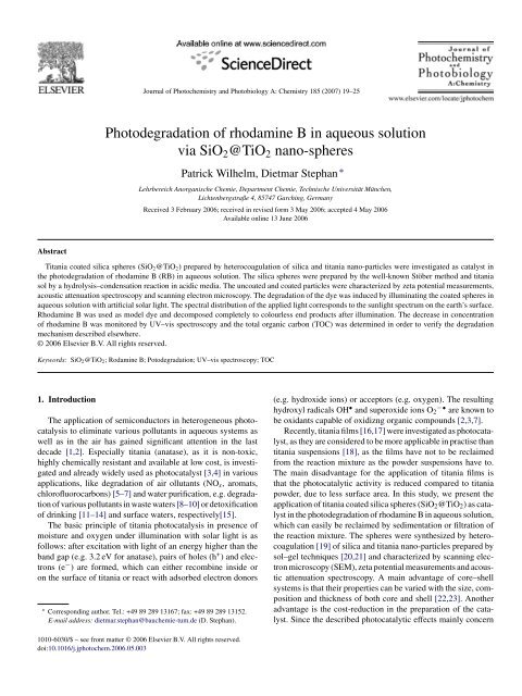
Journal <strong>of</strong> Photochemistry and Photobiology A: Chemistry 185 (2007) 19–25<br />
<strong>Photodegradation</strong> <strong>of</strong> <strong>rhodam<strong>in</strong>e</strong> B <strong>in</strong> <strong>aqueous</strong> <strong>solution</strong><br />
via SiO2@TiO2 nano-spheres<br />
Patrick Wilhelm, Dietmar Stephan ∗<br />
Lehrbereich Anorganische Chemie, Department Chemie, Technische Universität München,<br />
Lichtenbergstraße 4, 85747 Garch<strong>in</strong>g, Germany<br />
Received 3 February 2006; received <strong>in</strong> revised form 3 May 2006; accepted 4 May 2006<br />
Available onl<strong>in</strong>e 13 June 2006<br />
Titania coated silica spheres (SiO2@TiO2) prepared by heterocoagulation <strong>of</strong> silica and titania nano-particles were <strong>in</strong>vestigated as catalyst <strong>in</strong><br />
the photodegradation <strong>of</strong> <strong>rhodam<strong>in</strong>e</strong> B (RB) <strong>in</strong> <strong>aqueous</strong> <strong>solution</strong>. The silica spheres were prepared by the well-known Stöber method and titania<br />
sol by a hydrolysis–condensation reaction <strong>in</strong> acidic media. The uncoated and coated particles were characterized by zeta potential measurements,<br />
acoustic attenuation spectroscopy and scann<strong>in</strong>g electron microscopy. The degradation <strong>of</strong> the dye was <strong>in</strong>duced by illum<strong>in</strong>at<strong>in</strong>g the coated spheres <strong>in</strong><br />
<strong>aqueous</strong> <strong>solution</strong> with artificial solar light. The spectral distribution <strong>of</strong> the applied light corresponds to the sunlight spectrum on the earth’s surface.<br />
Rhodam<strong>in</strong>e B was used as model dye and decomposed completely to colourless end products after illum<strong>in</strong>ation. The decrease <strong>in</strong> concentration<br />
<strong>of</strong> <strong>rhodam<strong>in</strong>e</strong> B was monitored by UV–vis spectroscopy and the total organic carbon (TOC) was determ<strong>in</strong>ed <strong>in</strong> order to verify the degradation<br />
mechanism described elsewhere.<br />
© 2006 Elsevier B.V. All rights reserved.<br />
Keywords: SiO2@TiO2; Rodam<strong>in</strong>e B; Potodegradation; UV–vis spectroscopy; TOC<br />
1. Introduction<br />
The application <strong>of</strong> semiconductors <strong>in</strong> heterogeneous photocatalysis<br />
to elim<strong>in</strong>ate various pollutants <strong>in</strong> <strong>aqueous</strong> systems as<br />
well as <strong>in</strong> the air has ga<strong>in</strong>ed significant attention <strong>in</strong> the last<br />
decade [1,2]. Especially titania (anatase), as it is non-toxic,<br />
highly chemically resistant and available at low cost, is <strong>in</strong>vestigated<br />
and already widely used as photocatalyst [3,4] <strong>in</strong> various<br />
applications, like degradation <strong>of</strong> air ollutants (NOx, aromats,<br />
chlor<strong>of</strong>luorocarbons) [5–7] and water purification, e.g. degradation<br />
<strong>of</strong> various pollutants <strong>in</strong> waste waters [8–10] or detoxification<br />
<strong>of</strong> dr<strong>in</strong>k<strong>in</strong>g [11–14] and surface waters, respectively[15].<br />
The basic pr<strong>in</strong>ciple <strong>of</strong> titania photocatalysis <strong>in</strong> presence <strong>of</strong><br />
moisture and oxygen under illum<strong>in</strong>ation with solar light is as<br />
follows: after excitation with light <strong>of</strong> an energy higher than the<br />
band gap (e.g. 3.2 eV for anatase), pairs <strong>of</strong> holes (h + ) and electrons<br />
(e − ) are formed, which can either recomb<strong>in</strong>e <strong>in</strong>side or<br />
on the surface <strong>of</strong> titania or react with adsorbed electron donors<br />
∗ Correspond<strong>in</strong>g author. Tel.: +49 89 289 13167; fax: +49 89 289 13152.<br />
E-mail address: dietmar.stephan@bauchemie-tum.de (D. Stephan).<br />
1010-6030/$ – see front matter © 2006 Elsevier B.V. All rights reserved.<br />
doi:10.1016/j.jphotochem.2006.05.003<br />
(e.g. hydroxide ions) or acceptors (e.g. oxygen). The result<strong>in</strong>g<br />
hydroxyl radicals OH • and superoxide ions O2 −• are known to<br />
be oxidants capable <strong>of</strong> oxidizng organic compounds [2,3,7].<br />
Recently, titania films [16,17] were <strong>in</strong>vestigated as photocatalyst,<br />
as they are considered to be more applicable <strong>in</strong> practise than<br />
titania suspensions [18], as the films have not to be reclaimed<br />
from the reaction mixture as the powder suspensions have to.<br />
The ma<strong>in</strong> disadvantage for the application <strong>of</strong> titania films is<br />
that the photocatalytic activity is reduced compared to titania<br />
powder, due to less surface area. In this study, we present the<br />
application <strong>of</strong> titania coated silica spheres (SiO2@TiO2) as catalyst<br />
<strong>in</strong> the photodegradation <strong>of</strong> <strong>rhodam<strong>in</strong>e</strong> B <strong>in</strong> <strong>aqueous</strong> <strong>solution</strong>,<br />
which can easily be reclaimed by sedimentation or filtration <strong>of</strong><br />
the reaction mixture. The spheres were synthesized by heterocoagulation<br />
[19] <strong>of</strong> silica and titania nano-particles prepared by<br />
sol–gel techniques [20,21] and characterized by scann<strong>in</strong>g electron<br />
microscopy (SEM), zeta potential measurements and acoustic<br />
attenuation spectroscopy. A ma<strong>in</strong> advantage <strong>of</strong> core–shell<br />
systems is that their properties can be varied with the size, composition<br />
and thickness <strong>of</strong> both core and shell [22,23]. Another<br />
advantage is the cost-reduction <strong>in</strong> the preparation <strong>of</strong> the catalyst.<br />
S<strong>in</strong>ce the described photocatalytic effects ma<strong>in</strong>ly concern
20 P. Wilhelm, D. Stephan / Journal <strong>of</strong> Photochemistry and Photobiology A: Chemistry 185 (2007) 19–25<br />
the surface <strong>of</strong> the catalyst, the SiO2@TiO2 core–shell system is<br />
cheaper than pure titania as the <strong>in</strong>ert silica core is well available<br />
as <strong>in</strong>dustrial by-product.<br />
Rhodam<strong>in</strong>e B is used as model organic dye, as it is the most<br />
important xanthene dye and dye pollutants from the textile <strong>in</strong>dustry<br />
are an important factor <strong>in</strong> environmental pollution and its<br />
degradation mechanism has been studied quite well [24–26].<br />
The photodegradation <strong>of</strong> <strong>rhodam<strong>in</strong>e</strong> B <strong>in</strong> <strong>aqueous</strong> <strong>solution</strong> via<br />
coated spheres was <strong>in</strong>vestigated by UV–vis spectroscopy and<br />
determ<strong>in</strong>ation <strong>of</strong> the total organic carbon (TOC).<br />
2. Experimental<br />
2.1. Preparation <strong>of</strong> SiO2@TiO2<br />
SiO2@TiO2 [19,27,28] was synthesized through heterocoagulation<br />
<strong>of</strong> silica spheres and titania nanosol. The silica particles<br />
were synthesized accord<strong>in</strong>g to the method described by Stöber et<br />
al. [20]. To obta<strong>in</strong> particles <strong>in</strong> different sizes, the concentration<br />
<strong>of</strong> the used reagents was varied. Titania sol was prepared by a<br />
hydrolysis–condensation reaction <strong>of</strong> tetrapropylorthotitanate <strong>in</strong><br />
ethanol as solvent. To peptize the <strong>in</strong>termediate gel, hydrochloric<br />
acid was added to the <strong>solution</strong> [21]. The coat<strong>in</strong>g was performed<br />
by gradual addition <strong>of</strong> titania sol to silica particles dispersed<br />
<strong>in</strong> ultra-pure water. The coat<strong>in</strong>g progress was monitored by<br />
zeta potential measurements. After complete coat<strong>in</strong>g <strong>of</strong> the silica<br />
particles, excess titania was removed through centrifugation<br />
(10,000 g, 10 m<strong>in</strong>) and discard<strong>in</strong>g the supernatant. The coated<br />
particles were then redispersed <strong>in</strong> ultra-pure deionized water.<br />
More details about the preparation are given elsewhere [19].<br />
2.2. Characterization <strong>of</strong> SiO2@TiO2<br />
The size <strong>of</strong> titania, silica and coated silica was measured<br />
us<strong>in</strong>g acoustic attenuation spectroscopy. The zeta potential <strong>of</strong><br />
the particles was calculated from the colloidal vibration current<br />
(CVI) determ<strong>in</strong>ed by electroacoustic measurements [29]. Both<br />
methods were performed by us<strong>in</strong>g a DT 1200 from Dispersion<br />
Technology. The phase composition <strong>of</strong> titania nano-particles<br />
was determ<strong>in</strong>ed us<strong>in</strong>g Rietveld ref<strong>in</strong><strong>in</strong>g techniques with the fundamental<br />
parameter approach (Bruker AXS, Topas 2.1). The<br />
morphologies <strong>of</strong> silica and SiO2@TiO2 were <strong>in</strong>vestigated by<br />
scann<strong>in</strong>g electron microscopy studies (SEM) (Philips XL 30<br />
ESEM FEG).<br />
2.3. <strong>Photodegradation</strong> <strong>of</strong> <strong>rhodam<strong>in</strong>e</strong> B<br />
The photodegradation <strong>of</strong> <strong>rhodam<strong>in</strong>e</strong> B (RB) was <strong>in</strong>vestigated<br />
us<strong>in</strong>g a Suntest CPS+ from Atlas Material Test<strong>in</strong>g Solutions<br />
as source for artificial sunlight. The applied radiation <strong>in</strong>tensity<br />
for the experiments was 550 W/m 2 , which is equivalent to the<br />
solar <strong>in</strong>tensity at high noon <strong>in</strong> the equatorial region. The test<br />
was performed with 100 mL <strong>of</strong> water conta<strong>in</strong><strong>in</strong>g 10 −5 mol/L<br />
RB and 1.5·10 −3 wt% titania and dispersed SiO2@TiO2, respectively.<br />
The mixture was irradiated at the described <strong>in</strong>tensity and<br />
the progress <strong>of</strong> the degradation <strong>of</strong> <strong>rhodam<strong>in</strong>e</strong> B was monitored<br />
every hour by UV–vis spectroscopy (Varian Cary 50). In order<br />
to quantify the concentration <strong>of</strong> <strong>rhodam<strong>in</strong>e</strong> B, the UV–vis spectrometer<br />
was calibrated to a concentration range between 10 −7<br />
and 10 −5 mol/L at a wavelength <strong>of</strong> 554 nm, which corresponds<br />
to the absorption maximum <strong>of</strong> RB. As a reference measurement<br />
100 mL <strong>of</strong> water with 10 −5 mol/L RB was also irradiated and<br />
monitored by UV–vis spectroscopy to <strong>in</strong>vestigate if degradation<br />
also occurs <strong>in</strong> the absence <strong>of</strong> the photocatalyst.<br />
Additionally, TOC <strong>in</strong> the mixture was determ<strong>in</strong>ed by us<strong>in</strong>g a<br />
highTOC II from Elementar Analysensysteme <strong>in</strong> order to <strong>in</strong>vestigate<br />
if the dye is only photobleached or completely degraded.<br />
For these <strong>in</strong>vestigations, a <strong>solution</strong> <strong>of</strong> 75 mL 4·10 −5 mol/L RB<br />
plus 75 mL titania sol was used. This <strong>solution</strong> was also irradiated<br />
at the described conditions except for the fact that the UV<br />
filter was removed from the Suntest CPS+ <strong>in</strong> order to accelerate<br />
the degradation reaction as UV rays up to a wavelength <strong>of</strong><br />
250 nm are applied to the system. Samples at the beg<strong>in</strong>n<strong>in</strong>g, <strong>in</strong><br />
the middle and at the end <strong>of</strong> the experiment were taken for TOC<br />
analysis.<br />
3. Results and discussion<br />
3.1. Characteristics <strong>of</strong> SiO2@TiO2<br />
The size <strong>of</strong> the titania nanosol was 10 nm accord<strong>in</strong>g to acoustic<br />
attenuation measurements. Its isoelectric po<strong>in</strong>t (IEP) determ<strong>in</strong>ed<br />
by zeta potential measurements is at pH 6.7 (Fig. 1).<br />
Besides that X-ray diffraction (XRD) studies were made to determ<strong>in</strong>e<br />
the phase composition via Rietveld quantification. These<br />
studies revealed anatase as major phase and rutile as m<strong>in</strong>or<br />
phase. Exact quantification was not possible due to the lack <strong>of</strong><br />
sharp reflection signals <strong>in</strong> the diffraction pattern, as consequence<br />
<strong>of</strong> the nano-crystall<strong>in</strong>e character <strong>of</strong> titania.<br />
Silica spheres with a diameter <strong>of</strong> 220, 470 and 590 nm accord<strong>in</strong>g<br />
to acoustic attenuation spectroscopy were synthesized. Zeta<br />
potential measurements showed that <strong>in</strong> the <strong>in</strong>vestigated pH range<br />
(3.0–11.0) the zeta potential is negative. The IEP for the particles<br />
is at pH 1.0–2.0 depend<strong>in</strong>g on the particle size. Fig. 1 shows the<br />
zeta potential versus the pH value for 220 nm sized silica particles.<br />
In order to <strong>in</strong>vestigate the morphology <strong>of</strong> the synthesized<br />
Fig. 1. Change <strong>in</strong> zeta potential with the pH <strong>of</strong> silica (220 nm) (a), titania nanosol<br />
(10 nm) (b) and SiO2@TiO2 (470 nm) (c).
P. Wilhelm, D. Stephan / Journal <strong>of</strong> Photochemistry and Photobiology A: Chemistry 185 (2007) 19–25 21<br />
Fig. 2. SEM image <strong>of</strong> the synthesized silica particles.<br />
particles, SEM studies were performed. Fig. 2 shows a SEM<br />
image <strong>of</strong> the silica particles. The image illustrates that the particles<br />
are spherical and have a smooth surface.<br />
The coat<strong>in</strong>g was carried out by gradual addition <strong>of</strong> titania sol<br />
to the <strong>aqueous</strong> silica dispersion, which leads to heterocoagulation.<br />
In terms <strong>of</strong> this experiment, the smaller titania particles are<br />
bound on the surface <strong>of</strong> the silica spheres (Fig. 3). Important for<br />
this approach is the control <strong>of</strong> the pH value, as the particles have<br />
to be oppositionally charged for the coat<strong>in</strong>g to take place. A pH<br />
<strong>of</strong> 7.5 for silica and 2.0 for titania was used, as it appeared to<br />
be a good compromise between colloidal stability and surface<br />
charge.<br />
The progress <strong>of</strong> coat<strong>in</strong>g was monitored by zeta potential measurements.<br />
In the course <strong>of</strong> the experiment, the pH drops from<br />
7.5 to 2.5–3.0 after the last addition <strong>of</strong> titania sol and the zeta<br />
potential becomes more and more positive, which is a consequence<br />
<strong>of</strong> the decreas<strong>in</strong>g pH and the coat<strong>in</strong>g with titania, until<br />
a plateau is reached. At this plateau, where the zeta potential<br />
does not change anymore (except for small changes related to<br />
fluctuations <strong>in</strong> pH), the coat<strong>in</strong>g process is complete and the silica<br />
spheres are fully covered by titania. To verify the success <strong>of</strong><br />
the coat<strong>in</strong>g, the zeta potential <strong>of</strong> the coated silica spheres was<br />
measured <strong>in</strong> dependency <strong>of</strong> the pH. As can be seen <strong>in</strong> Fig. 1,<br />
Fig. 4. SEM image <strong>of</strong> the titania coated silica spheres.<br />
the coated particles exhibit almost the same pH dependency as<br />
pure titania, which leads to the conclusion that the silica spheres<br />
were successfully coated with titania. Furthermore, SEM studies<br />
<strong>of</strong> the coated spheres were performed, which illustrate that<br />
the particles are still monodisperse and spherical, but the surface<br />
is not smooth anymore (Fig. 4). This rough surface is related to<br />
the coat<strong>in</strong>g with titania.<br />
3.2. <strong>Photodegradation</strong> <strong>of</strong> <strong>rhodam<strong>in</strong>e</strong> B <strong>in</strong> the absence <strong>of</strong><br />
TiO2<br />
Fig. 5 shows the decrease <strong>in</strong> RB concentration determ<strong>in</strong>ed<br />
by UV–vis spectroscopy versus illum<strong>in</strong>ation time <strong>in</strong> presence <strong>of</strong><br />
titania nano-particles, coated silica spheres and absence <strong>of</strong> both.<br />
The reference sample is not photodegraded to a great extend. The<br />
concentration only decreases by about 10% dur<strong>in</strong>g the experiment.<br />
The reaction mechanism <strong>in</strong> absence <strong>of</strong> titania and presence<br />
<strong>of</strong> oxygen <strong>in</strong>cludes the follow<strong>in</strong>g possible steps [16,17,24,26]:<br />
RB + h → RB ∗<br />
Fig. 3. Schematic draw<strong>in</strong>g <strong>of</strong> the coat<strong>in</strong>g process by heterocoagulation.<br />
RB ∗ + O2 → RB +• + O2 −• (2)<br />
O2 −• + H + → OOH • (3)<br />
(1)
22 P. Wilhelm, D. Stephan / Journal <strong>of</strong> Photochemistry and Photobiology A: Chemistry 185 (2007) 19–25<br />
Fig. 5. Decrease <strong>in</strong> <strong>rhodam<strong>in</strong>e</strong> B concentration over the time <strong>of</strong> pure <strong>rhodam<strong>in</strong>e</strong><br />
B <strong>aqueous</strong> <strong>solution</strong> (a) and <strong>in</strong> presence <strong>of</strong> SiO2@TiO2 (590 nm) (b), SiO2@TiO2<br />
(470 nm) (c), SiO2@TiO2 (220 nm) (d) and titania nanosol (e).<br />
+• O2<br />
RB −→Rhodam<strong>in</strong>e → Products (4)<br />
The excitation <strong>of</strong> RB (Eq. (1)) and irradiation with visible<br />
light/UV rays is followed by the reduction <strong>of</strong> O2 to O2 −• by<br />
RB * (Eq. (2)). O2 −• reacts with a proton (from the autoprotolysis<br />
<strong>of</strong> the solvent water) to OOH • (Eq. (3)). In total, the cationic<br />
dye radical is degraded to carbon dioxide, water and m<strong>in</strong>eral<br />
acids via <strong>rhodam<strong>in</strong>e</strong> as <strong>in</strong>termediate (Eq. (4)). In case <strong>of</strong> our<br />
experiment, the <strong>rhodam<strong>in</strong>e</strong> <strong>in</strong>termediate could not be detected<br />
<strong>in</strong> the UV–vis spectroscopy (Fig. 6), only a decrease <strong>in</strong> RB concentration<br />
was found. Qu et al. [26] reported that OOH • and<br />
OH • are necessary for the N-deethylation <strong>of</strong> RB, which <strong>in</strong> turn<br />
is necessary for the complete degradation <strong>of</strong> the dye. Qu et al.<br />
also reported that Eq. (2) is a very slow reaction compared to<br />
the reaction <strong>of</strong> the excited dye with titania (Eq. (5)). The very<br />
slow N-deethylation followed by a much faster degradation <strong>of</strong><br />
RB through OOH • is a possible explanation for the decrease <strong>in</strong><br />
absorbance <strong>of</strong> the dye. The fact that the degradation without a<br />
detectable <strong>rhodam<strong>in</strong>e</strong> <strong>in</strong>termediate occurs was also reported by<br />
Fig. 6. Absorption spectra <strong>of</strong> <strong>rhodam<strong>in</strong>e</strong> B <strong>aqueous</strong> <strong>solution</strong> dur<strong>in</strong>g illum<strong>in</strong>ation.<br />
Wu and Zhang [17]. Pure photobleach<strong>in</strong>g, with aromatic hydrocarbons<br />
as f<strong>in</strong>al product is no possible explanation, as no rise<br />
<strong>in</strong> absorption at 225 nm – typical for aromatic r<strong>in</strong>gs – can be<br />
found.<br />
3.3. <strong>Photodegradation</strong> <strong>of</strong> <strong>rhodam<strong>in</strong>e</strong> B <strong>in</strong> the presence <strong>of</strong><br />
TiO2<br />
In case <strong>of</strong> titania nanosol, the degradation is fastest and the<br />
concentration follows an exponential decay. After an irradiation<br />
time <strong>of</strong> about 4 h, the <strong>rhodam<strong>in</strong>e</strong> is completely decomposed<br />
accord<strong>in</strong>g to UV–vis spectroscopy (Figs. 5 and 7) and a colourless<br />
<strong>solution</strong> is obta<strong>in</strong>ed. The degradation <strong>of</strong> the dye assisted by<br />
SiO2@TiO2. Fig. 5 shows almost a l<strong>in</strong>ear decay for the decrease<br />
<strong>in</strong> concentration for all particle sizes. Compar<strong>in</strong>g differently<br />
sized coated silica particles, the degradation slows down with<br />
ris<strong>in</strong>g particle diameter. This is related to the fact that smaller<br />
particles have a higher specific surface area than larger ones;<br />
hence, the mass proportion <strong>of</strong> titania as coat<strong>in</strong>g on the silica<br />
surface is <strong>in</strong>creas<strong>in</strong>g with decreas<strong>in</strong>g particle diameter.<br />
The illum<strong>in</strong>ation time for complete degradation doubles if<br />
pure titania sol is compared with 220 nm sized SiO2@TiO2,<br />
but the difference between 220 and 470 nm sized SiO2@TiO2<br />
is quite small. After 8 h <strong>of</strong> illum<strong>in</strong>ation, the RB concentration<br />
<strong>in</strong> case <strong>of</strong> the 470 nm sized particles is only slightly higher<br />
than the one <strong>of</strong> the 220 nm sized particles. The 590 nm sized<br />
SiO2@TiO2 behave different, the degradation is much slower.<br />
Compared with the 470 nm sized particles, it takes about additional<br />
3.5 h until complete degradation <strong>of</strong> the dye accord<strong>in</strong>g to<br />
UV–vis spectroscopy. For the largest particles, the degradation<br />
reaction slows down significantly at longer irradiation times.<br />
This is not observed <strong>in</strong> case <strong>of</strong> the smaller particles. The slowdown<br />
<strong>of</strong> the reaction is possibly due to the faster sedimentation<br />
<strong>of</strong> the 590 nm sized particles. Without stirr<strong>in</strong>g, the particles do<br />
not stay <strong>in</strong> suspension as long as the smaller particles do and<br />
therefore the diffusion path for unreacted molecules towards the<br />
active titania surface <strong>in</strong>creases, result<strong>in</strong>g <strong>in</strong> a slowdown <strong>of</strong> the<br />
overall degradation process.<br />
Fig. 7. Absorption spectra <strong>of</strong> <strong>rhodam<strong>in</strong>e</strong> B <strong>aqueous</strong> <strong>solution</strong> <strong>in</strong> presence <strong>of</strong><br />
titania nanosol dur<strong>in</strong>g illum<strong>in</strong>ation.
P. Wilhelm, D. Stephan / Journal <strong>of</strong> Photochemistry and Photobiology A: Chemistry 185 (2007) 19–25 23<br />
Fig. 8. Absorption spectra <strong>of</strong> <strong>rhodam<strong>in</strong>e</strong> B <strong>aqueous</strong> <strong>solution</strong> <strong>in</strong> presence <strong>of</strong><br />
SiO2@TiO2 (590 nm) dur<strong>in</strong>g illum<strong>in</strong>ation.<br />
In Fig. 8, UV–vis spectra after different illum<strong>in</strong>ation times<br />
<strong>of</strong> a <strong>rhodam<strong>in</strong>e</strong> B <strong>solution</strong> with the 590 nm sized SiO2@TiO2<br />
spheres are shown. As can be seen from the figure, the maximum<br />
absorption peak at 554 nm gradually decreases dur<strong>in</strong>g<br />
the illum<strong>in</strong>ation. Like <strong>in</strong> the case <strong>of</strong> RB without any additives,<br />
no blue-shift <strong>of</strong> the absorption maximum to 498 nm could be<br />
observed at any time <strong>of</strong> illum<strong>in</strong>ation. Accord<strong>in</strong>g to Watanabe<br />
et al. [24] the blue-shift <strong>in</strong> the absorption maximum is related<br />
to the N-deethylation <strong>of</strong> <strong>rhodam<strong>in</strong>e</strong> B to <strong>rhodam<strong>in</strong>e</strong> (Fig. 9),<br />
the major <strong>in</strong>termediate <strong>in</strong> the degradation <strong>of</strong> <strong>rhodam<strong>in</strong>e</strong> B. As<br />
mentioned above, the absence <strong>of</strong> the <strong>rhodam<strong>in</strong>e</strong> peak does not<br />
necessarily mean that the N-deethylation does not take place <strong>in</strong><br />
presence <strong>of</strong> TiO2 or SiO2@TiO2. S<strong>in</strong>ce there are no additional<br />
peaks appear<strong>in</strong>g <strong>in</strong> the UV–vis spectra <strong>in</strong> the course <strong>of</strong> the experiment<br />
either us<strong>in</strong>g titania nano-particles or SiO2@TiO2, the dye<br />
is completely degraded and not only photobleached. In presence<br />
<strong>of</strong> titania or SiO2@TiO2 under visible light/UV illum<strong>in</strong>ation<br />
additional reactions can occur [16,17,24,26]:<br />
TiO2 + RB ∗ → RB +• + TiO2(e − ) (5)<br />
TiO2(e − ) + O2 → TiO2 + O2 −• (6)<br />
TiO2(e − ) + O2 −• + H + → HO2 − + TiO2<br />
HO2 − + H + → H2O2<br />
H2O2 + e − → OH • + OH −<br />
(7)<br />
(8)<br />
(9)<br />
These equations refer to the case <strong>of</strong> visible light illum<strong>in</strong>ation,<br />
under UV irradiation; titania is excited directly, lead<strong>in</strong>g to the<br />
follow<strong>in</strong>g reactions:<br />
2TiO2 hν<br />
−→TiO2(e − ) + TiO2(h + ) (10)<br />
RB + TiO2(h + ) → RB +• + TiO2<br />
H2O + TiO2(h + ) → OH • + H + + TiO2<br />
Fig. 9. N-deethylation <strong>of</strong> <strong>rhodam<strong>in</strong>e</strong> B to <strong>rhodam<strong>in</strong>e</strong> [23].<br />
(11)<br />
(12)<br />
Titania can be excited directly or <strong>in</strong>directly through RB. If<br />
excited <strong>in</strong>directly, the reaction with oxygen leads to the formation<br />
<strong>of</strong> O2 −• (Eq. (6)). From subsequent reactions after the<br />
formation <strong>of</strong> the superoxide ion (Eqs. (7)–(9)) hydroxyl radicals<br />
are formed. The direct excitation <strong>of</strong> titania through UV rays also<br />
leads to the cationic dye radical RB +• and OH • (Eqs. (10)–(12)).<br />
For the reaction <strong>of</strong> water respectively RB with the electron holes<br />
<strong>of</strong> the excited titania, the adsorption <strong>of</strong> both on the titania surface<br />
is necessary. As already mentioned above, the formation <strong>of</strong> OH •<br />
is necessary for the N-deethylation <strong>of</strong> RB and therefore required<br />
for the complete degradation <strong>of</strong> RB.<br />
Accord<strong>in</strong>g to UV–vis spectroscopy after illum<strong>in</strong>ation <strong>of</strong> the<br />
samples RB has disappeared and no other products detectable by<br />
UV–vis spectroscopy are left <strong>in</strong> the reaction mixture. To prove if<br />
the concentration <strong>of</strong> the organic carbon decreases <strong>in</strong> course <strong>of</strong> the<br />
experiment, TOC measurements were carried out. A complete<br />
disappearance <strong>of</strong> organic carbon would confirm the complete<br />
degradation <strong>of</strong> RB, whereas only a small decrease <strong>in</strong> TOC would<br />
lead to the assumption that only photobleach<strong>in</strong>g occurs. Fig. 10<br />
shows the normalized TOC <strong>in</strong> dependency <strong>of</strong> the illum<strong>in</strong>ation<br />
time for the RB degradation assisted by titania sol. After 3 h<br />
illum<strong>in</strong>ation time, no RB could be detected by UV–vis spectroscopy<br />
and the carbon concentration decreased by about 70%<br />
<strong>of</strong> the <strong>in</strong>itial concentration. This demonstrates that no RB is left<br />
<strong>in</strong> the reaction mixture, but it also seems to be certa<strong>in</strong> that after<br />
the decolourization <strong>of</strong> the dye, there is still some organic carbon<br />
left – likely aldehydes/carboxylic acids – but no aromatic<br />
r<strong>in</strong>gs, s<strong>in</strong>ce these also could not be detected. So the reaction<br />
time for the complete degradation is longer than for complete<br />
disappearance <strong>of</strong> RB <strong>in</strong> UV–vis spectra.<br />
Additionally, the rate constants for the degradation <strong>of</strong> RB<br />
accord<strong>in</strong>g to UV–vis spectroscopy were determ<strong>in</strong>ed, assum<strong>in</strong>g<br />
pseudo-first-order reaction k<strong>in</strong>etics [17]:<br />
<br />
c<br />
ln =−kt (13)<br />
c0
24 P. Wilhelm, D. Stephan / Journal <strong>of</strong> Photochemistry and Photobiology A: Chemistry 185 (2007) 19–25<br />
Fig. 10. Decrease <strong>in</strong> TOC <strong>of</strong> <strong>rhodam<strong>in</strong>e</strong> B <strong>aqueous</strong> <strong>solution</strong> <strong>in</strong> presence <strong>of</strong> titania<br />
nanosol dur<strong>in</strong>g illum<strong>in</strong>ation.<br />
where c/c0 is the normalized RB concentration and k is the rate<br />
constant. In comparison to the data <strong>of</strong> Wu and Zhang [17] who<br />
determ<strong>in</strong>ed k as 0.01173 m<strong>in</strong> −1 , our determ<strong>in</strong>ed rate constants<br />
for pure titania and SiO2@TiO2 were 0.01719 m<strong>in</strong> −1 for the titania<br />
sol, respectively 0.00348 m<strong>in</strong> −1 for SiO2@TiO2 (590 nm),<br />
0.00465 m<strong>in</strong> −1 for SiO2@TiO2 (470 nm) and 0.00473 m<strong>in</strong> −1 for<br />
SiO2@TiO2 (220 nm) (Fig. 11). This confirms the fact mentioned<br />
<strong>in</strong> the beg<strong>in</strong>n<strong>in</strong>g that the sol is more reactive than the<br />
film, but cannot be reclaimed. The coated spheres show a significant<br />
lower reactivity compared to titania sol or film, but can<br />
be easily reclaimed.<br />
All experiments demonstrated that the titania coated silica<br />
particles have a significant photocatalytic activity towards<br />
degrad<strong>in</strong>g the dye <strong>in</strong> <strong>aqueous</strong> <strong>solution</strong>. Only the large<br />
SiO2@TiO2 particles do not perform very well <strong>in</strong> this experimental<br />
setup due to sedimentation effects. For further experiments<br />
and practical applications this can be prevented by stirr<strong>in</strong>g<br />
or pump<strong>in</strong>g the suspension. The experiments also showed<br />
that RB was completely degraded and not only photobleached<br />
accord<strong>in</strong>g to the known degradation mechanisms.<br />
Fig. 11. ln (c/c0) <strong>of</strong> <strong>rhodam<strong>in</strong>e</strong> B concentration versus time for titania sol (a),<br />
SiO2@TiO2 (220 nm) (b), SiO2@TiO2 (470 nm) (c) and SiO2@TiO2 (590 nm)<br />
(d). The slope refers to the rate constant k.<br />
It must be stated that the experimental conditions do not represent<br />
the solar radiation <strong>in</strong> middle Europe, as the solar radiation<br />
<strong>in</strong> middle Europe is not as high as the one applied <strong>in</strong> the experiment,<br />
so degradation under European solar conditions should be<br />
slower. Even though the particles have their advantages as they<br />
are non-toxic and can be reclaimed and therefore can be used<br />
<strong>in</strong> cycle. The fast sedimentation <strong>of</strong> coarse particles could be utilized<br />
for example <strong>in</strong> water purification plants. In comparison<br />
with the titania nano-particles, which have a higher photocatalytic<br />
activity, the coated particles could be used as one step<br />
<strong>in</strong> water purification as they easily can be separated from the<br />
purified water through sedimentation, e.g. simply stopp<strong>in</strong>g to<br />
stir or pump the water. This separation is not possible when<br />
us<strong>in</strong>g the titania sol, as it will not sediment without flocculation<br />
aids due to its colloidal particle size. Before the application <strong>of</strong><br />
the SiO2@TiO2 particles for real waste waters, more <strong>in</strong>vestigations<br />
have to be made <strong>in</strong> order to test the photocatalytic activity<br />
regard<strong>in</strong>g other organic as well as biological pollutants, get more<br />
experience <strong>in</strong> recover<strong>in</strong>g the catalyst and collect data about the<br />
activity <strong>of</strong> recovered particles.<br />
4. Summary<br />
The experiments showed that the titania coated silica particles<br />
have a significant photocatalytic activity regard<strong>in</strong>g the degradation<br />
<strong>of</strong> <strong>rhodam<strong>in</strong>e</strong> B <strong>in</strong> <strong>aqueous</strong> <strong>solution</strong> under solar light<br />
irradiation and that the dye was completely degraded and not<br />
only photobleached. The coated particles are easily available<br />
through well-known one-step synthesis followed by a heterocoagulation.<br />
Further <strong>in</strong>vestigations will be made <strong>in</strong> order to<br />
determ<strong>in</strong>e the photocatalytic activity when us<strong>in</strong>g other hydrocarbons<br />
as pollutants <strong>in</strong> water.<br />
Acknowledgements<br />
The authors thank Pr<strong>of</strong>. Dr. J. Plank for the cont<strong>in</strong>uous support<br />
<strong>of</strong> the work and helpful discussions, Dr. P. Chatziagorastou<br />
for the SEM images and Pr<strong>of</strong>. Dr. C. Zetzsch for the h<strong>in</strong>ts and<br />
discussions regard<strong>in</strong>g the application <strong>of</strong> the Suntest for our purposes.<br />
References<br />
[1] S. Mills, J. Le Hunte, Photochem. Photobiol. A 108 (1997) 1.<br />
[2] M.R. H<strong>of</strong>fmann, S.T. Mart<strong>in</strong>, W. Choi, D.W. Bahnemann, Chem. Rev. 95<br />
(1995) 69.<br />
[3] A. Fujishima, T.N. Rao, D.A. Tryk, Photochem. Photobiol. C 1 (2000) 1.<br />
[4] O. Carp, C.L. Huisman, A. Reller, Prog. Solid State Chem. 32 (2004) 33.<br />
[5] J.S. Dalton, P.A. James, N.G. Jones, J.A. Nicholson, K.R. Hallam, G.C.<br />
Allen, Environ. Pollut. 120 (2002) 415.<br />
[6] A. Str<strong>in</strong>i, S. Cassese, L. Schiavi, Appl. Catal. B 61 (2005) 94.<br />
[7] K. Tennakone, K.G.U. Wijayantha, Appl. Catal. B 57 (2005) 9.<br />
[8] R. Franke, C. Franke, Chemosphere 39 (1999) 2651.<br />
[9] J.M. Hermann, C. Guillard, J. Disdier, C. Lehaut, S. Malato, J. Blanco,<br />
Appl. Catal. B 35 (2002) 281.<br />
[10] E. Basso, G.L. Sant’Anna Jr., M. Dezotti, J. Adv. Oxid. Technol. 4 (1999)<br />
196.<br />
[11] B. Berns, F.C. Jent<strong>of</strong>t, R. Schlögl, Appl. Catal. B 20 (1999) 155.<br />
[12] C. Sr<strong>in</strong>ivasan, N. Somasundram, Curr. Sci. 85 (2003) 1431.
P. Wilhelm, D. Stephan / Journal <strong>of</strong> Photochemistry and Photobiology A: Chemistry 185 (2007) 19–25 25<br />
[13] D. Ljubas, Energy 30 (2005) 1699.<br />
[14] P.S.M. Dunlop, J.A. Byrne, N. Manga, B.R. Egg<strong>in</strong>s, J. Photochem. Photobiol.<br />
A 148 (2002) 355.<br />
[15] M. Lackh<strong>of</strong>f, R. Niessner, Environ. Sci. Technol. 36 (2002) 5342.<br />
[16] Y. Ma, J.-N. Yao, J. Photochem. Photobiol. A 116 (1998) 167.<br />
[17] J.-M. Wu, T.-W. Zhang, J. Photochem. Photobiol. A 162 (2004) 171.<br />
[18] F. Zhang, J. Zhao, L. Zang, T. Shen, H. Hidaka, E. Pelizzetti, N. Serpone,<br />
J. Mol. Catal. A 120 (1997) 173.<br />
[19] P. Wilhelm, D. Stephan, J. Colloid Interface Sci. 293 (2006) 88.<br />
[20] W. Stöber, A. F<strong>in</strong>k, E. Bohn, J. Colloid Interface Sci. 26 (1968)<br />
62.<br />
[21] J.R. Bartlett, J.L. Woolfrey, Ceram. Trans. 81 (1997) 3.<br />
[22] F. Caruso, Colloids and Colloid Assemblies, Wiley-VCH, We<strong>in</strong>heim, 2004.<br />
[23] G. Schmid, Nanoparticles, Wiley-VCH, We<strong>in</strong>heim, 2004.<br />
[24] T. Watanabe, T. Takizawa, K. Honda, J. Phys. Chem. 81 (1977) 1845.<br />
[25] T. Takizawa, T. Watanabe, K. Honda, J. Phys. Chem. 82 (1978) 1391.<br />
[26] P. Qu, J. Zhao, T. Shen, H. Hidaka, J. Mol. Catal. A 129 (1998) 257.<br />
[27] W.P. Hsu, R. Yu, E. Matijevic, J. Colloid Interface Sci. 156 (1994) 56.<br />
[28] M. Holgado, A. C<strong>in</strong>tas, M. Ibisate, C.J. Serna, C. Lopez, F. Meseguer, J.<br />
Colloid Interface Sci. 229 (2000) 6.<br />
[29] A.S. Dukh<strong>in</strong>, P.J. Goetz, Ultrasound for Characteriz<strong>in</strong>g Colloids, Elsevier,<br />
Amsterdam, 2002.