Cell-based tools for interrogating the pI3K/Akt/mTor pathway
Cell-based tools for interrogating the pI3K/Akt/mTor pathway
Cell-based tools for interrogating the pI3K/Akt/mTor pathway

You also want an ePaper? Increase the reach of your titles
YUMPU automatically turns print PDFs into web optimized ePapers that Google loves.
PrACtICAL APPLICAtIONS<br />
A B<br />
C<br />
460 nm/530 nm<br />
low [ FOXO3 ]<br />
(green cells)<br />
1.60<br />
1.40<br />
1.20<br />
1.00<br />
0.80<br />
0.60<br />
0.40<br />
0.20<br />
– Insulin<br />
+ Insulin<br />
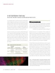
Figure 2—<strong>Cell</strong>Sensor® reporter gene readout <strong>for</strong> PI3K/<strong>Akt</strong> <strong>pathway</strong> analysis. (A) The T-REx FOXO3 DBE-bla HeLa cell line (Cat. no. K1468) has a FOXO3<br />
response element (driving beta-lactamase (BLA) expression (DBE-bla)), tetracycline repressor, and tetracycline-inducible Foxo3 constructs. Addition of doxycycline<br />
(DOX, a tetracycline analog) upregulates FOXO3-driven BLA expression. Activation of <strong>the</strong> endogenous PI3K/<strong>Akt</strong> signaling cascade with insulin (Cat.<br />
no. 12585-014) leads to phosphorylation/inactivation of FOXO3 and concomitant suppression of BLA. Interruption of <strong>the</strong> <strong>pathway</strong> with inhibitors restores<br />
FOXO3 transcriptional activity and thus BLA expression (green to blue cells). (B, top) The cellular response to growth factor stimulation was tested with IGF-1<br />
and insulin, and <strong>the</strong> EC 50 values obtained were 1.6 nM and 5 nM, respectively. (B, bottom) The small-molecule inhibitors PI-103 and PI3Kα inhibitor IV were<br />
evaluated, and <strong>the</strong> IC 50 values obtained were 110 nM and 330 nM, respectively. (C) RNAi knockdown experiments of FOXO3 and AKT <strong>for</strong> target validation<br />
were per<strong>for</strong>med. <strong>Cell</strong>s were reverse-transfected with Lipofectamine RNAiMAX (Cat. no. 13778-075) and 20 nM of a panel of RNAi duplexes, including <strong>the</strong><br />
FOXO3 Validated Stealth RNAi DuoPak (Cat. no. 12937-07), <strong>the</strong> AKT1 Validated Stealth RNAi DuoPak (Cat. no. 12935-001), Stealth RNAi oligonucleotides<br />
against beta-lactamase, and <strong>the</strong> medium GC control from <strong>the</strong> Stealth RNAi Negative Control Kit (Cat. no. 12935-100).<br />
transduction <strong>pathway</strong>s upon exposure to drug candidates or o<strong>the</strong>r stim-<br />
uli. 2 The growing portfolio of cell-<strong>based</strong> beta-lactamase reporter assays<br />
addresses (as part of <strong>the</strong> SelectScreen cell-<strong>based</strong> profiling service) more<br />
than 20 different signaling <strong>pathway</strong>s, as well as specific protein targets<br />
(e.g., kinases) that are involved in <strong>the</strong> endogenous <strong>pathway</strong>s.<br />
To build a PI3K/<strong>Akt</strong> <strong>pathway</strong>–specific <strong>Cell</strong>Sensor® cell line, we stably<br />
engineered <strong>the</strong> beta-lactamase reporter under <strong>the</strong> control of a FOXO3<br />
response element (DBE-bla) into HeLa cervical cancer cells. Additionally,<br />
we fur<strong>the</strong>r designed this cell line to feature tetracycline-inducible FOXO3<br />
expression (via <strong>the</strong> T-REx mechanism), because overexpression of FOXO3<br />
is capable of triggering apoptosis through transcription of cell death<br />
genes (e.g., FAS-L), and precise regulation of FOXO3 levels was <strong>the</strong>re<strong>for</strong>e<br />
necessary. When cells are left untreated and <strong>the</strong>n loaded with LiveBLAzer<br />
substrate, <strong>the</strong> FRET-<strong>based</strong> beta-lactamase substrate remains green (no BLA<br />
present) (Figure 2A). Expression of BLA (induced by tetracycline and driven<br />
by FOXO3), however, results in cleavage of <strong>the</strong> fluorescent substrate mol-<br />
ecule, disrupting <strong>the</strong> energy transfer, and cells turn blue. Activation of PI3K<br />
signaling upon growth factor binding (e.g., insulin or IGF-1; Figure 2B) to a<br />
cell-surface receptor tyrosine kinase leads to increased downstream activity<br />
18 | BioProbes 56 | June 2008<br />
[ FOXO3 ]<br />
(blue cells)<br />
+ DOX + insulin<br />
FOXO3–<br />
(green cells)<br />
<strong>pathway</strong> inhibitor (e.g., PI-103)<br />
RNAi against AKT1<br />
enhances -lac readout<br />
RNAi against FOXO3<br />
diminishes -lac readout<br />
0<br />
no Tfxn mock neg -lac FOXO3 FOXO3<br />
control<br />
#1 #2<br />
AKT1<br />
#1<br />
AKT1<br />
#2<br />
Blue/green ratio (460 nm/530 nm)<br />
Blue/green ratio (460 nm/530 nm)<br />
1.50<br />
1.25<br />
1.00<br />
0.75<br />
0.50<br />
0.25<br />
10<br />
[Agonist] (nM)<br />
–1 100 10 –2 101 102 103 0<br />
1.0<br />
0.9<br />
0.8<br />
0.7<br />
0.6<br />
0.5<br />
0.4<br />
0.3<br />
PI3K inhibitor IV<br />
PI-103<br />
10<br />
[Compound] (nM)<br />
0 101 10 –1 102 103 104 0.2<br />
of AKT and subsequent phosphorylation of <strong>the</strong> FOXO3 transcription factor<br />
(among o<strong>the</strong>r targets) to promote cell survival and oppose apoptosis. This<br />
modification at Thr32 inactivates FOXO3, leading to its translocation out of<br />
<strong>the</strong> nucleus and concomitant suppression of BLA expression to yield green<br />
cells. Application of <strong>pathway</strong> inhibitors (e.g., PI-103 or PI3Kα Inhibitor IV;<br />
Figure 2B) or Stealth RNAi (e.g., against AKT1; Figure 2C) restores FOXO3<br />
activity and BLA expression, turning <strong>the</strong> cells back to blue.<br />
Advantages of <strong>the</strong> <strong>Cell</strong>Sensor® assay system include <strong>the</strong> use of a<br />
sensitive fluorescent reporter readout that is amenable to low-volume<br />
HTS <strong>for</strong>mats, ratiometric FRET-<strong>based</strong> data analysis that improves data<br />
quality, and a live-cell signal (blue/green cells) that is highly suitable<br />
<strong>for</strong> microscopic imaging and cell sorting. The T-REx FOXO3 DBE-bla<br />
HeLa cell line provides an effective endpoint readout of PI3K/<strong>Akt</strong>/<br />
Foxo3 <strong>pathway</strong> alteration upon exposure to a variety of ligands.<br />
LanthaScreen GFP cellular assays<br />
LanthaScreen assays use a technology <strong>based</strong> on time-resolved FRET<br />
(TR-FRET) to monitor phosphorylation of a specific kinase substrate in<br />
© 2008 Invitrogen Corporation. All rights reserved. These products may be covered by one or more Limited Use Label Licenses (see Invitrogen catalog or www.invitrogen.com). By use of <strong>the</strong>se products<br />
you accept <strong>the</strong> terms and conditions of all applicable Limited Use Label Licenses. For research use only. Not intended <strong>for</strong> any animal or human <strong>the</strong>rapeutic or diagnostic use, unless o<strong>the</strong>rwise stated.<br />
Insulin<br />
IGF-1