Cell-based tools for interrogating the pI3K/Akt/mTor pathway
Cell-based tools for interrogating the pI3K/Akt/mTor pathway
Cell-based tools for interrogating the pI3K/Akt/mTor pathway

Create successful ePaper yourself
Turn your PDF publications into a flip-book with our unique Google optimized e-Paper software.
PrACtICAL APPLICAtIONS<br />
160<br />
110<br />
80<br />
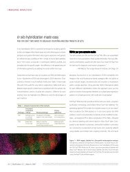
Figure 5—Validation of LanthaScreen and <strong>Cell</strong>Sensor® cell lines via western blot. (A) LanthaScreen GFP-AKT HEK 293E cells were treated under <strong>the</strong><br />
indicated conditions, including IGF-1 (Cat. no. PHG9071), LY294002 (Cat. no. PHZ1144), and wortmannin (Cat. no. PHZ1301). <strong>Cell</strong> extracts were resolved by<br />
SDS-PAGE and transferred to nitrocellulose with <strong>the</strong> iBlot® Gel Transfer Device (Cat. no. IB1001). The membrane was <strong>the</strong>n probed with <strong>the</strong> anti-AKT [PS473] PSSA<br />
(Cat. no. 44-621G), and <strong>the</strong> signal was detected using a WesternBreeze® Chromogenic Kit (Cat. no. WB7105). (B) <strong>Cell</strong>Sensor® T-REx FOXO3 DBE-bla HeLa cell<br />
lysates were generated by treatment with doxycycline to induce FOXO3 expression, followed by incubation with <strong>the</strong> indicated stimulants or inhibitors and lysis.<br />
The nitrocellulose membrane was prepared and developed as in (A), except that <strong>the</strong> PSSA used in this case was anti-FOXO3 [PT32] (Cat. no. 44-1240G).<br />
LY294002, and wortmannin) but is insensitive to rapamycin (Figure 4B).<br />
Interestingly, <strong>the</strong> mTOR kinase is able to phosphorylate PRAS40 directly<br />
at Ser183 in a rapamycin-sensitive manner, which fur<strong>the</strong>r complicates<br />
<strong>the</strong> role of this protein within <strong>the</strong> <strong>mTor</strong> <strong>pathway</strong>.<br />
To monitor <strong>pathway</strong> activation downstream of <strong>the</strong> mTORC1<br />
complex, a cell line was developed that contains a GFP fusion of<br />
programmed cell death protein 4 (PDCD4). This protein is a tumor<br />
suppressor that inhibits translation initiation by binding to eIF4A. In<br />
response to insulin stimulation, PDCD4 is phosphorylated by p70 S6<br />
kinase (at Ser457), which is immediately downstream of (and activated<br />
by) mTORC1. This phosphorylation event marks PDCD4 <strong>for</strong> SCF βTRCP -<br />
mediated ubiquitination and subsequent degradation. 6 Inactivation<br />
of PDCD4 is necessary <strong>for</strong> efficient protein syn<strong>the</strong>sis, and ultimately<br />
<strong>for</strong> cell growth and proliferation. Because PDCD4 is part of <strong>the</strong> PI3K/<br />
<strong>Akt</strong> <strong>pathway</strong> and is linked to mTORC1 activity, its phosphorylation is<br />
sensitive to both PI-103 and rapamycin (Figure 4C).<br />
To examine <strong>pathway</strong> activation downstream of <strong>the</strong> multiprotein<br />
mTOR complex with rictor (mTORC2), a cell line containing a GFP<br />
fusion of AKT was constructed. Also known as protein kinase B (PKB),<br />
AKT has emerged as one of <strong>the</strong> most important and most actively<br />
studied kinases due to its versatility in regulating protein function<br />
and influencing human disease. Although AKT is often placed at <strong>the</strong><br />
“beginning” of <strong>the</strong> <strong>pathway</strong>, it has also been shown to be a substrate<br />
of mTORC2, which phosphorylates AKT at Ser473. 7 This modification is<br />
required <strong>for</strong> full activation of AKT, and it leads to <strong>the</strong> repression of pro-<br />
apoptotic events. The precise mechanism and consequences of Ser473<br />
phosphorylation remain controversial, yet this site has been shown to<br />
be insensitive to acute treatment with rapamycin (Figure 4D).<br />
20 | BioProbes 56 | June 2008<br />
60<br />
50<br />
MW<br />
Untreated<br />
+ Insulin<br />
+ Wortmannin<br />
+ LY290042<br />
+ Rapamycin<br />
A B<br />
GFP–AKT P<br />
AKT P<br />
115<br />
82<br />
+ PI-103<br />
+ LY290042<br />
The future of PI3K/<strong>Akt</strong>/<strong>mTor</strong> <strong>pathway</strong> analysis<br />
Each of <strong>the</strong> cell lines described here is a clonal population isolated by<br />
flow cytometry. The corresponding HTS assays have been optimized<br />
in 384-well <strong>for</strong>mat by testing a variety of parameters (e.g., DMSO<br />
tolerance, cell number, and stimulation time) and provide excellent<br />
statistical data (Z´-factor ≥0.5). Additionally, we have validation data<br />
<strong>for</strong> several known agonists and commercially available ligands, and<br />
<strong>the</strong> observed pharmacology (EC 50 or IC 50 values) is in agreement<br />
with reported literature data. Most importantly, we have compared<br />
<strong>the</strong>se data to those obtained using alternative technologies from<br />
Invitrogen (e.g., western blotting with PSSA (Figure 5); phosphoELISA<br />
and Mercator Phospho-AKT Pathway Array; data not shown), with<br />
excellent correlation seen. Taken toge<strong>the</strong>r, <strong>the</strong>se cell lines constitute<br />
a validated set of <strong>tools</strong> <strong>for</strong> studying a complex <strong>pathway</strong> in a simpli-<br />
fied <strong>for</strong>mat that complement our existing biochemical assays. These<br />
cell-<strong>based</strong> assays will facilitate <strong>the</strong> identification of novel modulators<br />
of PI3K/<strong>Akt</strong>/<strong>mTor</strong> signaling. Learn more about <strong>Akt</strong> <strong>pathway</strong>–related<br />
products at www.invitrogen.com/bioprobes56. ■<br />
References<br />
+ Wortmannin<br />
+ AKT inhibitor II<br />
+ PI3K inhibitor II<br />
+ AKT inhibitor IV<br />
+ AKT inhibitor VIII<br />
+ Insulin<br />
<strong>Cell</strong>s: GFP-AKT 293E WB: anti-AKT [pS473] <strong>Cell</strong>s: T-REx FOXO3 DBE-bla HeLa WB: anti-FOXO3 [pT32]<br />
1. Manning, B.D. and Cantley, L.C. (2007) <strong>Cell</strong> 129:1261–1274.<br />
2. Zlokarnik, G. et al. (1998) Science 279:84–88.<br />
3. Robers, M.B. et al. (2008) Anal Biochem 372:189–197.<br />
4. Fonesca, B.D. et al. (2007) J Biol Chem 282:24514–24524.<br />
5. Vander Haar, E. et al. (2007) Nat <strong>Cell</strong> Biol 9:316–323.<br />
6. Dorrello, N.V. et al. (2006) Science 314:467–471.<br />
7. Sarbassov, D.D. et al. (2005) Science 307:1098–1101.<br />
FOXO3 P<br />
© 2008 Invitrogen Corporation. All rights reserved. These products may be covered by one or more Limited Use Label Licenses (see Invitrogen catalog or www.invitrogen.com). By use of <strong>the</strong>se products<br />
you accept <strong>the</strong> terms and conditions of all applicable Limited Use Label Licenses. For research use only. Not intended <strong>for</strong> any animal or human <strong>the</strong>rapeutic or diagnostic use, unless o<strong>the</strong>rwise stated.<br />
+ IGF-1<br />
+ Growth medium<br />
No DOX; + insulin