Fungi Fimicoli Italici - Mycosphere-online journal
Fungi Fimicoli Italici - Mycosphere-online journal
Fungi Fimicoli Italici - Mycosphere-online journal

You also want an ePaper? Increase the reach of your titles
YUMPU automatically turns print PDFs into web optimized ePapers that Google loves.
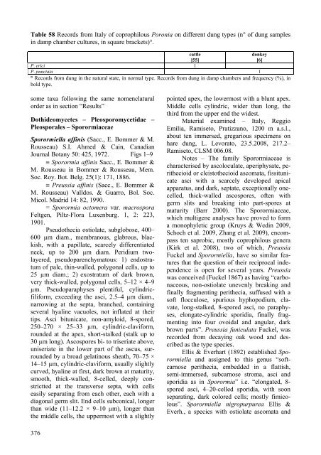
Table 58 Records from Italy of coprophilous Poronia on different dung types (n° of dung samples<br />
in damp chamber cultures, in square brackets)°.<br />
cattle<br />
donkey<br />
[55]<br />
[6]<br />
P. erici 1<br />
P. punctata 1<br />
° Records from dung in the natural state, in normal type. Records from dung in damp chambers and frequency (%), in<br />
bold type.<br />
some taxa following the same nomenclatural<br />
order as in section “Results”<br />
Dothideomycetes – Pleosporomycetidae –<br />
Pleosporales – Sporormiaceae<br />
Sporormiella affinis (Sacc., E. Bommer & M.<br />
Rousseau) S.I. Ahmed & Cain, Canadian<br />
Journal Botany 50: 425, 1972. Figs 1–9<br />
Sporormia affinis Sacc., E. Bommer &<br />
M. Rousseau in Bommer & Rousseau, Mem.<br />
Soc. Roy. Bot. Belg. 25(1): 171, 1886.<br />
Preussia affinis (Sacc., E. Bommer &<br />
M. Rousseau) Valldos. & Guarro, Bol. Soc.<br />
Micol. Madrid 14: 82, 1990.<br />
= Sporormia octomera var. macrospora<br />
Feltgen, Piltz-Flora Luxenburg. 1, 2: 223,<br />
1901.<br />
Pseudothecia ostiolate, subglobose, 400–<br />
600 µm diam., membranous, glabrous, blackish,<br />
with a papillate, scarcely differentiated<br />
neck, up to 200 µm diam. Peridium twolayered,<br />
pseudoparenchymatous: 1) endostratum<br />
of pale, thin-walled, polygonal cells, up to<br />
25 µm diam.; 2) exostratum of dark brown,<br />
very thick-walled, polygonal cells, 5–12 × 4–9<br />
µm. Pseudoparaphyses plentiful, cylindricfiliform,<br />
exceeding the asci, 2.5–4 µm diam.,<br />
narrowing at the septa, branched, containing<br />
several hyaline vacuoles, not inflated at their<br />
tips. Asci bitunicate, non-amyloid, 8-spored,<br />
250–270 × 25–33 µm, cylindric-claviform,<br />
rounded at the apex, short-stalked (stalk up to<br />
30 µm long). Ascospores bi- to triseriate above,<br />
uniseriate in the lower part of the ascus, surrounded<br />
by a broad gelatinous sheath, 70–75 ×<br />
14–15 µm, cylindric-claviform, usually slightly<br />
curved, hyaline at first, dark brown at maturity,<br />
smooth, thick-walled, 8-celled, deeply constrictted<br />
at the transverse septa, with cells<br />
easily separating from each other, each with a<br />
diagonal germ slit. End cells subconical, longer<br />
than wide (11–12.2 × 9–10 µm), longer than<br />
the middle cells, the uppermost with a slightly<br />
376<br />
pointed apex, the lowermost with a blunt apex.<br />
Middle cells cylindric, wider than long, the<br />
third from the upper end the widest.<br />
Material examined – Italy, Reggio<br />
Emilia, Ramiseto, Pratizzano, 1200 m a.s.l.,<br />
about ten immersed, gregarious specimens on<br />
hare dung, L. Levorato, 23.5.2008, 217.2–<br />
Ramiseto, CLSM 006.08.<br />
Notes – The family Sporormiaceae is<br />
characterised by ascoloculate, aperiphysate, perithecioid<br />
or cleistothecioid ascomata, fissitunicate<br />
asci with a scarcely developed apical<br />
apparatus, and dark, septate, exceptionally onecelled,<br />
thick-walled ascospores, often with<br />
germ slits and breaking into part-spores at<br />
maturity (Barr 2000). The Sporormiaceae,<br />
which multigene analyses have proved to form<br />
a monophyletic group (Kruys & Wedin 2009,<br />
Schoch et al. 2009, Zhang et al. 2009), encompass<br />
ten saprobic, mostly coprophilous genera<br />
(Kirk et al. 2008), two of which, Preussia<br />
Fuckel and Sporormiella, have so similar features<br />
that the question of their reciprocal independence<br />
is open for several years. Preussia<br />
was conceived (Fuckel 1867) as having “carbonaceous,<br />
non-ostiolate unevenly breaking and<br />
finally fragmenting perithecia, suffused with a<br />
soft flocculose, spurious hyphopodium, clavate,<br />
long-stalked, 8-spored asci, no paraphyses,<br />
elongate-cylindric sporidia, finally fragmenting<br />
into four ovoidal and angular, dark<br />
brown parts”. Preussia funiculata Fuckel, was<br />
recorded from decaying oak wood and described<br />
as the type species.<br />
Ellis & Everhart (1892) established Sporormiella<br />
and assigned to this genus “softcarnose<br />
perithecia, embedded in a flattish,<br />
semi-immersed, subcarnose stroma, asci and<br />
sporidia as in Sporormia” i.e. “elongated, 8spored<br />
asci, 4–20-celled sporidia, with soon<br />
separating, dark colored cells; mostly fimicolous”.<br />
Sporormiella nigropurpurea Ellis &<br />
Everh., a species with ostiolate ascomata and