Radial osteomyelitis as a complication of venous cannulation
Radial osteomyelitis as a complication of venous cannulation
Radial osteomyelitis as a complication of venous cannulation
You also want an ePaper? Increase the reach of your titles
YUMPU automatically turns print PDFs into web optimized ePapers that Google loves.
408<br />
Department <strong>of</strong><br />
Pediatrics C,<br />
Schneider Children’s<br />
Medical Center <strong>of</strong><br />
Israel, Petah Tikva,<br />
Israel 49202<br />
R Straussberg<br />
L Harel<br />
J Amir<br />
Department <strong>of</strong><br />
Nuclear Imaging,<br />
Schneider Children’s<br />
Medical Center <strong>of</strong><br />
Israel, Petah Tikva and<br />
Sackler School <strong>of</strong><br />
Medicine, Tel Aviv<br />
University, Tel Aviv,<br />
Israel<br />
Z Bar-Sever<br />
Correspondence to:<br />
Dr Straussberg<br />
rachelst@post.tau.ac.il<br />
Accepted 20 July 2001<br />
Downloaded from<br />
adc.bmj.com on April 5, 2013 - Published by group.bmj.com<br />
<strong>Radial</strong> <strong>osteomyelitis</strong> <strong>as</strong> a <strong>complication</strong> <strong>of</strong> <strong>venous</strong><br />
<strong>cannulation</strong><br />
R Straussberg, L Harel, Z Bar-Sever, J Amir<br />
Abstract<br />
Venepuncture <strong>of</strong> the superficial veins in<br />
the forearm is considered a relatively safe<br />
procedure. We report two patients who<br />
presented with <strong>osteomyelitis</strong> <strong>of</strong> the proximal<br />
radius following <strong>venous</strong> <strong>cannulation</strong><br />
<strong>of</strong> the median cubital vein, and one patient<br />
who developed <strong>osteomyelitis</strong> <strong>of</strong> the distal<br />
radius after <strong>cannulation</strong> <strong>of</strong> the cephalic<br />
vein. Osteomyelitis developing in proximity<br />
to a venepuncture site should raise the<br />
suspicion that a pathogen causing superficial<br />
thrombophlebitis h<strong>as</strong> spread through<br />
the deep veins <strong>of</strong> the arm into the adjacent<br />
bone, thus causing <strong>osteomyelitis</strong>.<br />
(Arch Dis Child 2001;85:408–410)<br />
Keywords: <strong>osteomyelitis</strong>; <strong>venous</strong> <strong>cannulation</strong>;<br />
thrombophlebitis<br />
Venous <strong>cannulation</strong> <strong>of</strong> the superficial forearm<br />
veins is a common procedure. The visible and<br />
accessible median cubital vein is a preferred<br />
site for insertion <strong>of</strong> an intra<strong>venous</strong> line. In the<br />
majority <strong>of</strong> c<strong>as</strong>es, the procedure is without<br />
<strong>complication</strong>s, although superficial thrombophlebitis<br />
is sometimes a sequela. Rarely,<br />
infection <strong>of</strong> the median cubital vein spreads to<br />
the deep <strong>venous</strong> system <strong>of</strong> the arm.<br />
We report a rare <strong>complication</strong> <strong>of</strong> catheterisation<br />
<strong>of</strong> the superficial veins <strong>of</strong> the forearm (the<br />
median cubital and cephalic veins). This<br />
caused proximal and distal radial <strong>osteomyelitis</strong>,<br />
respectively. The infection developed at the site<br />
<strong>of</strong> venepuncture, suggesting that it spread<br />
locally through an<strong>as</strong>tomoses between the<br />
superficial medial cubital and cephalic veins<br />
and the deep radial vein. Whenever point<br />
tenderness develops over a long bone, over the<br />
underlying skin, after venepuncture, <strong>osteomyelitis</strong><br />
should be suspected.<br />
C<strong>as</strong>e reports<br />
CASE 1<br />
A 13 year old boy w<strong>as</strong> admitted because <strong>of</strong> pain<br />
in the left elbow. He had been diagnosed <strong>as</strong><br />
having familial Mediterranean fever eight years<br />
previously, on the b<strong>as</strong>is <strong>of</strong> a history <strong>of</strong> bouts <strong>of</strong><br />
fever accompanied by arthritis <strong>of</strong> the hip, knee,<br />
and ankle joints. He w<strong>as</strong> treated regularly with<br />
colchicine 1.5 mg/day. Seven days prior to<br />
admission to our hospital, he w<strong>as</strong> hospitalised<br />
elsewhere with pneumonia. Treatment consisted<br />
<strong>of</strong> cefuroxime administered through a<br />
“Quickcath” inserted in the left median cubital<br />
vein. Blood culture w<strong>as</strong> negative. He w<strong>as</strong><br />
discharged after four days and prescribed oral<br />
roxithromycin 150 mg twice daily.<br />
www.archdischild.com<br />
Arch Dis Child 2001;85:408–410<br />
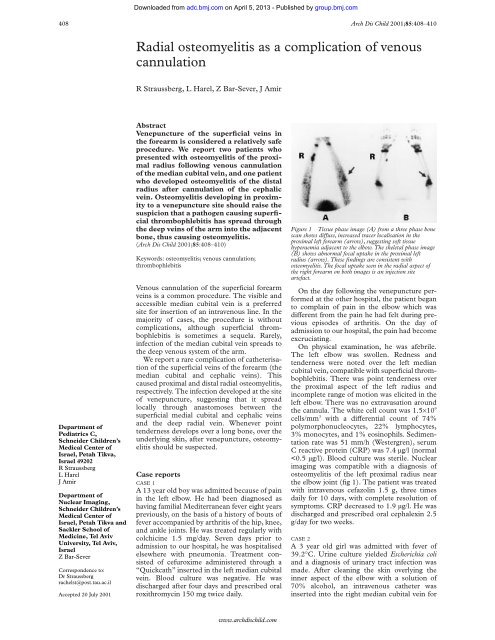
Figure 1 Tissue ph<strong>as</strong>e image (A) from a three ph<strong>as</strong>e bone<br />
scan shows diVuse, incre<strong>as</strong>ed tracer localisation in the<br />
proximal left forearm (arrow), suggesting s<strong>of</strong>t tissue<br />
hyperaemia adjacent to the elbow. The skeletal ph<strong>as</strong>e image<br />
(B) shows abnormal focal uptake in the proximal left<br />
radius (arrow). These findings are consistent with<br />
<strong>osteomyelitis</strong>. The focal uptake seen in the radial <strong>as</strong>pect <strong>of</strong><br />
the right forearm on both images is an injection site<br />
artefact.<br />
On the day following the venepuncture performed<br />
at the other hospital, the patient began<br />
to complain <strong>of</strong> pain in the elbow which w<strong>as</strong><br />
diVerent from the pain he had felt during previous<br />
episodes <strong>of</strong> arthritis. On the day <strong>of</strong><br />
admission to our hospital, the pain had become<br />
excruciating.<br />
On physical examination, he w<strong>as</strong> afebrile.<br />
The left elbow w<strong>as</strong> swollen. Redness and<br />
tenderness were noted over the left median<br />
cubital vein, compatible with superficial thrombophlebitis.<br />
There w<strong>as</strong> point tenderness over<br />
the proximal <strong>as</strong>pect <strong>of</strong> the left radius and<br />
incomplete range <strong>of</strong> motion w<strong>as</strong> elicited in the<br />
left elbow. There w<strong>as</strong> no extrav<strong>as</strong>ation around<br />
the cannula. The white cell count w<strong>as</strong> 1.5×10 9<br />
cells/mm 3 with a diVerential count <strong>of</strong> 74%<br />
polymorphonucleocytes, 22% lymphocytes,<br />
3% monocytes, and 1% eosinophils. Sedimentation<br />
rate w<strong>as</strong> 51 mm/h (Westergren), serum<br />
C reactive protein (CRP) w<strong>as</strong> 7.4 µg/l (normal<br />
Downloaded from<br />
adc.bmj.com on April 5, 2013 - Published by group.bmj.com<br />
<strong>Radial</strong> <strong>osteomyelitis</strong> <strong>as</strong> a <strong>complication</strong> <strong>of</strong> <strong>venous</strong> <strong>cannulation</strong> 409<br />
administration <strong>of</strong> gentamicin. The next day<br />
body temperature returned to normal. The<br />
intra<strong>venous</strong> line w<strong>as</strong> transferred to the contralateral<br />
side after three days. However, after five<br />
days <strong>of</strong> treatment, the temperature rose again,<br />
and the parents noted the refusal <strong>of</strong> the child to<br />
move her right arm.<br />
On physical examination the skin overlying<br />
the right elbow w<strong>as</strong> warmer than the contralateral<br />
side, and there w<strong>as</strong> point tenderness<br />
over the medial <strong>as</strong>pect <strong>of</strong> the proximal radius.<br />
There w<strong>as</strong> no extrav<strong>as</strong>ation around the cannula.<br />
White blood cell count w<strong>as</strong> 1.7×10 9 cells/<br />
mm 3 with a diVerential count <strong>of</strong> 68% polymorphonucleocytes,<br />
20% lymphocytes, 3%<br />
eosinophils, 8% monocytes, and 1% b<strong>as</strong>ophils.<br />
An x ray examination <strong>of</strong> the arm w<strong>as</strong><br />
interpreted <strong>as</strong> normal. Sedimentation rate w<strong>as</strong><br />
72 mm/h (Westergren). Blood culture w<strong>as</strong> sterile.<br />
Radionuclide imaging showed pathological<br />
uptake <strong>of</strong> the isotope in the right proximal<br />
radius at the late ph<strong>as</strong>e <strong>of</strong> the examination. A<br />
diagnosis <strong>of</strong> <strong>osteomyelitis</strong> w<strong>as</strong> made. The<br />
patient w<strong>as</strong> treated with cloxacillin 1.5 g per<br />
day intra<strong>venous</strong>ly for three weeks with complete<br />
resolution <strong>of</strong> symptoms. At discharge she<br />
w<strong>as</strong> prescribed oral cephalexin 1.5 g/day for an<br />
additional two weeks.<br />
CASE 3<br />
An 18 year old female with insulin dependent<br />
diabetes mellitus (diagnosed at the age <strong>of</strong> 8<br />
years and treated with subcutaneous insulin),<br />
w<strong>as</strong> admitted to our hospital with fever, sore<br />
throat, and abdominal pain; there w<strong>as</strong> laboratory<br />
evidence <strong>of</strong> ketoacidosis and pharyngitis<br />
w<strong>as</strong> diagnosed. After cleaning the skin over the<br />
distal end <strong>of</strong> the forearm with a solution <strong>of</strong> 70%<br />
alcohol, an intra<strong>venous</strong> line w<strong>as</strong> inserted into<br />
the cephalic vein; treatment with intra<strong>venous</strong><br />
fluids and insulin w<strong>as</strong> initiated. Initial leucocyte<br />
count w<strong>as</strong> 0.8×10 9 cells/mm 3 with 63%<br />
polymorphonucleocytes, 20% lymphocytes,<br />
5% monocytes, 10% eosinophils, and 2%<br />
b<strong>as</strong>ophils. The pharyngitis w<strong>as</strong> believed to be<br />
viral. On the third day her temperature<br />
returned to normal; she w<strong>as</strong> treated with<br />
subcutaneous insulin, but continued to be hospitalised<br />
because <strong>of</strong> unstable blood glucose<br />
concentrations.<br />
On the fifth day her temperature rose to<br />
39.6°C and she complained <strong>of</strong> pain in the distal<br />
<strong>as</strong>pect <strong>of</strong> the radius. On examination there<br />
were signs <strong>of</strong> phlebitis—the skin over the intra<strong>venous</strong><br />
insertion w<strong>as</strong> warm and red but point<br />
tenderness w<strong>as</strong> elicited only secondary to<br />
strong pressure on the area <strong>of</strong> the styloid process<br />
<strong>of</strong> the radius. There w<strong>as</strong> no extrav<strong>as</strong>ion<br />
around the cannula. Blood count w<strong>as</strong> 1.3×10 9<br />
leucocytes per mm 3 with a diVerential count <strong>of</strong><br />
63% polymorphonucleocytes, 17% lymphocytes,<br />
11% monocytes, 6% eosinophils,<br />
and 3% b<strong>as</strong>ophils. Sedimentation rate w<strong>as</strong> 94<br />
mm/h (Westergren); serum CRP w<strong>as</strong> 11.1<br />
µg/dl. Blood culture w<strong>as</strong> negative. The intra<strong>venous</strong><br />
line w<strong>as</strong> transferred to another site. A<br />
technetium bone scan in the bony ph<strong>as</strong>e<br />
suggested incre<strong>as</strong>ed uptake <strong>of</strong> the colloid in the<br />
styloid process <strong>of</strong> the distal radius, supporting a<br />
diagnosis <strong>of</strong> <strong>osteomyelitis</strong> (fig 2).<br />
www.archdischild.com<br />
Figure 2 Dynamic images (A) from the angiographic<br />
ph<strong>as</strong>e <strong>of</strong> a bone scan show incre<strong>as</strong>ed blood flow to the left<br />
wrist. Dorsal (B) and palmar (C) views <strong>of</strong> the hands from<br />
the skeletal ph<strong>as</strong>e <strong>of</strong> the study show diVuse, incre<strong>as</strong>ed<br />
uptake in the region <strong>of</strong> the left wrist and carpus, consistent<br />
with cellulitis. There is some prominence in the appearance<br />
<strong>of</strong> the styloid process <strong>of</strong> the radius, suggesting <strong>osteomyelitis</strong>.<br />
Treatment with vancomycin 50 mg/kg/day in<br />
three divided doses and garamicin 5 mg/kg/day<br />
<strong>as</strong> a single dose, w<strong>as</strong> commenced. Temperature<br />
returned to normal after three days and the<br />
patient ce<strong>as</strong>ed to complain <strong>of</strong> pain after two<br />
days.<br />
Discussion<br />
The veins <strong>of</strong> the upper arm are frequently used<br />
for drawing blood for intra<strong>venous</strong> injections<br />
and infusions. The median cubital vein is commonly<br />
used for venepuncture and for <strong>cannulation</strong><br />
because it is e<strong>as</strong>ily accessible and allows<br />
communication between the b<strong>as</strong>ilic and cephalic<br />
veins, through which superficial <strong>venous</strong><br />
drainage <strong>of</strong> the forearm occurs. The b<strong>as</strong>ilic vein<br />
penetrates the deep f<strong>as</strong>cia on the medial side <strong>of</strong><br />
the middle part <strong>of</strong> the arm and then joins the<br />
brachial veins to form the axillary vein.<br />
Numerous deep veins drain the structures <strong>of</strong><br />
the forearm. They arise from a deep <strong>venous</strong><br />
arcade (a series <strong>of</strong> an<strong>as</strong>tomosing <strong>venous</strong><br />
arches) in the hand. The deep veins <strong>as</strong>cend the<br />
forearm along the sides <strong>of</strong> the corresponding<br />
arteries, receiving tributaries from veins leaving<br />
the related muscles and communicating with<br />
superficial veins. The deep interosseous veins<br />
that accompany the respective arteries unite<br />
with the accompanying veins <strong>of</strong> the radial and<br />
ulnar arteries. The deep veins in the cubital<br />
fossa are connected to the median cubital vein<br />
and unite with the accompanying veins <strong>of</strong> the<br />
respective artery. 1<br />
In the first two patients, <strong>osteomyelitis</strong> <strong>of</strong> the<br />
proximal radius developed after venepuncture<br />
and insertion <strong>of</strong> an intra<strong>venous</strong> line through<br />
the median cubital vein. In the third patient,<br />
<strong>osteomyelitis</strong> <strong>of</strong> the distal radius developed <strong>as</strong> a<br />
consequence <strong>of</strong> cannulating the cephalic vein.<br />
Although the bacterial aetiology w<strong>as</strong> not<br />
defined, we believe that the diagnosis <strong>of</strong> <strong>osteomyelitis</strong><br />
w<strong>as</strong> established b<strong>as</strong>ed on the clinical<br />
findings and the results <strong>of</strong> the nuclear imaging.<br />
We were able to find several previous reports <strong>of</strong><br />
<strong>osteomyelitis</strong> <strong>as</strong> a <strong>complication</strong> <strong>of</strong> venepuncture.<br />
2–6 The site <strong>of</strong> <strong>osteomyelitis</strong> w<strong>as</strong> the<br />
clavicle in all c<strong>as</strong>es, and the infection developed<br />
following subclavian vein catheterisation. The
Downloaded from<br />
adc.bmj.com on April 5, 2013 - Published by group.bmj.com<br />
410 Straussberg, Harel, Bar-Sever, Amir<br />
investigators <strong>as</strong>sumed that the causative organisms<br />
were inoculated directly into the clavicular<br />
periosteum and did not propagate from distant<br />
foci. 5 We have found two additional reports, <strong>of</strong><br />
<strong>osteomyelitis</strong> secondary to multiple punctures<br />
<strong>of</strong> the great toe for draining blood in a premature<br />
infant, 7 and following needle puncture in<br />
two neonates. 8 We suggest that in our three<br />
c<strong>as</strong>es, bacteria, possibly Staphylococcus aureus<br />
from the overlying skin inoculated during the<br />
venepuncture, caused superficial thrombophlebitis<br />
<strong>of</strong> the median cubital vein and the cephalic<br />
vein. From there, the infection spread to the<br />
deep <strong>venous</strong> system <strong>of</strong> the forearm, via the<br />
radial veins through their an<strong>as</strong>tomoses with the<br />
median cubital vein and the cephalic vein, and<br />
then to the adjacent bone itself. The fact that<br />
<strong>osteomyelitis</strong> developed in the site <strong>of</strong> the<br />
venepuncture and not in a distant locus<br />
suggests that there w<strong>as</strong> a local infection and not<br />
bacteraemia with distant dissemination. The<br />
blood cultures in our three patients were negative,<br />
thus ruling out a pneumonia causing agent<br />
or E coli bacteraemia <strong>as</strong> aetiologies for the<br />
<strong>osteomyelitis</strong>. There were no signs <strong>of</strong> superinfection<br />
around the cannula site which extended<br />
down into deep tissues with direct continuous<br />
Rapid responses<br />
spread. The inflamed area appeared to be<br />
thrombophlebitis and not local cellulitis.<br />
We believe that this <strong>complication</strong> <strong>of</strong> intra<strong>venous</strong><br />
line insertion h<strong>as</strong> not been reported<br />
previously in the English literature. We conclude<br />
that <strong>osteomyelitis</strong> should be suspected<br />
whenever point tenderness over a long bone<br />
develops after venepuncture.<br />
1 Moore KI, Dalley AF. Forearm. In: Kelly PJ, ed. Clinically<br />
oriented anatomy, 4th edn. Baltimore: Lippincott Williams<br />
& Wilkins, 1999:734–57.<br />
2 Lee Y-H, Kerstein MD. Osteomyelitis and septic arthritis: a<br />
<strong>complication</strong> <strong>of</strong> subclavian <strong>venous</strong> catheterization. N Engl J<br />
Med 1971;285:79–80.<br />
3 Manny J, Haruzi I, Yosipovitch Z. Osteomyelitis <strong>of</strong> the clavicle<br />
following subclavian vein catheterization. Arch Surg<br />
1973;106:342–3.<br />
4 Steein J, Pederson JHJ. Osteomyelitis <strong>of</strong> the clavicle following<br />
percutaneous subclavian catheterization. Dan Med Bull<br />
1978;25:260–1.<br />
5 Klein B, Mittelman M, Katz R, Djaldetti M. Osteomyelitis<br />
<strong>of</strong> both clavicles <strong>as</strong> a <strong>complication</strong> <strong>of</strong> subclavian venipuncture.<br />
Chest 1983;83:143–4.<br />
6 Garcia S, Combalia A, Segur JM, Llovera AJ. Osteomyelitis<br />
<strong>of</strong> the clavicle. A c<strong>as</strong>e report. Acta Orthop Belg 1999;65:<br />
369–71.<br />
7 Puczynski MS, Dvonch VM, Menendez CE, Caldwell CC.<br />
Osteomyelitis <strong>of</strong> the great toe secondary to phlebotomy.<br />
Clin Orthop 1984;190:239–40.<br />
8 Nelson DL, Hable KA, Matsen JM. Proteus mirabilis <strong>osteomyelitis</strong><br />
in two neonates following needle puncture.<br />
Successful treatment with ampicilin. Am J Dis Child 1973;<br />
125:109–10.<br />
Letters on the following papers have been published recently <strong>as</strong> rapid responses on the ADC website. To read<br />
these letters visit www.archdischild.com and click on “Read eLetters”<br />
Diagnostic <strong>as</strong>sessment <strong>of</strong> haemorrhagic r<strong>as</strong>h and fever. H E Nielsen, E A Andersen, J Andersen, et al.<br />
Arch Dis Child 2001;85:160–5.<br />
The child with a non-blanching r<strong>as</strong>h: how likely is meningococcal dise<strong>as</strong>e? L C Wells, J C Smith,<br />
V C Weston, et al. Arch Dis Child 2001;85:218–22.<br />
Me<strong>as</strong>urement <strong>of</strong> urinary constituents and output using disposable napkins. SB Roberts, A Luc<strong>as</strong>. Arch<br />
Dis Child 1985;60:1021–4.<br />
Accidents and resulting injuries in premobile infants: data from the ALSPAC study. S A Warrington,<br />
C M Wright, ALSPAC Study Team. Arch Dis Child 2001;85:104–7.<br />
If you would like to post an electronic response to these or any other articles published in the journal, ple<strong>as</strong>e<br />
go to the website, access the article in which you are interested, and click on “eLetters: Submit a response to<br />
this article” in the box in the top right hand corner.<br />
www.archdischild.com
References<br />
Email alerting<br />
service<br />
Topic<br />
Collections<br />
Notes<br />
<strong>Radial</strong> <strong>osteomyelitis</strong> <strong>as</strong> a <strong>complication</strong> <strong>of</strong><br />
<strong>venous</strong> <strong>cannulation</strong><br />
R Straussberg, L Harel, Z Bar-Sever, et al.<br />
Arch Dis Child 2001 85: 408-410<br />
doi: 10.1136/adc.85.5.408<br />
Updated information and services can be found at:<br />
http://adc.bmj.com/content/85/5/408.full.html<br />
These include:<br />
To request permissions go to:<br />
http://group.bmj.com/group/rights-licensing/permissions<br />
To order reprints go to:<br />
http://journals.bmj.com/cgi/reprintform<br />
To subscribe to BMJ go to:<br />
http://group.bmj.com/subscribe/<br />
Downloaded from<br />
adc.bmj.com on April 5, 2013 - Published by group.bmj.com<br />
This article cites 7 articles<br />
http://adc.bmj.com/content/85/5/408.full.html#ref-list-1<br />
Receive free email alerts when new articles cite this article. Sign up in the<br />
box at the top right corner <strong>of</strong> the online article.<br />
Articles on similar topics can be found in the following collections<br />
Clinical diagnostic tests (534 articles)<br />
Radiology (458 articles)<br />
Bone and joint infections (14 articles)<br />
Rheumatology (289 articles)


