Radial osteomyelitis as a complication of venous cannulation
Radial osteomyelitis as a complication of venous cannulation
Radial osteomyelitis as a complication of venous cannulation

Create successful ePaper yourself
Turn your PDF publications into a flip-book with our unique Google optimized e-Paper software.
Downloaded from<br />
adc.bmj.com on April 5, 2013 - Published by group.bmj.com<br />
<strong>Radial</strong> <strong>osteomyelitis</strong> <strong>as</strong> a <strong>complication</strong> <strong>of</strong> <strong>venous</strong> <strong>cannulation</strong> 409<br />
administration <strong>of</strong> gentamicin. The next day<br />
body temperature returned to normal. The<br />
intra<strong>venous</strong> line w<strong>as</strong> transferred to the contralateral<br />
side after three days. However, after five<br />
days <strong>of</strong> treatment, the temperature rose again,<br />
and the parents noted the refusal <strong>of</strong> the child to<br />
move her right arm.<br />
On physical examination the skin overlying<br />
the right elbow w<strong>as</strong> warmer than the contralateral<br />
side, and there w<strong>as</strong> point tenderness<br />
over the medial <strong>as</strong>pect <strong>of</strong> the proximal radius.<br />
There w<strong>as</strong> no extrav<strong>as</strong>ation around the cannula.<br />
White blood cell count w<strong>as</strong> 1.7×10 9 cells/<br />
mm 3 with a diVerential count <strong>of</strong> 68% polymorphonucleocytes,<br />
20% lymphocytes, 3%<br />
eosinophils, 8% monocytes, and 1% b<strong>as</strong>ophils.<br />
An x ray examination <strong>of</strong> the arm w<strong>as</strong><br />
interpreted <strong>as</strong> normal. Sedimentation rate w<strong>as</strong><br />
72 mm/h (Westergren). Blood culture w<strong>as</strong> sterile.<br />
Radionuclide imaging showed pathological<br />
uptake <strong>of</strong> the isotope in the right proximal<br />
radius at the late ph<strong>as</strong>e <strong>of</strong> the examination. A<br />
diagnosis <strong>of</strong> <strong>osteomyelitis</strong> w<strong>as</strong> made. The<br />
patient w<strong>as</strong> treated with cloxacillin 1.5 g per<br />
day intra<strong>venous</strong>ly for three weeks with complete<br />
resolution <strong>of</strong> symptoms. At discharge she<br />
w<strong>as</strong> prescribed oral cephalexin 1.5 g/day for an<br />
additional two weeks.<br />
CASE 3<br />
An 18 year old female with insulin dependent<br />
diabetes mellitus (diagnosed at the age <strong>of</strong> 8<br />
years and treated with subcutaneous insulin),<br />
w<strong>as</strong> admitted to our hospital with fever, sore<br />
throat, and abdominal pain; there w<strong>as</strong> laboratory<br />
evidence <strong>of</strong> ketoacidosis and pharyngitis<br />
w<strong>as</strong> diagnosed. After cleaning the skin over the<br />
distal end <strong>of</strong> the forearm with a solution <strong>of</strong> 70%<br />
alcohol, an intra<strong>venous</strong> line w<strong>as</strong> inserted into<br />
the cephalic vein; treatment with intra<strong>venous</strong><br />
fluids and insulin w<strong>as</strong> initiated. Initial leucocyte<br />
count w<strong>as</strong> 0.8×10 9 cells/mm 3 with 63%<br />
polymorphonucleocytes, 20% lymphocytes,<br />
5% monocytes, 10% eosinophils, and 2%<br />
b<strong>as</strong>ophils. The pharyngitis w<strong>as</strong> believed to be<br />
viral. On the third day her temperature<br />
returned to normal; she w<strong>as</strong> treated with<br />
subcutaneous insulin, but continued to be hospitalised<br />
because <strong>of</strong> unstable blood glucose<br />
concentrations.<br />
On the fifth day her temperature rose to<br />
39.6°C and she complained <strong>of</strong> pain in the distal<br />
<strong>as</strong>pect <strong>of</strong> the radius. On examination there<br />
were signs <strong>of</strong> phlebitis—the skin over the intra<strong>venous</strong><br />
insertion w<strong>as</strong> warm and red but point<br />
tenderness w<strong>as</strong> elicited only secondary to<br />
strong pressure on the area <strong>of</strong> the styloid process<br />
<strong>of</strong> the radius. There w<strong>as</strong> no extrav<strong>as</strong>ion<br />
around the cannula. Blood count w<strong>as</strong> 1.3×10 9<br />
leucocytes per mm 3 with a diVerential count <strong>of</strong><br />
63% polymorphonucleocytes, 17% lymphocytes,<br />
11% monocytes, 6% eosinophils,<br />
and 3% b<strong>as</strong>ophils. Sedimentation rate w<strong>as</strong> 94<br />
mm/h (Westergren); serum CRP w<strong>as</strong> 11.1<br />
µg/dl. Blood culture w<strong>as</strong> negative. The intra<strong>venous</strong><br />
line w<strong>as</strong> transferred to another site. A<br />
technetium bone scan in the bony ph<strong>as</strong>e<br />
suggested incre<strong>as</strong>ed uptake <strong>of</strong> the colloid in the<br />
styloid process <strong>of</strong> the distal radius, supporting a<br />
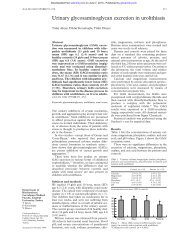
diagnosis <strong>of</strong> <strong>osteomyelitis</strong> (fig 2).<br />
www.archdischild.com<br />
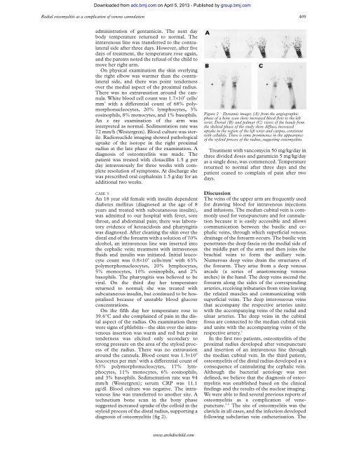
Figure 2 Dynamic images (A) from the angiographic<br />
ph<strong>as</strong>e <strong>of</strong> a bone scan show incre<strong>as</strong>ed blood flow to the left<br />
wrist. Dorsal (B) and palmar (C) views <strong>of</strong> the hands from<br />
the skeletal ph<strong>as</strong>e <strong>of</strong> the study show diVuse, incre<strong>as</strong>ed<br />
uptake in the region <strong>of</strong> the left wrist and carpus, consistent<br />
with cellulitis. There is some prominence in the appearance<br />
<strong>of</strong> the styloid process <strong>of</strong> the radius, suggesting <strong>osteomyelitis</strong>.<br />
Treatment with vancomycin 50 mg/kg/day in<br />
three divided doses and garamicin 5 mg/kg/day<br />
<strong>as</strong> a single dose, w<strong>as</strong> commenced. Temperature<br />
returned to normal after three days and the<br />
patient ce<strong>as</strong>ed to complain <strong>of</strong> pain after two<br />
days.<br />
Discussion<br />
The veins <strong>of</strong> the upper arm are frequently used<br />
for drawing blood for intra<strong>venous</strong> injections<br />
and infusions. The median cubital vein is commonly<br />
used for venepuncture and for <strong>cannulation</strong><br />
because it is e<strong>as</strong>ily accessible and allows<br />
communication between the b<strong>as</strong>ilic and cephalic<br />
veins, through which superficial <strong>venous</strong><br />
drainage <strong>of</strong> the forearm occurs. The b<strong>as</strong>ilic vein<br />
penetrates the deep f<strong>as</strong>cia on the medial side <strong>of</strong><br />
the middle part <strong>of</strong> the arm and then joins the<br />
brachial veins to form the axillary vein.<br />
Numerous deep veins drain the structures <strong>of</strong><br />
the forearm. They arise from a deep <strong>venous</strong><br />
arcade (a series <strong>of</strong> an<strong>as</strong>tomosing <strong>venous</strong><br />
arches) in the hand. The deep veins <strong>as</strong>cend the<br />
forearm along the sides <strong>of</strong> the corresponding<br />
arteries, receiving tributaries from veins leaving<br />
the related muscles and communicating with<br />
superficial veins. The deep interosseous veins<br />
that accompany the respective arteries unite<br />
with the accompanying veins <strong>of</strong> the radial and<br />
ulnar arteries. The deep veins in the cubital<br />
fossa are connected to the median cubital vein<br />
and unite with the accompanying veins <strong>of</strong> the<br />
respective artery. 1<br />
In the first two patients, <strong>osteomyelitis</strong> <strong>of</strong> the<br />
proximal radius developed after venepuncture<br />
and insertion <strong>of</strong> an intra<strong>venous</strong> line through<br />
the median cubital vein. In the third patient,<br />
<strong>osteomyelitis</strong> <strong>of</strong> the distal radius developed <strong>as</strong> a<br />
consequence <strong>of</strong> cannulating the cephalic vein.<br />
Although the bacterial aetiology w<strong>as</strong> not<br />
defined, we believe that the diagnosis <strong>of</strong> <strong>osteomyelitis</strong><br />
w<strong>as</strong> established b<strong>as</strong>ed on the clinical<br />
findings and the results <strong>of</strong> the nuclear imaging.<br />
We were able to find several previous reports <strong>of</strong><br />
<strong>osteomyelitis</strong> <strong>as</strong> a <strong>complication</strong> <strong>of</strong> venepuncture.<br />
2–6 The site <strong>of</strong> <strong>osteomyelitis</strong> w<strong>as</strong> the<br />
clavicle in all c<strong>as</strong>es, and the infection developed<br />
following subclavian vein catheterisation. The