Klinefelter Syndrome in Adolescence - The Journal of Clinical ...
Klinefelter Syndrome in Adolescence - The Journal of Clinical ...
Klinefelter Syndrome in Adolescence - The Journal of Clinical ...

You also want an ePaper? Increase the reach of your titles
YUMPU automatically turns print PDFs into web optimized ePapers that Google loves.
2266 J Cl<strong>in</strong> Endocr<strong>in</strong>ol Metab, May 2004, 89(5):2263–2270 Wikström et al. Testicular Degeneration <strong>in</strong> Adolescent KS Boys<br />
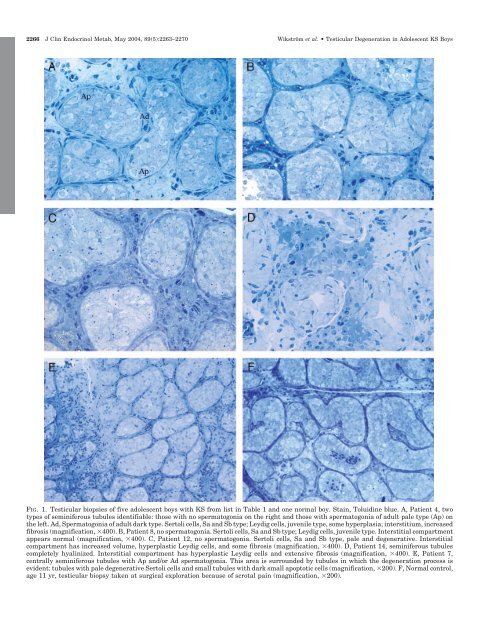
FIG. 1. Testicular biopsies <strong>of</strong> five adolescent boys with KS from list <strong>in</strong> Table 1 and one normal boy. Sta<strong>in</strong>, Toluid<strong>in</strong>e blue. A, Patient 4, two<br />
types <strong>of</strong> sem<strong>in</strong>iferous tubules identifiable: those with no spermatogonia on the right and those with spermatogonia <strong>of</strong> adult pale type (Ap) on<br />
the left. Ad, Spermatogonia <strong>of</strong> adult dark type. Sertoli cells, Sa and Sb type; Leydig cells, juvenile type, some hyperplasia; <strong>in</strong>terstitium, <strong>in</strong>creased<br />
fibrosis (magnification, 400). B, Patient 8, no spermatogonia. Sertoli cells, Sa and Sb type; Leydig cells, juvenile type. Interstitial compartment<br />
appears normal (magnification, 400). C, Patient 12, no spermatogonia. Sertoli cells, Sa and Sb type, pale and degenerative. Interstitial<br />
compartment has <strong>in</strong>creased volume, hyperplastic Leydig cells, and some fibrosis (magnification, 400). D, Patient 14, sem<strong>in</strong>iferous tubules<br />
completely hyal<strong>in</strong>ized. Interstitial compartment has hyperplastic Leydig cells and extensive fibrosis (magnification, 400). E, Patient 7,<br />
centrally sem<strong>in</strong>iferous tubules with Ap and/or Ad spermatogonia. This area is surrounded by tubules <strong>in</strong> which the degeneration process is<br />
evident: tubules with pale degenerative Sertoli cells and small tubules with dark small apoptotic cells (magnification, 200). F, Normal control,<br />
age 11 yr, testicular biopsy taken at surgical exploration because <strong>of</strong> scrotal pa<strong>in</strong> (magnification, 200).