Klinefelter Syndrome in Adolescence - The Journal of Clinical ...
Klinefelter Syndrome in Adolescence - The Journal of Clinical ...
Klinefelter Syndrome in Adolescence - The Journal of Clinical ...

Create successful ePaper yourself
Turn your PDF publications into a flip-book with our unique Google optimized e-Paper software.
Wikström et al. Testicular Degeneration <strong>in</strong> Adolescent KS Boys J Cl<strong>in</strong> Endocr<strong>in</strong>ol Metab, May 2004, 89(5):2263–2270 2267<br />
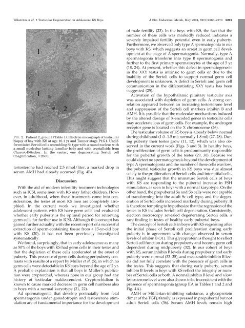
FIG. 2. Patient 2, group I (Table 1). Electron micrograph <strong>of</strong> testicular<br />
biopsy <strong>of</strong> boy with KS at age 10.1 yr and Tanner stage P1G1. Undifferentiated<br />
Sertoli cells resembl<strong>in</strong>g Sa type with a round nucleus with<br />
a small nucleolus lack<strong>in</strong>g lamellar body and with crystalloids from<br />
Charcot-Bötscher. In the center, one degenerat<strong>in</strong>g Sertoli cell<br />
(magnification, 2500).<br />
testosterone had reached 2.5 nmol/liter, a marked drop <strong>in</strong><br />
serum AMH had already occurred (Fig. 4B).<br />
Discussion<br />
With the aid <strong>of</strong> modern <strong>in</strong>fertility treatment technologies<br />
such as ICSI, some men with KS may father children. However,<br />
<strong>in</strong> adulthood, when these treatments come <strong>in</strong>to consideration,<br />
the testes <strong>of</strong> most KS men are completely atrophied.<br />
In the current work we <strong>in</strong>vestigated whether<br />
adolescent patients with KS have germ cells and evaluated<br />
whether early puberty is the optimal period for retriev<strong>in</strong>g<br />
germ cells for further use <strong>in</strong> ICSI. Although this concept has<br />
ga<strong>in</strong>ed further actuality with the recent report <strong>of</strong> a successful<br />
extraction <strong>of</strong> sperm-conta<strong>in</strong><strong>in</strong>g tissue from a 15-yr-old boy<br />
with KS (20), it has not been previously <strong>in</strong>vestigated<br />
systematically.<br />
We found, surpris<strong>in</strong>gly, that <strong>in</strong> early adolescence as many<br />
as 50% <strong>of</strong> the boys with KS had germ cells <strong>in</strong> their testes and<br />
that the depletion <strong>of</strong> these cells accelerated at the onset <strong>of</strong><br />
puberty. This presence <strong>of</strong> germ cells dur<strong>in</strong>g peripuberty contrasts<br />
with results <strong>of</strong> a report by Müller et al. (5), <strong>in</strong> which no<br />
germ cells were detectable <strong>in</strong> KS boys beyond the age <strong>of</strong> 2 yr.<br />
A probable explanation is that all boys <strong>in</strong> Müller’s publication<br />
were cryptorchid, whereas none <strong>in</strong> our group had any<br />
history <strong>of</strong> testicular maldescendent. Cryptorchidism is<br />
known to cause marked decrease <strong>in</strong> germ cell numbers also<br />
<strong>in</strong> boys with a normal karyotype (21, 22).<br />
Ad spermatogonia that develop postnatally from fetal<br />
spermatogonia under gonadotrop<strong>in</strong> and testosterone stimulation<br />
are <strong>of</strong> fundamental importance for the development<br />
<strong>of</strong> male fertility (23). In the boys with KS, the fact that the<br />
number <strong>of</strong> these cells was markedly reduced <strong>in</strong>dicates a<br />
severely impaired fertility potential even <strong>in</strong> early puberty.<br />
Furthermore, we observed only type A spermatogonia <strong>in</strong> our<br />
boys with KS, which suggests an arrest <strong>in</strong> germ cell development<br />
at the stage <strong>of</strong> A spermatogonia. Normally, type A<br />
spermatogonia transform <strong>in</strong>to type B spermatogonia and<br />
further to the first primary spermatocytes at the age <strong>of</strong> 5 yr<br />
(18, 24). At present, whether this defect <strong>in</strong> spermatogenesis<br />
<strong>in</strong> the XXY testis is <strong>in</strong>tr<strong>in</strong>sic to germ cells or due to the<br />
<strong>in</strong>ability <strong>of</strong> the Sertoli cells to support normal germ cell<br />
development is unknown. A defect <strong>in</strong> Sertoli and germ cell<br />
communication <strong>in</strong> the differentiat<strong>in</strong>g XXY testis has been<br />
suggested (25).<br />
Activation <strong>of</strong> the hypothalamic pituitary testicular axis<br />
was associated with depletion <strong>of</strong> germ cells. A strong correlation<br />
appeared between an <strong>in</strong>creas<strong>in</strong>g testosterone level<br />
and suppression <strong>of</strong> the Sertoli cell markers <strong>in</strong>hib<strong>in</strong> B and<br />
AMH. It is possible that the molecular mechanisms <strong>in</strong>duced<br />
by the altered dosage <strong>of</strong> X-encoded genes <strong>in</strong> testicular cells<br />
may accelerate loss <strong>of</strong> germ cells. For example, the androgen<br />
receptor gene is located on the X chromosome (26).<br />
<strong>The</strong> testicular volume <strong>of</strong> KS boys is already below normal<br />
dur<strong>in</strong>g childhood (1.0–1.5 ml; normally 1.8 ml) (27, 28). Dur<strong>in</strong>g<br />
puberty their testes grow (11, 12), which was also observed<br />
<strong>in</strong> the current work (Figs. 3 and 5). In healthy boys,<br />
the proliferation <strong>of</strong> germ cells is predom<strong>in</strong>antly responsible<br />
for the pubertal growth <strong>of</strong> the testes (29, 30). Because we<br />
could detect no spermatogenesis beyond the development <strong>of</strong><br />
type A spermatogonia and the number <strong>of</strong> these cells was low,<br />
the pubertal testicular growth <strong>in</strong> KS boys was due almost<br />
solely to the proliferation <strong>of</strong> Sertoli cells and <strong>in</strong>terstitial cells.<br />
This might suggest that the immature Sertoli cells <strong>of</strong> boys<br />
with KS are respond<strong>in</strong>g to the pubertal <strong>in</strong>crease <strong>in</strong> FSH<br />
stimulation, as seen <strong>in</strong> boys with a normal karyotype. On the<br />
other hand, the prepubertal Sa and Sb cells were not capable<br />
<strong>of</strong> transform<strong>in</strong>g <strong>in</strong>to the adult Sc cell type, and the degeneration<br />
<strong>of</strong> Sertoli cells <strong>in</strong>creased markedly dur<strong>in</strong>g puberty. It<br />
is therefore tempt<strong>in</strong>g to hypothesize that the regression <strong>of</strong> the<br />
testes <strong>in</strong> KS <strong>in</strong>cludes Sertoli cell degeneration. Consistently,<br />
electron microscopy revealed degenerat<strong>in</strong>g Sertoli cells, a<br />
rare f<strong>in</strong>d<strong>in</strong>g <strong>in</strong> testes <strong>of</strong> healthy early pubertal boys.<br />
<strong>The</strong> concept <strong>of</strong> Sertoli cells <strong>in</strong> boys with KS regress<strong>in</strong>g after<br />
the <strong>in</strong>itial phase <strong>of</strong> Sertoli cell proliferation dur<strong>in</strong>g early<br />
puberty is <strong>in</strong> agreement with changes observed <strong>in</strong> serum<br />
levels <strong>of</strong> <strong>in</strong>hib<strong>in</strong> B (31). This glycoprote<strong>in</strong> is thought to reflect<br />
Sertoli cell function dur<strong>in</strong>g prepuberty and become germ cell<br />
dependent dur<strong>in</strong>g midpuberty (32). In our cohort <strong>of</strong> boys<br />
with KS, serum <strong>in</strong>hib<strong>in</strong> B levels dur<strong>in</strong>g prepuberty and early<br />
puberty were normal (33–35), and measurable <strong>in</strong>hib<strong>in</strong> B levels<br />
did not fully correlate with the presence <strong>of</strong> germ cells <strong>in</strong><br />
the testes. This suggests that dur<strong>in</strong>g early puberty, serum<br />
<strong>in</strong>hib<strong>in</strong> B levels <strong>in</strong> boys with KS reflect the <strong>in</strong>tegrity or number<br />
<strong>of</strong> Sertoli cells or both. A normal <strong>in</strong>hib<strong>in</strong> B level and a low<br />
testosterone level were also shown to be <strong>in</strong>consistent with the<br />
presence <strong>of</strong> spermatogonia (group IIA <strong>in</strong> Tables 1 and 2 and<br />
Fig. 4A).<br />
AMH or Müllerian-<strong>in</strong>hibit<strong>in</strong>g substance, a glycoprote<strong>in</strong><br />
dimer <strong>of</strong> the TGF family, is expressed <strong>in</strong> prepubertal but not<br />
adult Sertoli cells (36). Serum AMH levels rema<strong>in</strong> high