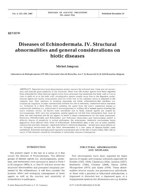
Diseases of Echinodermata. IV. Structural ... - Inter Research
Diseases of Echinodermata. IV. Structural ... - Inter Research
Diseases of Echinodermata. IV. Structural ... - Inter Research

You also want an ePaper? Increase the reach of your titles
YUMPU automatically turns print PDFs into web optimized ePapers that Google loves.
Vol. 3: 221-229, 1987<br />
REVIEW<br />
DISEASES OF AQUATIC ORGANISMS<br />
Dis. aquat. Org.<br />
Published December 31<br />
<strong>Diseases</strong> <strong>of</strong> <strong>Echinodermata</strong>. <strong>IV</strong>. <strong>Structural</strong><br />
abnormalities and general considerations on<br />
biotic diseases<br />
Michel Jangoux<br />
Laboratoire de Biologie marine (CP 160), Universite Libre de Bruxelles. Ave F. D. Roosevelt 50. B-1050 Bruxelles, Belgium<br />
ABSTRACT: Reported structural abnormalities mostly concern the echinoid test; these are not uncommon<br />
and indicate great plasticity <strong>of</strong> the echinoids. More than 400 animal agents have been reported<br />
from echinoderms. Most external agents either have processes that penetrate the body wall or live in<br />
cysts or galls on or in the body wall; intradigestive agents usually occur free in the digestive cavity;<br />
internal agents are mostly intracoelomic and live either free in the coelomic cavity or attached to the<br />
coelomic wall. Host reactions to invading organisms are either inflammatory-like reactions (i.e.<br />
intratissular migration <strong>of</strong> some coelomocytes towards the site <strong>of</strong> infection), connective-tissue reactions<br />
(i e. formation <strong>of</strong> a thick fibrous sheet isolating the agent from the host's connective tissue) or<br />
coelomocyte reactions (i.e. endocytosis <strong>of</strong> microorganisms or walling <strong>of</strong>f <strong>of</strong> anlmal agents entering the<br />
host's coelomic cavity). All known mass mortalities due to biobc disease agents are caused by<br />
microorganisms. With very few exceptions, animal agents do not kill their ech~noderm host: most <strong>of</strong><br />
them are well tolerated and do not appear to result in major consequences for the hosts concerned.<br />
Echinozoa (Holothuroidea and Echinoidea) and Asterozoa (Asteroidea and Ophiuroidea) exhibit a<br />
different degree <strong>of</strong> sensitivity to animal pathogens, the asterozoans' defensive mechanisms being<br />
apparently more efficient than those <strong>of</strong> echinozoans. Echinoderms play a key role in many benthic<br />
communities and their diseases - especially lethal or castrating deseases - should have major effects on<br />
the biological environment (this has been demonstrated in a few cases <strong>of</strong> disease-related mass<br />
rnortalities). Echinoids and ophiuroids represent a prominent part <strong>of</strong> the diet <strong>of</strong> many fishes; their role as<br />
vector <strong>of</strong> fish diseases caused by trematodes or nematodes requires investigation.<br />
INTRODUCTION<br />
The present paper is the last <strong>of</strong> a series <strong>of</strong> 4 that<br />
review the diseases <strong>of</strong> <strong>Echinodermata</strong>. The different<br />
groups <strong>of</strong> disease agents (i.e. microorganisms, protis-<br />
tans, and metazoans) were surveyed in detail in Parts I<br />
to 111 (Jangoux 1987a, b, c). Part <strong>IV</strong> will first review the<br />
structural abnormalities and presumed neoplasia pre-<br />
sented by the echinoderms; it will then consider the<br />
location, effect and ecological consequence <strong>of</strong> biotic<br />
agents as well as the reaction and sensitivity <strong>of</strong><br />
echinoderms to pathogens.<br />
O <strong>Inter</strong>-<strong>Research</strong>lPrinted in F. R. Germany<br />
STRUCTURAL ABNORMALITIES<br />
AND NEOPLASIA<br />
Test abnormalities have been reported for many<br />
species <strong>of</strong> regular and irregular echlnoids especially by<br />
Koehler (1922, 1924), Chadwick (1924), Jackson (1927),<br />
Brattstrom (1946), Chesher (1969), Moore (1974),<br />
Hinegardner (1975) and Allain (1978). These abnor-<br />
malities involve mainly non-pentamerous individuals;<br />
or those with a pinched or bifurcated ambulacrum, a<br />
depressed or distorded test, a depressed apex, or a<br />
depigmented epiderm (for illustrations see e.g. Koehler
222 Dis. aquat. Org. 3: 221-229, 1987<br />
1922, Moore 1974). Test abnormalities may have differ-<br />
ent causes: external injuries, genetic malformations,<br />
critical environmental conditions, or biotic or nutri-<br />
tional diseases. That test abnormalities may be caused<br />
by malnutrition was emphasized by Koehler (1922).<br />
While Moore (1974) considered they resulted mainly<br />
from metabolic upset, Hinegardner (1975) attributed<br />
the loss <strong>of</strong> pentamerous symmetry in laboratory-reared<br />
echinoids to as yet undetermined genetic factors.<br />
Whatever the cause, that these abnormalities are not<br />
uncommon indicates great plasticity <strong>of</strong> the echinoids.<br />
Conspicuous test deformations in echinoids living in<br />
polluted areas (Dafni 1980, 1983) suggest that many <strong>of</strong><br />
the abnormalities reported may be induced by particu-<br />
lar environmental conditions (see also Allain 1978).<br />
Body shape abnormalities (e.g. abnormal arm<br />
number in normally pentamerous species) were<br />
reported also for asteroids. According to Hotchkiss<br />
(1979), these abnormalities appear to be generally the<br />
consequence <strong>of</strong> regeneration following predator injury.<br />
Watts et al. (1983), however, present evidence that ray-<br />
number abnormalities in asteroids can be caused by<br />
high salinities during early development.<br />
A review <strong>of</strong> early literature by Wellings (1969) does<br />
not reveal any definite case <strong>of</strong> neoplasia among<br />
echinoderms. According to Sparks (1972) the only pos-<br />
sible neoplasms recorded in echinoderms are the<br />
tumor-like epidermal lesions reported by Fontaine<br />
(1969) in the ophiuroid Ophiocoma nigra. Ophiuroid<br />
tumors consist <strong>of</strong> densely packed cells (mostly<br />
melanocytes and spherulocytes with Light-brown<br />
granules); they lack connective tissue elements. These<br />
tumors may grow. Late-stage lesions are roughly<br />
divided into cortical and medullary regions, the latter<br />
having undifferentiated and fibroblast-like cells.<br />
According to Sparks (1972) tumors <strong>of</strong> 0. nigra are<br />
apparent neoplasms and present evidence <strong>of</strong> metas-<br />
tases. However, similar kinds <strong>of</strong> 'tumors', i.e. clots <strong>of</strong><br />
densely packed cells with brownish pigmented granu-<br />
les, frequently occur within echinoderm tissues. These<br />
clots are <strong>of</strong>ten located either in the hemal system or<br />
within epithelia] tissues. Possibly, they correspond to<br />
unwanted material, mostly degenerating coelomocy-<br />
tes, in the process <strong>of</strong> being eliminated. As for the tumor<br />
observed by Smith et al. (1973) in the intestine <strong>of</strong><br />
Holothuria leucopilota, the reviewer believes it to be<br />
simply an unusual outgrowth <strong>of</strong> the ventral hemal<br />
vessel <strong>of</strong> the holothuroid gut.<br />
GENERAL CONSIDERATIONS<br />
ON BIOTIC DISEASES<br />
As discussed in Part I (Jangoux 1987a). I have<br />
adopted the definition <strong>of</strong> parasites proposed by Lnne<br />
(1980, p. 19) and used it in a very broad sense, considering<br />
disease agents (parasites sensu lafo) to represent<br />
any kind <strong>of</strong> a harmful associate which affects, if even<br />
slightly, the echinoderm's tissues or internal fluids (i.e<br />
coelomic and hemal fluids). I consequently considered<br />
all external and internal associates whose detrimental<br />
effects were demonstrated or documented, as well as<br />
intradigestive symbiotes that act or may potentially act<br />
as parasites. The latter still require critical investigation<br />
as to their role - neutral or detrimental - in relation to<br />
the echinoderm's life, i.e. its ecological potential and<br />
health status.<br />
Immune defense mechanisms <strong>of</strong> echinoderms have<br />
been studied mostly during the past 10 yr. Cellular<br />
defense mechanisms act through phagocytic<br />
coelomocytes whose ability to phagocytose and/or to<br />
wall <strong>of</strong>f the unwanted material entering the<br />
echinoderm body cavities is well known (Smith 1981,<br />
Bang 1982, Karp & C<strong>of</strong>faro 1982, Dybas & Fankboner<br />
1986). Cell-types similar to coelomocytes have been<br />
seen also in experimentally altered echinoderm body<br />
walls (pathological alteration or reaction to allografts:<br />
Hobaus 1980, Karp & C<strong>of</strong>faro 1982, Gilles & Pearse<br />
1986, Maes et al. 1986). These cells massively invade<br />
the altered areas producing an inflammatory-like reaction.<br />
Moreover, coelomic fluids <strong>of</strong> echinoderms possess<br />
naturally-occurring humoral factors such as hemolysin<br />
and hemagglutinin (Ryoyama 1973, 1974; see also<br />
Bang 1982) as well as bactericidal substances (Wardlaw<br />
& Unkles 1978, Service & Wardlaw 1984, 1985). All<br />
this demonstrates that echinoderms are well armed to<br />
Animal agents associated with echinoderms are<br />
summarized in Table 1. More than one-third <strong>of</strong> these<br />
agents live on or in holothuroids (Fig. 1). Crinoids,<br />
regular echinoids and asteroids harbor a rather similar<br />
number <strong>of</strong> harmful associates. As for irregular<br />
echinoids. agents mostly infest spatangoids (18<br />
species), the remaining species affecting clypeas-<br />
teroids. Gastropods, turbellarians and copepods com-<br />
prise the most numerous agents affecting the phylum<br />
<strong>Echinodermata</strong>. However, myzostomids, ascothoracids<br />
and sporozoans are also well represented. Holothuroids<br />
appear to provide a most suitable substrate for para-<br />
sites <strong>of</strong> many kinds, except for myzostomids and<br />
ascothoracids. These latter 2 groups infest specifically<br />
crinoids or asteroids. Detailed information on each<br />
species <strong>of</strong> disease agents (its taxonomical position and<br />
relations with the host) are given in this review Parts 1<br />
to I11 (Jangoux 1987a, b, c). Although a great deal <strong>of</strong><br />
echinoderm parasites have been known for many<br />
years, definite information on the biotic diseases <strong>of</strong><br />
echinoderms is still rare. <strong>Inter</strong>estingly, echinoderms<br />
-while rather intensively parasitized - never act as<br />
parasites themselves.
Jangoux: <strong>Diseases</strong> <strong>of</strong> <strong>Echinodermata</strong>: structural abnormalities and general considerations 223<br />
Table 1. Number <strong>of</strong> species <strong>of</strong> animal agents living with echinoderms. Only identified and documented agents are considered<br />
(detrimental effects demonstrated or probable). (Original)<br />
Hosts v,<br />
Agents<br />
Crinoidea 1 1 - 4 - 4 2 - 1 - 1 9 7 - - 2 1 0 2 2 - 6 - 6 1<br />
Holothuroidea 15 - 1 1 - 39 3 - 1 2 - 34 3 1 22 - - 10 2 4 7 145<br />
Echinoidea<br />
(Regularia)<br />
- - 1 1 4 7 - - 2 4 1 1 6 5 2 - - 76<br />
Echinoidea<br />
(Irregularia)<br />
5 6 - - 1 1 - 2 2 1 0 - - 29<br />
Asteroidea - - - l 6 - 2 2 1 9 - 3 - 2 5 1 2 3 65<br />
Ophiuroidea - - 1 1 1 1 4 1 1 1 3 6 - - 1 6 - 6 - 1 - - 43<br />
Total 23 5 2 8 1 70 16 6 3 3 24 90 4 2 54 18 35 28 7 10 10<br />
' Non-sporozoan protozoans<br />
Miscellaneous arthropod groups (acar~ans, pycnogonida, insects)<br />
HOLO<br />
Fig. 1. Relative percentage <strong>of</strong> ammal agent species living in<br />
each <strong>of</strong> the echinoderm groups ~ndicated. CRIN: Crinoidea;<br />
HOLO: Holothuroidea; ECHI: Echinoidea (I: irregular; R: reg-<br />
ular); ASTE: Asteroidea; OPHI: Ophiuroidea. Original<br />
Location and effects <strong>of</strong> animal agents<br />
A classification according to host sites <strong>of</strong> harmful<br />
associates (on or in echinoderms) is presented in Table<br />
2 (see also Fig. 2). Agents mostly inhabit 3 different<br />
sites: body wall, digestive cavity, and coelomic cavity.<br />
This corresponds to an external (A), intradigestive (B)<br />
or internal (C) location.<br />
External agents (A-agents)<br />
Among external agents (A1 to A3-agents; Table 2),<br />
Al-agents mostly occur on holothuroids and echinoids.<br />
They comprise strictly ectoparasitic species such as, for<br />
example, eulimid gastropods and pinnotherid crabs<br />
infesting the external body surface. Al-agents presum-<br />
ably include many more species than considered here.<br />
This is because most unattached associates, while <strong>of</strong>ten<br />
said to be ectoparasitic, have received only taxonomi-<br />
cal attention.<br />
A2-agents can firmly attach themselves to the host's<br />
external body surface in spite <strong>of</strong> the presence <strong>of</strong> a<br />
cutaneous epithelium. Such associates have rarely<br />
been observed on asteroids and irregular echinoids;<br />
they have been reported only casually on holothuroids,<br />
regular echinoids and ophiuroids. However, crinoids<br />
appear to be rather sensitive to A2-agents. Possibly,<br />
crinoid sensitivity to these agents results from a weak<br />
defensive capacity <strong>of</strong> their epidermal barrier.<br />
A3-agents are by far the most common external<br />
echinoderm associates. Although they all live, in one<br />
way or another, within the echinoderms' body wall,<br />
they show conspicuous differences in their feeding<br />
habits. Several A3-agents feed independently <strong>of</strong> their<br />
host. They may be, for instance, suspension-feeders<br />
simply sheltering in galls or cysts (i. e. most <strong>of</strong> the<br />
harmful myzostomids). Independent feeding also<br />
occurs in a few gastropods and copepods causing galls<br />
in echinoid spines or body wall. This could also be the<br />
case in some organisms inhabiting ophiuroid branchial<br />
bursae. However, most AS-agents feed at the expense<br />
<strong>of</strong> their host, either by ingesting body-wall tissues or by<br />
sucking up internal fluids. Body-wall feeders are<br />
mainly gallicole gastropods, such as asteroid-associ-<br />
ated Stilifer spp. A few copepods also feed in this<br />
manner (e. g. Scottornyzon gibberum). External fluid-<br />
feeders are also mainly gastropod molluscs. Several<br />
species use their proboscis to penetrate the body wall
224 Dis. aquat. Org. 3: 221-229, 1987<br />
Table 2 Classification <strong>of</strong> animal agents affecting echinoderms according to location. (Original)<br />
Hosts Agent<br />
External Intradigestlve <strong>Inter</strong>nal<br />
A l A2 A3 B1 B2 C1 C2 C3<br />
Crinoidea 23 28 6 - 7 1 1<br />
Holothuroidea 8 7 10 43 11 53 12 2<br />
Echinoidea (Regularia) 6 8 24 29 7 12 - 6<br />
Echinoidea (Irregularia) 3 10 - 6 - 9 - -<br />
Asteroidea 3 1 2 1 4 1 35 - 1<br />
Ophiuroidea 1 6 24 2 l 4 - 6<br />
Total 2 1 55 107 90 20 120 13 16<br />
A agents: 283 B agents: 110 C agents: 149<br />
Al-agents live free or simply cling to outer host-body surface<br />
A2-agents attach to outer host-body surface (viz. epithelium-covered body surface)<br />
A3-agents have processes that permanently penetrate or cross the body wall, or live in cysts or galls on or in the body wall<br />
(including spines), or live permanently In naturally-occurring ectoderrnal lnvaginations (viz. genital bursae <strong>of</strong> ophiuroids)<br />
B1-agents live free in digestive cavity<br />
B2-agents attach to or bury in the digestive wall<br />
Cl-agents inhabit coelomic cavity or ambulacral system (live free, or attached to coelomic wall, or embedded in rnesenteries)<br />
C2-agents live in hemal system<br />
C3-agents live in gonads<br />
Fig. 2. Relative percentage <strong>of</strong> each category <strong>of</strong> echinoderm-<br />
infesting animal agents; for explanations see Table 2. (Orig-<br />
inal)<br />
in order to suck up coelomic, ambulacral, or hemal fluid<br />
(Euchineulima spp., Thyca spp., Pisolamia brychius,<br />
respectively). It has been suggested that the proboscis<br />
<strong>of</strong> parasitic eulimid gastropods would secrete particular<br />
material that brings about a rapid loosening <strong>of</strong> connec-<br />
tive tissue <strong>of</strong> the echinoderm body wall, thereby<br />
facilitating penetration <strong>of</strong> the host's integument (Smith<br />
1984). Ectoparasitic gatropods sucking up hemal fluid<br />
occur mostly in holothuroids whose hemal fluid has an<br />
high energy content. Possibly, a similar feeding habit<br />
has been developed by other ectoparasitic gastropods<br />
infesting crinoids (e. g. Balcis devians and Melanella<br />
comatulicola) and asteroids (Thyca spp.). The probos-<br />
cises <strong>of</strong> these gastropods have been seen to be inserted<br />
into the host's coelomic or radial canals which are<br />
closely associated with a well-developed hemal lacuna.<br />
Coelomic-fluid feeders appear to be a rather paradoxi-<br />
cal adaptation, since the coelomic fluid is not particu-<br />
larly energy-rich. Such a feeding habit implies that the<br />
agents feed on coelomocytes or 'browse' on internal<br />
tissues, however this is poorly documented for A3-<br />
agents. Can ingestion <strong>of</strong> coelomocytes alone meet the<br />
energy requirements <strong>of</strong> the parasites? If so, such infes-<br />
tation should be rather harmless since echinoderms<br />
produce new coelomocytes almost continuously.<br />
Finally, many A3-agents <strong>of</strong> ophiuroids, especially crus-<br />
taceans, inhabit the genital bursae <strong>of</strong> their host. Genital<br />
bursae are easily accessible and well sheltered.<br />
Moreover, gonads open into the bursae which means a<br />
potential supply <strong>of</strong> energy-rich material. Yet this does<br />
not imply that bursal-inhabiting associates necessarily<br />
feed on the host's gonads.<br />
Detrimental effects <strong>of</strong> external agents are not<br />
restricted to their feeding. Some induce conspicuous<br />
modifications <strong>of</strong> the host's skeleton. There are 3 kinds<br />
<strong>of</strong> such modifications: (1) hypertrophy <strong>of</strong> skeletal ossi-<br />
cles (caused for example by gallicole myzostomids and<br />
spine-inhabiting gastropods or copepods); (2) develop-<br />
ment <strong>of</strong> supernumerary skeletal ossicles (e. g. around<br />
subcutaneous cysts <strong>of</strong> some crinoid-infesting myzo-<br />
stomids; around cysts <strong>of</strong> some copepods living in<br />
ophiuroid genital bursae); (3) inhibition <strong>of</strong> the develop-<br />
ment <strong>of</strong> skeletal ossicles (such as that caused by the<br />
asteroid-associated gastropod Parvioris equestris).<br />
Skeletal modifications have also been reported for dis-<br />
eases caused by microorganisms, i.e. the bald-sea-<br />
urchin disease (see Jangoux 1987a). A3-agents may
Jangoux: <strong>Diseases</strong> <strong>of</strong> <strong>Echinodermata</strong>: structural abnormalities and general considerations 225<br />
also cause dermal outgrowths, mostly in gastropods<br />
and copepods.<br />
Echinoderm ectoparasites do not castrate their host,<br />
except for some forms <strong>of</strong> Ascothoracida which live in<br />
the genital bursae <strong>of</strong> ophiuroid. These, however, do not<br />
cause typical castration - none <strong>of</strong> them is said to feed<br />
on gonadal tissues - but are considered to inhibit both<br />
gonadal growth and germinal development. The bur-<br />
sal-inhabiting copepod Amphiurophilus amphiurae,<br />
which infests the brooding ophiuroid Amphipholis<br />
squamata, presumably suppresses incubating embryos<br />
by diverting part <strong>of</strong> their food supply without affecting<br />
the host's gonads.<br />
Intradigestive agents (B-agents)<br />
Agents inhabiting the digestive system <strong>of</strong><br />
echinoderms (B1 and B2-agents; see Table 2) mainly<br />
infest holothuroids and regular echinoids. While most<br />
B1-agents are turbellarian worms, a few gastropods<br />
and copepods also live freely in the digestive cavity.<br />
Presumed detrimental effects <strong>of</strong> these associates are<br />
poorly documented. However, some <strong>of</strong> them exhibit a<br />
typical parasitic behavior by feeding on digestive host<br />
epithelium. In contrast, B2-agents, while living within<br />
the digestive wall and producing conspicuous deforma-<br />
tions, seem to be rather independent from host tissues<br />
as far as feeding is concerned. Thus gut-associated<br />
crabs <strong>of</strong> echinoids ingest primarily the food pellets <strong>of</strong><br />
the host. Similarily, bivalve molluscs inhabiting gut<br />
outgrowths are basically suspension feeders. A detri-<br />
mental effect on host nutrition might <strong>of</strong> course occur.<br />
This depends greatly on the location <strong>of</strong> the associates,<br />
those living in the holothuroid cloaca or echinoid anal<br />
tube being supposedly harmless. B2-agents feeding at<br />
the expense <strong>of</strong> host structures are almost exclusively<br />
gastropod molluscs. They generally do not feed on<br />
digestive tissues; in most species the proboscis pene-<br />
trates hemal lacunae, gonads or the body wall.<br />
<strong>Inter</strong>nal agents (C-agents)<br />
<strong>Inter</strong>nal agents (C1 to C3-agents; Table 2) live either<br />
within the coelom, the hemal system or the gonads <strong>of</strong><br />
the host. Echinoderms harbor a highly diverse<br />
intracoelomic fauna (Cl-agents): sporozoans, turbella-<br />
rians, aberrant gastropods, copepods, ascothoracids<br />
and fishes are not uncommon in their body cavities.<br />
Turbellarians and copepods are the main free-living<br />
Cl-agents. Their feeding habits require special atten-<br />
tion by researchers. Only Changeux (1961) reported<br />
intracoelomic copepods 'browsing' on holothuroid<br />
mesothelium. The assumption <strong>of</strong> Jennings & Mettrick<br />
(1968) that intracoelomic turbellarians feed essentially<br />
on intracoelomic ciliates, although not impossible,<br />
appears improbable (so far, intracoelomic turbellarians<br />
have been recorded much more <strong>of</strong>ten than intra-<br />
coelomic ciliates). Other free-living Cl-agents are<br />
fishes and motile stages <strong>of</strong> sporozoans, viz, trophozoites<br />
and gamonts. These latter are almost exclusively found<br />
in the coelon~ <strong>of</strong> spatangoid echinoids. (Trophozoites<br />
and gamonts <strong>of</strong> sporozoans <strong>of</strong> holothuroids inhabit<br />
primarily hemal lacunae.) Presumably they feed via<br />
direct intramembranous absorption <strong>of</strong> nutrients from<br />
the coelomic fluid. Fishes either behave as strict para-<br />
sites in ingesting the host's gonads or respiratory trees,<br />
or they only shelter in the echinoderm coelom and prey<br />
upon shrimp and other free-living crustaceans they<br />
catch outside.<br />
A second category <strong>of</strong> Cl-agents are simply floating<br />
or deposited in the host's body cavity, i. e. sporozoan<br />
cysts and most ascothoracids. Intrategumentary<br />
absorption <strong>of</strong> coelomic nutrients has usually been sug-<br />
gested as the feeding method <strong>of</strong> intracoelomic<br />
ascothoracids. Wagin (1976). however, concluded that<br />
they feed mostly on coelomocytes.<br />
The last category <strong>of</strong> Cl-agents includes organisms<br />
attached to the coelomic wall and hanging into the<br />
body cavity: spatangoid ascothoracids and aberrant<br />
gastropods from holothuroids (Enteroxenos and allied<br />
genera). The body wall <strong>of</strong> the latter is always<br />
surrounded by a host-produced envelope consisting <strong>of</strong><br />
an inner connective tissue layer and an outer mesothe-<br />
lial layer. As indicated by Jangoux (1987b, p. 227)<br />
hemal lacunae within the host's envelope would facili-<br />
tate parasite nutrition. In the absence <strong>of</strong> hemal lacunae<br />
- an absence which would be worth demonstrating -<br />
intrategumentary nutrition from the coelomic fluid<br />
must be assumed. The only intracoelomic gastropod<br />
that is definitely a hemal-fluid feeder, Gasterosiphon<br />
deimatis, has been recorded in the body cavity <strong>of</strong> a<br />
deep-sea holothuroid.<br />
C2-agents spend most <strong>of</strong> their life cycle in the host's<br />
hemal lacunae. So far, this group comprises only<br />
holothuroid-infesting sporozoans.<br />
Animal agents parasitizing echinoderm gonads (C3-<br />
agents) are not very numerous. They include species <strong>of</strong><br />
protozoans, mesozoan, trematodes, nematodes and<br />
myzostomids. Most <strong>of</strong> them feed directly on gonadal<br />
tissues.<br />
While internal agents <strong>of</strong>ten castrate their host, the<br />
method <strong>of</strong> castration may differ considerably. Classical<br />
castration occurs with agents feeding directly on ger-<br />
minal tissues, e. g. gonad-infesting ciliates <strong>of</strong> asteroids<br />
and gonad-infesting myzostomids <strong>of</strong> ophiuroids. Less<br />
drastic castration may be caused by encysted organ-<br />
isms invading the gonad and supposedly blocking the<br />
passage <strong>of</strong> hormonal substances needed by
226 Dis aquat Org. 3: 221-229, 1987<br />
echinoderms for gametogenesis (Pearse & Timm 1971).<br />
The 'passive castration' (Wagin 1946) induced by some<br />
intracoelomic and intrabursal associates is <strong>of</strong> great<br />
interest. It should be more properly termed 'competi-<br />
tive castration' as it appears to result from nutritional<br />
competition between associate(s) and host's gonads. It<br />
causes either slight or total regression <strong>of</strong> the gonads,<br />
depending on the number <strong>of</strong> agents, and is presumably<br />
reversible when the agents are dislodged from their<br />
host. Competitive castration may be inferred for<br />
Mesozoa and some intracoelomic gastropods inhabit-<br />
ing holothuroids (e.g. Entocolax schwanwitschi and<br />
Paedophorus dicoelobius) as well as for intracoelomic<br />
ascothoracids <strong>of</strong> spatangoids (Ulophysema spp.).<br />
Infestation routes, host reactions<br />
and virulence<br />
Infestation <strong>of</strong> echinoderms by internal agents takes<br />
place mainly through body openings (i.e. mouth, anus,<br />
gonopores; also branchial bursae <strong>of</strong> ophiuroids), or<br />
through thin areas <strong>of</strong> the body-wall (e.g. tube feet,<br />
respiratory papulae). Intracoelomic copepods <strong>of</strong><br />
Holothuna spp. enter the host's body cavity by pene-<br />
trating the anterior part <strong>of</strong> its gut. A similar behavior<br />
prevails in most sporozoans inhabiting deposit-feeding<br />
echinoderms. Cloaca and respiratory trees <strong>of</strong> holo-<br />
thuroids also allow the passage <strong>of</strong> infesting stages <strong>of</strong><br />
internal agents (i.e. some sporozoans, turbellanans,<br />
and fishes). Larvae <strong>of</strong> most intracoelomic gastropods <strong>of</strong><br />
holothuroids (Enferoxenos and allied genera) also enter<br />
the host's body cavity through the digestive wall or,<br />
more rarely, through the body wall. These gastropods,<br />
however, live never totally free in the coelom; they are<br />
always surrounded by an envelope produced by the<br />
hosts (this envelope is continuous with the outer tissues<br />
<strong>of</strong> the host's digestive tract or body wall). Infesting<br />
larvae <strong>of</strong> asteroid-associated ascothoracids may enter<br />
their host through respiratory papulae, as do some<br />
intracoelomic copepods. Larvae <strong>of</strong> ascothoracids living<br />
in spatangoids enter the host through the genital pores.<br />
No report has come to the reviewer's attention that<br />
refer to the routes taken by nematodes and digenic<br />
trematodes which enter the internal tissues or body<br />
cavities <strong>of</strong> echinoderms.<br />
There are 3 kinds <strong>of</strong> host reactions counteracting<br />
invading organisms: (1) inflammatory-like reactions;<br />
(2) connective-tissue reactions; (3) coelomocyte reac-<br />
tions. Inflammatory-like reactions occur mostly in dis-<br />
eases caused by microorganisms and algae (see Jan-<br />
goux 1987a) or upon cutaneous wound repair (e.g.<br />
Menton & Eisen 1973). Basically these reactions consist<br />
<strong>of</strong> a migration <strong>of</strong> red spherule cells and phagocytic cells<br />
towards the site <strong>of</strong> infection (Johnson & Chapman 1970,<br />
Johnson 1971, Maes et al. 1986) These cells are <strong>of</strong><br />
coelomic origin. According to Johnson (1971; see also<br />
Service & Wardlaw 1984), red spherule cells would act<br />
as a 'general disinfectant'. Their presence in or close to<br />
a wounded or infested area prevents penetration or<br />
settlement <strong>of</strong> unwanted organisms. In holothuroids and<br />
echinoids red spherule cells always occur In every<br />
tissue or organ, although they are generally present in<br />
rather low densities. This cell type has not been<br />
reported in pathologically altered body areas <strong>of</strong><br />
asteroids or ophiuroids.<br />
Connective-tissue reactions counteract organisms<br />
which tend to stay within the connective tissue layer. A<br />
thick fibrous sheet is formed which surrounds and thus<br />
isolates the foreign organisms from the host's tissue.<br />
Such reactions are not an uncommon defense mechan-<br />
ism against sporozoans cysts, trematode metacercariae<br />
or invading nematodes. They also counteract some<br />
animal agents, such as copepods and ascothoracids,<br />
that infest either the genital bursae <strong>of</strong> ophiuroids or the<br />
coelomic cavity <strong>of</strong> holothuroids.<br />
Coelomocyte reactions counteract agents entering the<br />
host's coelomic cavity. Generally, they are inconspicu-<br />
ous reactions resulting in phagocyting or completely<br />
walling <strong>of</strong>f the foreign organism. A massive and very<br />
particular coelomocyte reaction is initiated by motile<br />
stages <strong>of</strong> spatangoid intracoelomic gregannes; each<br />
participating coelomocyte becomes pointed, the parasite<br />
talung on the appearence <strong>of</strong> a minute pin cushion.<br />
It was generally not yet possible to evaluate agent<br />
virulence, except in the case <strong>of</strong> microorganisms or<br />
algae. They produce conspicuous pathological altera-<br />
tions which <strong>of</strong>ten kill the host. All known mass mor-<br />
talities <strong>of</strong> echinoderms due to biotic disease agents are<br />
caused by microorganisms. So far, no animal agent has<br />
been reported to kill the host, except for a few small<br />
ectoparasitic crabs living on some echinoids. <strong>Diseases</strong><br />
caused by animal agents become obvious only if they<br />
result in particular changes in the echinoderm's body<br />
shape. As a rule, internal damage is not detectable<br />
from the outside, and echinoderms appear perfectly<br />
healthly even when massively infested (e.g. by some<br />
sporozoans or mesozoans).<br />
Relative sensitivity <strong>of</strong> echinoderms<br />
to animal pathogens<br />
The 'pathogenic index' for each echinoderm group has<br />
been tentatively estimated (Table 3). Although rather<br />
approximate, the indexes reveal that Echinozoa and<br />
Asterozoa exhibit a very different degree <strong>of</strong> sensitivity to<br />
animal pathogens. It is not possible to explain this<br />
difference on the grounds <strong>of</strong> morphological, ethological<br />
or ecological considerations. The reviewer supposes that
Jangoux. <strong>Diseases</strong> <strong>of</strong> <strong>Echinodermata</strong>: structur -a1 abnormal~t~es and general considerations 227<br />
Table 3. Pathogenic indexes <strong>of</strong> echinoderm classes. (Original)<br />
Hosts Estimated no. No. <strong>of</strong> Pathogen~c<br />
<strong>of</strong> recent species <strong>of</strong> anl- index'<br />
host species mal agents<br />
Echinozoa 2000 240 12.0<br />
Holothuro~dea 1100 141 12.8<br />
Echinoidea 900 99 11 0<br />
Asterozoa 3800 109 2 9<br />
Asteroidea 1800 66 3 7<br />
Ophiuroidea 2000 43 2 2<br />
'(No <strong>of</strong> species <strong>of</strong> animal agents / no. <strong>of</strong> echinoderm<br />
specles) x 100<br />
it results basically from physiological properties and<br />
implies that the asterozoans' defensive mechanisms are<br />
more efficient than those <strong>of</strong> echinozoans.<br />
ECOLOGICAL CONSEQUENCES OF<br />
ECHINODERM DISEASES<br />
<strong>Echinodermata</strong> constitute a large and highly distinctive<br />
group <strong>of</strong> mailne invertebrates. World-wide, they<br />
are found from the shoreline to the deepest ocean<br />
trencheS. As bottom-dwellers they have colonized all<br />
marine benthic biotopes and, characteristically, tend to<br />
occur in dense populations. Clearly, the ecological<br />
radiation <strong>of</strong> the Ech~nodermata has been considerable<br />
and they can be considered a major macrobenthic<br />
animal group. Many littoral echinoderms greatly affect<br />
the bioeconomics <strong>of</strong> both hard- and s<strong>of</strong>t-bottom com-<br />
munities. For. instance, they are frequently top preda-<br />
tors in their community (e.g. many asteroids) or con-<br />
trolling agents <strong>of</strong> seagrass or kelp beds (e. g. numerous<br />
regular echinoids). Some species have tentatively been<br />
classified as 'key species', i. e, species which by size or<br />
number, mode <strong>of</strong> life, or activity can functionally domi-<br />
nate a community. Moreover, echinoids and ophiuroids<br />
form part <strong>of</strong> the diet <strong>of</strong> many fishes and macroinverte-<br />
brates, such as crustaceans. Although the ecological<br />
consequences <strong>of</strong> echinoderm diseases have not been<br />
studied nor considered - except for a few cases <strong>of</strong><br />
spectacular and virulent diseases caused by microor-<br />
ganisms - these diseases should have prominent<br />
effects on the biological environment.<br />
The ultimate ecological consequence <strong>of</strong> diseases<br />
caused by microorganisms in echinoderm populations<br />
is a reduction or even elimination <strong>of</strong> the populations<br />
concerned. In contrast, most diseases caused by animal<br />
agents do not appear to result in major consequences<br />
for the echinoderms concerned and are well tolerated.<br />
One may consider each echinoderm, especially the<br />
holothuroids, as an animal substrate on/in which various<br />
other organisms live, either permanently or temporarily.<br />
Together, the echinoderm and its associates<br />
form a usually well-balanced biological complex. Some<br />
animal agents, however, may castrate echinoderms<br />
and consequently affect the renewal and long-term<br />
stability <strong>of</strong> the population concerned. Quantitative estimations<br />
<strong>of</strong> the effect <strong>of</strong> castrating agents have never<br />
been made. Of course, the effects will depend on infestation<br />
rates. Ecological effects <strong>of</strong> other non-castrating<br />
agents are almost impossible to assess at the population<br />
level. We can only imagine that, when numerous,<br />
agents such as arm-infesting myzostomids, bursalinhabiting<br />
copepods or coelom-sheltered fishes would<br />
'weaken' their host population.<br />
Effects <strong>of</strong> echinoderm diseases on echinoderms'<br />
predators mostly concern echinoderms affected by<br />
biotic communicable diseases, i. e, those caused by<br />
digenic treinatodes or by nematodes (see Jangoux<br />
198713). Since numerous echinoids and ophiuroids represent<br />
a prominent part <strong>of</strong> the diet <strong>of</strong> many fishes, the<br />
role <strong>of</strong> echinoderms as vectors <strong>of</strong> fish diseases requires<br />
investigation.<br />
Littoral echinoderms are frequently top predators in<br />
their community (many paxillosid and forcipulatid<br />
asteroids; for review see Menge 1982), or controlling<br />
agents <strong>of</strong> seagrass or kelp beds (numerous regular<br />
echinoids; for review see Lawrence & Sammarco 1982,<br />
Harrold & Pearse 1987). Experimental or natural<br />
removal <strong>of</strong> these predators or controlling agents produces<br />
major environmental changes (e. g. Paine 197 1,<br />
Estes et al. 1978, respectively). Catastrophic decline <strong>of</strong><br />
predatory echinoderms caused by disease was noted<br />
only by Dungan et al. (1982) in the asteroid Heliaster<br />
kubin~i; these authors, however, did not consider<br />
impacts on the biological environment. Mass mortalities<br />
by diseased Strongylocentrotus franciscanus<br />
were followed by rapid expansion <strong>of</strong> 4 species <strong>of</strong> brown<br />
algae (Pearse & Hines 1979). Subsequent con~petition<br />
among algal species was severe, and within 1 yr only 1<br />
algal species inhabited the area. Another lethal disease<br />
that affects the echinoid Paracentrotus lividus was<br />
studied by Boudouresque et al. (1981) who established<br />
that the decrease in echinoid density promoted an<br />
explosive growth <strong>of</strong> epithytes on leaves <strong>of</strong> the seagrass<br />
Posidonia oceanlca.<br />
Widespread disease-related mass mortalities <strong>of</strong><br />
Strongylocentrotus droebachie~~sis occurred from 1981<br />
along the coast <strong>of</strong> Nova Scotia, Canada (Miller & Colodey<br />
1983, Scheibling 1984). S. droebacl~iensis is the<br />
dominant herbivore <strong>of</strong> the kelp beds <strong>of</strong> Nova Scotia<br />
(e.g. Mann 1982); mass mortalities <strong>of</strong> the echinoids was<br />
expected to result in colonization by subtidal macroalgae<br />
followed by an increase <strong>of</strong> benthic primary producbon<br />
(Miller & Colodey 1983). Moore & Miller (1983)
228 Dis. aquat. Org. 3: 221-229, 1987<br />
reported that in areas where S. droebachiensis was<br />
absent for 1 yr the percentage algal cover was 2 to 14<br />
times higher than in areas with echinoids. In the ab-<br />
sence <strong>of</strong> echinoids, macroalgae expanded to deeper<br />
and more sheltered locations: the kelp Laminaria lon-<br />
gicruris gained a dominant status and fleshy seaweeds<br />
developed in the subtidal (Moore & Miller 1983, Miller<br />
1985, Johnson & Mann 1986). According to Scheibling<br />
(1984) and Scheibling & Stephenson (1984), mass mor-<br />
talities <strong>of</strong> S. droebachiensis would play a key role in<br />
determining the structure and stability <strong>of</strong> the rocky<br />
subtidal ecosystem <strong>of</strong>f Nova Scotia, by controlling the<br />
abundance <strong>of</strong> the dominant herbivore.<br />
The echinoid Diadema antillarum - the principal<br />
herbivore and the most effective bioeroder <strong>of</strong> the<br />
Caribbean region (e.g. Bak et al. 1984, Liddell &<br />
Ohlhorst 1986) - suffered a widespread and conspicu-<br />
ous mass mortality resulting in a drastic reduction <strong>of</strong><br />
the population densities to 1 to 7 O/O <strong>of</strong> their previous<br />
level (Lessios et al. 1984; see also Jangoux 1987a).<br />
Mass mortality was caused by a virulent biotic disease<br />
(Bak et al. 1984, Hughes et al. 1985). Elimination <strong>of</strong> D.<br />
antillarum from most Caribbean reefs resulted in a<br />
significant increase <strong>of</strong> fleshy and filamentous algae as<br />
well as <strong>of</strong> some macroalgae (Hughes et al. 1985, de<br />
Ruyter van Steveninck & Bak 1986, de Ruyter van<br />
Steveninck & Breeman 1987). This increase was<br />
achieved at the expense <strong>of</strong> other benthic organisms<br />
such as corals, crustose corallines and clionid sponges<br />
whose settlement has been considerably reduced (de<br />
Ruyter van Steveninck & Bak 1986, Liddell & Ohlhorst<br />
1986). Investigators generally believe the situation is<br />
irreversible and will fundamentally affect the Carib-<br />
bean reef ecosystem unless the populations <strong>of</strong> D. antil-<br />
larum are restored or those <strong>of</strong> other herbivores take<br />
over the role <strong>of</strong> the diadematids.<br />
LITERATURE CITED<br />
Allain, J. Y. (1978). Deformations du test chez l'oursin<br />
Lytechinus variegatus (Lamarck) (Echinoidea) de la Baie<br />
de Carthagene. Caldasia 12: 363-375<br />
Bak. R. P. M., Carpey, M. J. E., de Ruyter van Steveninck, E.<br />
D. (1984). Densities <strong>of</strong> the sea urchin Diadema antillarum<br />
before and after mass mortalities on the coral reef <strong>of</strong><br />
Curaqao. Mar Ecol. Prog. Ser. 17: 105-108<br />
Bang, F. B. (1982). Disease processes in seastar a Metchniko-<br />
vian challenge. Biol. Bull. mar. biol. Lab., Woods Hole 162:<br />
135-248<br />
Boudouresque, C. F., Nedelec, H., Shepherd, S. A. (1981) The<br />
decline <strong>of</strong> a population <strong>of</strong> the sea urchin Paracentrotus livi-<br />
dusin the Bay <strong>of</strong> Port-Cros (Var. France). Rapp. P.-v. Reun.<br />
Commn int. Explor. Scient. Mer Mediterr. 27: 223-224<br />
Brattstrom, H. (1946). Observations on Brissopsis lyrifera (For-<br />
bes) in the Gullmar Fjord. Ark. Zool. 37A (18): 1-25<br />
Changeux, J. P. (1961). Contribution a I'etude des animaux<br />
associes aux holothurides. Vie Milieu 10 (Suppl.): 1-124<br />
Chadwick, H. C. (1924). On some abnormal and imperfectly<br />
devc,loped specimens <strong>of</strong> the sea urchin Echinus esculentus.<br />
Proc. zool. Soc Lond. 94: 163-172<br />
Chesher, R. H. (1969). Contributions to the biology <strong>of</strong> Meoma<br />
ventricosa (Echinoidea. Spatangoida). Bull. mar Sci. 19:<br />
72-110<br />
Dafni, J. (1980). Abnormal growth patterns In the sea urchin<br />
Tripneustes cf. gratilla (L.) under pollution (<strong>Echinodermata</strong>,<br />
Echinoidea). J. exp, mar. Biol. Ecol. 47: 259-279<br />
Dafni, J. (1983). Aboral depressions in the test <strong>of</strong> the sea<br />
urchin Tripneustes cf. gratilla (L) in the Gulf <strong>of</strong> Ellat, Red<br />
Sea. J. exp. mar. Biol. Ecol. 67 1-15<br />
de Ruyter van Steveninck, E. D., Bak, R. P. M. (1986). Changes<br />
in abundance <strong>of</strong> coral-reef bottom components related to<br />
mass mortality <strong>of</strong> the sea urchln Diadema antillarum. Mar.<br />
Ecol. Prog. Ser 34: 87-94<br />
de Ruyter van Steveninck, E. D., Breeman, A. M. (1987). Deep<br />
water vegetations <strong>of</strong> Lobophora variegata (Phaeophyceae)<br />
in the coral reef <strong>of</strong> Curapo: population dynamics in relation<br />
to mass mortality <strong>of</strong> the sea urchin Dialdema antillarum.<br />
Mar. Ecol. Prog. Ser 36: 81-90<br />
Dungan, M. L., Miller, T E., Thomson, D. A. (1982). Catastrophic<br />
decline <strong>of</strong> a top carnivore in the Gulf <strong>of</strong> California<br />
rocky intertidal zone. Science 216: 989-991<br />
Dybas, L., Fankboner, P. v. (1986). Holothurian survival<br />
strategie: mechanisms for the maintenance <strong>of</strong> a bacteriostatic<br />
environment in the coelomic cavity <strong>of</strong> the sea<br />
cucumber, Parastichopus californicus. Dev. comp.<br />
Immunol. 10: 311-330<br />
Estes, J. A., Smith, N. S., Palmisano, J. F. (1978). Sea otter<br />
predation and community organization in the western<br />
Aleutian Islands, Alaska. Ecology 59: 822-833<br />
Fontaine, A. R. (1969). Pigmented tumor-like lesions in an<br />
ophiuroid echinoderm. In: Dawe, C. J.. Harshbarger, J. C.,<br />
(eds.) Neoplasia and related disorders <strong>of</strong> invertebrate and<br />
lower vertebrate animals. Natn. Cancer Inst., Monogr. 31:<br />
225-261<br />
Gilles, K. W., Pearse, J. S. (1986). Disease in sea urchins<br />
Strongylocentrotus purpuralus: experimental infection<br />
and bacterial virulence. Dis. aquat. Org 1: 105-114<br />
Harrold, C., Pearse, J. S. (1987). The ecological role <strong>of</strong><br />
echinoderm in kelp forests. In: Jangoux. M., Lawrence, J.<br />
M. (eds.) Echinoderm studies, Vol. 2. Balkema, Rotterdam<br />
(in press)<br />
Hinegardner, R. T (1975). Morphology and genetics <strong>of</strong> sea<br />
urchin development. Am. 2001. 15: 679-690<br />
Hobaus, E. 61980). Coelomocytes in normal and pathologically<br />
altered body walls <strong>of</strong> sea urchins. In: Jangoux, M,,<br />
(ed.) Echinoderms: present and past. Balkema, Rotterdam,<br />
p. 247-249<br />
Hotchkiss. F. M. (1979). Case studies in the teratology <strong>of</strong><br />
starfish Proc. Acad, nat. Scl Philad. 131: 139-153<br />
Hughes. T P., Keller, B. D., Jackson, J. B. C., Boyle, M. J.<br />
(1985). Mass mortality <strong>of</strong> the echinoid Diadema antillarum<br />
Philippi in Jamaica. Bull. mar Sci. 36: 377-384<br />
Jackson, R T (1927). Studies <strong>of</strong> Arbacia punctulata and allies,<br />
and <strong>of</strong> nonpentamerous echini. Mem. Boston Soc. nat.<br />
Hist. 8: 435-565<br />
Jangoux, M. (1987a). <strong>Diseases</strong> <strong>of</strong> <strong>Echinodermata</strong>. I. Agents<br />
microorganisms and protistans. Dis, aquat. Org. 2. 147-162<br />
Jangoux, M. (1987b). <strong>Diseases</strong> <strong>of</strong> <strong>Echinodermata</strong>. 11. Agents<br />
metazoans (Mesozoa to Bryozoa). Dis. aquat. Org. 2:<br />
205-234<br />
Jangoux, M. (1987~). <strong>Diseases</strong> <strong>of</strong> <strong>Echinodermata</strong>. 111. Agents<br />
metazoans (Annehda to Pisces). Dis. aquat. Org. 3: 59-83<br />
Jennings, J. B.. Mettrick, D. F. (1968). Observations on the<br />
ecology, morphology and nutntion <strong>of</strong> the rhabdocoel tur-
Jangoux <strong>Diseases</strong> <strong>of</strong> Echinodeimata structulal abnormalities and general considerations 229<br />
bellarian Syndesmis franc~scana (Lehman, 1946) in<br />
Jamaica. Caribb J. Sci. 8. 57-69<br />
Johnson, C. R., Mann, K N. (1986) The importance <strong>of</strong> plant<br />
defence abil~ties to the structure <strong>of</strong> subtidal seaweed conlmunities.<br />
the kelp Laniinaria longicrr~r~s de le Pylaie survives<br />
grazing by the snall Lacuna vincta (hdontagu) at high<br />
population densities. J exp mar. Ecol. B101 97. 231-267<br />
Johnson, P T, (1971). Studies on diseased urchins from Point<br />
Loma Ann. Rep. Kelp Habit Impr. Project (1970-1971).<br />
Calif Inst. Technol , Pasadena, p 82-90<br />
Johnson, P. T., Chapman, F. A (1970). Infection with diatoms<br />
and other microorganisms in sea urchin spines (Strongylocentrotus<br />
franc~scanus) J. Invert Pathol. 16: 268-276<br />
Karp, R D, C<strong>of</strong>faro, K A. (1982) Cellular defense systems <strong>of</strong><br />
the <strong>Echinodermata</strong>. In: The reticule-endothelial system. 3.<br />
Phylogeny and ontogeny Plenum Press, New York, p<br />
257-282<br />
Kinne, 0. (1980). <strong>Diseases</strong> <strong>of</strong> marine animals general aspects.<br />
In. 0. hnne, (ed.) <strong>Diseases</strong> <strong>of</strong> marine animals, Vol. I,<br />
General aspects, Protozoa to Gasti-opoda. Wiley, Chichester,<br />
p. 13-73<br />
Koehler, R. (1922) Anomalies et irregulantes du test des<br />
echintdes. Bull. Inst. oceanogr. Monaco 419: 1-158<br />
Koehler, R. (1924). Anomalies, irregularites et deformations du<br />
test chez les echinides. Annls Inst. oceanogr. 1 (5): 159480<br />
Lawrence, J. M , Sammarco, P W. (1982). Effects <strong>of</strong> feeding on<br />
the environment. Echinoidea. In: Jangoux, h{., Lawrence,<br />
J. h4 (eds.) Echinoderm nutrition. Balkema, Rotterdam, p<br />
499-519<br />
Lessios, H A., Robertson, D. R., Cubit, J. D. (1984). Spread <strong>of</strong><br />
Diadema mass mortality through the Canbbean. Science<br />
226. 335-337<br />
Liddell, W. D., Ohlhorst, S. L 61986) Changes in benthic<br />
community coinposition following the mass mortality <strong>of</strong><br />
Diadema at Jamaica. J exp. mar. Biol. Ecol 95 271-278<br />
Maes, P, Jangoux, M., Fenaux, L (1986). La maladie de<br />
l'oursin-chauve ultrastructure des lesions et caracterisation<br />
de leur pigmentation Annls Inst oceanogr. 62. 3745<br />
Mann, K H. (1982). Kelp, sea urchin und predators: a review<br />
<strong>of</strong> strong interactions in rocky ecosystems <strong>of</strong> eastern<br />
Canada, 1970-1980. Neth J. Sea Res. 16: 414423<br />
hlcnye. B A. (1982). Effects <strong>of</strong> feeding on the environment:<br />
Asteroidea In. Jangoux, M., Lawrence, J. h4 (eds.)<br />
Echinoderm nutrition. Balkema, Rotterdam, p. 521-551<br />
Menton, D. N., Eisen, A. 2. (1973). Cutaneous wound healing<br />
in the sea cucumber, Thyone brlareus. J. Morphol. 141:<br />
185-204<br />
Miller, R. J. (1985). Succession in sea urchin and seaweed<br />
abundance in Nova Scotia, Canada Mar Biol. 84: 275-286<br />
Miller, R. J., Colodey, A C. (1983). Widespread mass mortalities<br />
<strong>of</strong> the green sea urchin in Nova Scotia, Canada.<br />
Mar. Biol. 73: 263-267<br />
Moore, D. S., Miller, R. J (1983) Recovery <strong>of</strong> macro algae<br />
following widespread sea-urchin Strongylocentrotus<br />
droebachiensis mortality with a description <strong>of</strong> the nearshore<br />
hard bottom habitat on the Atlantic coast <strong>of</strong> Nova-<br />
Scotia, Canada. Can. tech. Rep. Fish. aquat. Sci. 1230: 1-94<br />
Moore, H. B. (1974) Irregularities in the test <strong>of</strong> regular sea<br />
urchins. Bull. mar. Sci. 24- 545-567<br />
Paine, R. T. (1971). A short-term experimental investigation <strong>of</strong><br />
resource partitioning in a New Zealand rocky intertidal<br />
habitat. Ecology 52: 1096-1 106<br />
Pearse, J S, Hines, A. H. (1979). Expansion <strong>of</strong> a central<br />
California kelp forest follow~ng the mass mortality <strong>of</strong> sea<br />
urchins Mar Biol 51 83-91<br />
Peal-se. J S, Timn~, T W. (1971) Juveniles nematodes<br />
(Echinoccphalus pseudouncinatus) in the gonads <strong>of</strong> sea<br />
urchins (Ccntrostephanus coronatus) and their effect on<br />
host gametogenesis. Biol. Bull. mar. biol. Lab , Woods Hole<br />
140 95-103<br />
Ryoyaina, K. (1973) Studies on the biological properties <strong>of</strong><br />
coelomic fluld <strong>of</strong> sea urchin 1 Naturally occurring hemolysin<br />
in sea urchin Biochim Biophys. Acta 320: 157-165<br />
Ryoyama, K (1974). Studies on the biological properties <strong>of</strong><br />
coelomic fluid <strong>of</strong> sea urchin 11. Naturally occurring hemaglutintn<br />
in sea urchin Biol. Bull mar biol Lab , Woods<br />
Hole 146: 404414<br />
Scheibling, R. E. (1984). Echinoids, epizootics and ecological<br />
stability in the rocky subtidal <strong>of</strong>f Nova Scotia, Canada.<br />
Helgolander Meeresunters. 37. 233-242<br />
Scheibllng, R. E., Stephenson, R L. (1984). Mass mortality <strong>of</strong><br />
Stronyylocentrotus droebachiensis (<strong>Echinodermata</strong>-<br />
Echinoidea) <strong>of</strong>f Nova Scotia, Canada Mar. Biol. 78:<br />
153-164<br />
Service, M,, Wardlaw, A. C. (1984). Echinochrome-A as a<br />
bactericidal substance in the coelomic fluid <strong>of</strong> Ecl~inus<br />
esculentus (L ) Comp Biochem. Physiol. 79B- 161-165<br />
Service, M., Wardlaw, A C. (1985). Bactericidal actimty <strong>of</strong><br />
coelomic fluid <strong>of</strong> the sea urchln, Echlnus esculentus, on<br />
different marine bacteria. J. mar. biol Ass U. K. 65<br />
133-139<br />
Smith. A C., Taylor, R L., Chun-Akana, H., Ramos, F. (1973).<br />
Intestinal tumor in the sea cucumber, Holothuria leucospllota.<br />
J invert. Pathol 22. 305-307<br />
Smith, T B. (1984). Ultrastructure and function <strong>of</strong> the proboscis<br />
<strong>of</strong> Melanella alba (Gastropoda: Eulimidae). J. mar biol.<br />
ASS. U. K. 64 503-512<br />
Smith, V J. (1981). The echinoderms In. Ratchffe, N A,<br />
Rowley, A. F. (eds.) Invertebrate blood cells, Vol. 2.<br />
Academic Press, London, p. 513-562<br />
Sparks, A. K (1972). Invertebrate pathology: noncommunicable<br />
bseases Academic Press, New York<br />
Wagin, V. L. (1946) Ascothol-as ophioctenis and the position<br />
<strong>of</strong> Ascothoracida Wagin in the system <strong>of</strong> the Entomostracea.<br />
Acta zool., Stockh. 27 155-267<br />
Wagin, V. L. (1976). Ascothoracida. Kazan Univ. Press, Kazan<br />
(Russian)<br />
Wardlaw, A. C., Unkles, S. S (1978). Bactericidal activity <strong>of</strong><br />
coelomic fluid from the sea urchin Echinus esculentus J<br />
invert. Pathol. 32: 25-34<br />
Watts, S. A., Scheibling, R. E., Marsh, A. G., McClintock. J. B<br />
(1983). Induction <strong>of</strong> aberrant ray numbers in Echinaster sp<br />
(<strong>Echinodermata</strong>: Asteroidea) by high salinity Fla Scient<br />
46: 120-125<br />
Wellings, S. R (1969). Neoplasia and prim~tive vertebrate<br />
phylogeny. echinoderms, prevertebrates and fishes - a<br />
review. In: Dawe, C. J, Harksbarger, J. C (eds.) Neoplasms<br />
and related disorders <strong>of</strong> invertebrates and lower<br />
vertebrate animals. Natn. Cancer Inst., Monogr. 31<br />
59-128<br />
Editorial responsibility: Managing Editor; accepted for pnnting on November 9, 1987