Diseases of Echinodermata. 11. Agents metazoans ... - Inter Research
Diseases of Echinodermata. 11. Agents metazoans ... - Inter Research
Diseases of Echinodermata. 11. Agents metazoans ... - Inter Research

Create successful ePaper yourself
Turn your PDF publications into a flip-book with our unique Google optimized e-Paper software.
206 Dis. aquat. Org. 2. 205-234, 1987<br />
l \ 'l' I<br />
FREE , PARASITIC<br />
I<br />
are completely surrounded by an epithelia1 layer pre-<br />
sumably formed by host mesothelium (Caullery & Mes-<br />
nil 1901, Rader 1982). Whether each plasmodium<br />
derived from a whole larva or from one or more cells <strong>of</strong><br />
that larva is not known. The plasmodia grow and some<br />
<strong>of</strong> them move along the coelomic lining. Fully<br />
developed plasmodia consist <strong>of</strong> an enlarged cytoplas-<br />
mic (?) mass surrounded by an epithelium <strong>of</strong> host on-<br />
gin. Each plasmodial mass contains numerous small<br />
nuclei (the 'plasmodic' or 'vegetative' nuclei), some<br />
germ cells (sometimes called 'agametes') and a few<br />
embryos at different developmental stages. These are<br />
either males or females, embryos <strong>of</strong> both sexes within<br />
the same plasmodium belng exceptional. When<br />
mature, the plasmodium presumably disintegrates and<br />
numerous adult R. ophiocomae are emitted into the<br />
outer medium through the host's bursal slit.<br />
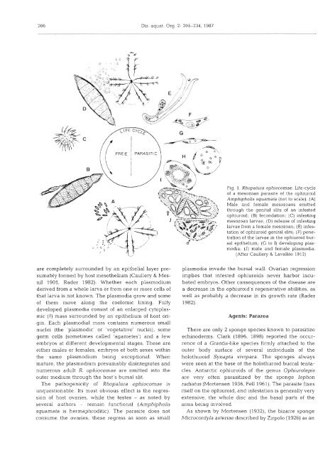
The pathogenicity <strong>of</strong> Rhopalura ophiocomae is<br />
unquestionable. Its most obvious effect is the regres-<br />
sion <strong>of</strong> host ovaries, while the testes - as noted by<br />
several authors - remain functional (Amphipholis<br />
squamata is hermaphroditic). The parasite does not<br />
consume the ovaries; these regress as soon as small<br />
' @ A,<br />
Fig. 1. Rhopalura ophiocomae. Ife-cycle<br />
<strong>of</strong> a mesozoan parasite <strong>of</strong> the ophiuroid<br />
AmphiphoLis squamata (not to scale). (A)<br />
Male and female mesozoans emitted<br />
through the genital slits <strong>of</strong> an infested<br />
ophiuroid; (B) fecondation; (C) infesting<br />
mesozoan larvae; (D) release <strong>of</strong> infesting<br />
larvae from a female mesozoan. (E) infes-<br />
tation <strong>of</strong> ophiuroid genital slits; (F) pene-<br />
tration <strong>of</strong> the larvae in the ophiuroid bur-<br />
sal epithelium; (G to I) developing plas-<br />
modia; (J) male and female plasmodia.<br />
(After Caullery & Lavallee 1912)<br />
plasmodia invade the bursal wall. Ovarian regression<br />
implies that infested ophiuroids never harbor incu-<br />
bated embryos. Other consequences <strong>of</strong> the disease are<br />
a decrease in the ophiuroid's regenerative abilities, as<br />
well as probably a decrease in its growth rate (Rader<br />
1982).<br />
<strong>Agents</strong>: Parazoa<br />
There are only 2 sponge species known to parasitize<br />
echinoderms. Clark (1896, 1898) reported the occur-<br />
rence <strong>of</strong> a Grantia-like species firmly attached to the<br />
outer body surface <strong>of</strong> several individuals <strong>of</strong> the<br />
holothuroid Synapta vivipara. The sponges always<br />
were seen at the base <strong>of</strong> the holothuroid buccal tenta-<br />
cles. Antarctic ophiuroids <strong>of</strong> the genus Ophiurolepis<br />
are very <strong>of</strong>ten parasitized by the sponge Iophon<br />
radiatus (Mortensen 1936, Fell 1961). The parasite fixes<br />
itself on the ophiuroid, and infestation is generally very<br />
extensive, the whole disc and the basal parts <strong>of</strong> the<br />
arms being involved.<br />
As shown by Mortensen (1932), the bizarre sponge<br />
Microcordyla asteriae described by Zirpolo (1926) as an