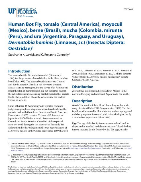
Human Bot Fly, torsalo (Central America), moyocuil - EDIS ...
Human Bot Fly, torsalo (Central America), moyocuil - EDIS ...
Human Bot Fly, torsalo (Central America), moyocuil - EDIS ...

You also want an ePaper? Increase the reach of your titles
YUMPU automatically turns print PDFs into web optimized ePapers that Google loves.
<strong>Human</strong> <strong>Bot</strong> <strong>Fly</strong>, <strong>torsalo</strong> (<strong>Central</strong> <strong>America</strong>), <strong>moyocuil</strong><br />
(Mexico), berne (Brasil), mucha (Colombia, mirunta<br />
(Peru), and ura (Argentina, Paraguay, and Uruguay),<br />
Dermatobia hominis (Linnaeus, Jr.) (Insecta: Diptera:<br />
Oestridae) 1<br />
Stephanie K. Larrick and C. Roxanne Connelly 2<br />
Introduction<br />
The human bot fly, Dermatobia hominis (Linnaeus Jr.,<br />
1781), is a large, densely haired fly that looks like a bumblebee<br />
(Kahn 1999). The human bot fly is native to <strong>Central</strong><br />
and South <strong>America</strong>. The fly is not known to transmit<br />
disease-causing pathogens, but the larvae of D. hominis will<br />
infest the skin of mammals and live out the larval stage in<br />
the subcutaneous layer, causing painful pustules that secrete<br />
fluids. The infestation of any fly larvae inside the body is<br />
known as myiasis.<br />
Cases of human D. hominis myiasis reported from nonindigenous<br />
people are diagnosed when travelers bring the<br />
parasite back with them from <strong>Central</strong> and South <strong>America</strong>.<br />
Haruki et al. (2005) reported 33 cases of D. hominis in<br />
Japan from 1974-2005 as a result of overseas travel to<br />
<strong>Central</strong> and South <strong>America</strong>. One third of the reported<br />
cases occurred during the last six years of the study. Six<br />
different studies have documented seven reported cases of<br />
D. hominis myiasis in the United States since 1999 (Lawson<br />
EENY 440<br />
et al. 2005, Liebert et al. 2004, Maier et al. 2004, Marty et al.<br />
2005, Millikan 1999, Sampson et al. 2002). All the patients<br />
with confirmed D. hominis myiasis had recently been to<br />
<strong>Central</strong> or South <strong>America</strong>.<br />
Distribution<br />
Dermatobia hominis is indigenous from Mexico in the<br />
north to Paraguay and northeast Argentina in the south.<br />
Description<br />
Adult: The adult bot fly is 12 to 18 mm long with a wide<br />
array of colors (Kahn 1999, Sampson et al. 2001). The face<br />
is yellow with a metallic blue abdomen and orange legs and<br />
each body segment is covered with hairs which give the fly<br />
a bumblebee appearance (Khan 1999).<br />
Egg: The egg of the bot fly is creamy colored and oval in<br />
shape, and is attached to different species of blood-feeding<br />
insects captured by the female bot fly. The eggs, usually<br />
1. This document is EENY 440 (IN775), one of a series of Featured Creatures from the Entomology and Nematology Department, Florida Cooperative<br />
Extension Service, Institute of Food and Agricultural Sciences, University of Florida. Original publication date September 2008. Reviewed: December<br />
2011. This document is also available on Featured Creatures website at http://entomology.ifas.ufl.edu/creatures. Please visit the <strong>EDIS</strong> website at http://<br />
edis.ifas.ufl.edu.<br />
2. C. Roxanne Connelly, Associate professor and extension medical entomologist, Department of Entomology, at the Florida Medical Entomology Lab,<br />
200 9th St. SE, Vero Beach Florida 32962: and Stephanie K. Larrick, graduate assistant, Department of Entomology, at the Florida Medical Entomology<br />
Lab, 200 9th St. SE, Vero Beach Florid; Cooperative Extension Service Institute of Food and Agricultural Sciences, University of Florida, Gainesville,<br />
32611.<br />
The Institute of Food and Agricultural Sciences (IFAS) is an Equal Opportunity Institution authorized to provide research, educational information and other services only to<br />
individuals and institutions that function with non-discrimination with respect to race, creed, color, religion, age, disability, sex, sexual orientation, marital status, national<br />
origin, political opinions or affiliations. U.S. Department of Agriculture, Cooperative Extension Service, University of Florida, IFAS, Florida A&M University Cooperative<br />
Extension Program, and Boards of County Commissioners Cooperating. Millie Ferrer-Chancy, Interim Dean
Figure 1. The geographical distribution of the human bot fly,<br />
Dermatobia hominis (Linnaeus f.).<br />
Credits: C. Roxanne Connelly, University of Florida<br />
Figure 2. Lateral view of an adult human bot fly, Dermatobia hominis<br />
(Linnaeus Jr.).<br />
Credits: Lyle J. Buss, University of Florida<br />
attached to the ventral side of the body, hatch when the<br />
insect carrying the eggs begins to blood feed on a warmblooded<br />
host.<br />
Larva: The larva, or white maggot, goes through three<br />
instars once in the mammalian host. Each instar develops<br />
a distinctive shape. The first instar is worm-like with a bulbous<br />
end. The second instar larva has a bottle-neck shape.<br />
The third instar is cylinder shaped. Each instar possesses<br />
backward projecting spines that encircle the thorax.<br />
Pupa: The puparium may exhibit the prominent anterior<br />
spiracles of the third instar larva.<br />
Figure 3. Frontal view of an adult human bot fly, Dermatobia hominis<br />
(Linnaeus Jr.).<br />
Credits: Lyle J. Buss, University of Florida<br />
Figure 4. Dorsal view of an adult human bot fly, Dermatobia hominis<br />
(Linnaeus Jr.).<br />
Credits: Lyle J. Buss, University of Florida<br />
Life Cycle<br />
Eggs: Female D. hominis adults deposit their mature eggs<br />
on a blood-feeding arthropod, usually a mosquito or a<br />
tick, that is captured by the bot fly in flight. This behavior<br />
is known as phoresy (Safdar et al. 2003). As the vector<br />
takes a blood meal, the bot fly eggs react to the change in<br />
temperature and hatch.<br />
Larvae: The larvae enter the skin through the bite wound<br />
or hair follicles, where it then burrows into the skin. The<br />
larvae breathe through two posterior spiracles which lie<br />
flush with the skin of the host. A study by Pereira et al.<br />
(2001) showed that in rats, it took two days for the larvae<br />
to reach the subcutaneous tissue. After the seventh day of<br />
infestation, the larvae molt to the second instars, and then<br />
2
Figure 5. Left to right – 1st, 2nd, early 3rd, and late 3rd instar larva of<br />
the human bot fly, Dermatobia hominis (Linnaeus Jr.).<br />
Credits: Francisco M. Marty, M.D. and Kristen R. Whiteside, B.S.,<br />
Brigham and Women’s Hospital, Boston, MA<br />
Figure 6. Third instar larva of the human bot fly, Dermatobia hominis<br />
(Linnaeus Jr.), lateral view.<br />
Credits: Lyle J. Buss, University of Florida<br />
to third instar after eighteen days. After approximately<br />
thirty days, the third instar larvae, which can grow to be<br />
relatively large (Fig. 7), crawl out of the host to pupate in<br />
the soil. In general, the life of the larvae inside the host is<br />
five to 12 weeks (Acha and Szyfres 1994). The larva feed on<br />
tissue exudates (Haruki et al 2005 and Kahn 1999).<br />
Pupae: Pupation takes place in the ground and the pupae<br />
do not feed. Adults will emerge after two to three weeks.<br />
Adults: <strong>Bot</strong> flies emerge from the pupal cases, and their<br />
extremely sensitive antennae (making up for poor vision)<br />
allow the males and females to find each other quickly<br />
(Fernandes et al. 2004). During the adult stage, D. hominis<br />
does not feed.<br />
Figure 7. Third instar larva of the human bot fly, Dermatobia hominis<br />
(Linnaeus Jr.), frontal view<br />
Credits: Lyle J. Buss, University of Florida<br />
Figure 8. Backward projecting spines on larva of the human bot fly,<br />
Dermatobia hominis (Linnaeus Jr.).<br />
Credits: C. Roxanne Connelly, University of Florida<br />
Figure 9. Pupa of the human bot fly, Dermatobia hominis (Linnaeus<br />
Jr.).<br />
Credits: Marcelo de Campos Pereira, University of Sao Paulo<br />
3
Figure 10. Posterior spiracles of the larva of the human bot fly,<br />
Dermatobia hominis (Linnaeus Jr.).<br />
Credits: C. Roxanne Connelly, University of Florida<br />
Figure 11. Side view of posterior spiracles of the larva of the human<br />
bot fly, Dermatobia hominis (Linnaeus Jr.).<br />
Credits: C. Roxanne Connelly, University of Florida<br />
Hosts<br />
The most common hosts for the human bot fly are cattle<br />
and dogs. However, they are found in many warm-blooded<br />
animals including buffalo, cattle, cats, dogs, humans,<br />
monkeys, pigs, rabbits, and sheep.<br />
Symptoms and Treatment<br />
Dermatobia hominis larvae cause a raised lesion in the skin<br />
that becomes hard and sometimes painful. In some cases<br />
Figure 12. Third instar larva of the human bot fly, Dermatobia hominis<br />
(Linnaeus Jr.).<br />
Credits: C. Roxanne Connelly, University of Florida<br />
the patients can feel the larvae moving when they shower or<br />
cover the wound (Haruki et al. 2005, Sampson et al. 2001).<br />
The host reacts with elevated white cell counts and a high<br />
amount of macrophages can be found around the wound.<br />
For this reason, the lesion often secretes pus.<br />
Figure 13. Raised lesion on the skin caused by the presence of a larva<br />
of the human bot fly, Dermatobia hominis (Linnaeus Jr.).<br />
Credits: Francisco M. Marty, M.D. and Kristen R. Whiteside, B.S.,<br />
Brigham and Women’s Hospital, Boston, MA<br />
There are several treatment options for treatment of D.<br />
hominis myiasis. The most conventional way of removing<br />
the larvae is with a simple surgical procedure that includes<br />
4
local anesthesia. Using a scalpel to cut a slit to enlarge the<br />
wound, the larvae can be taken out.<br />
Dermatobia hominis survives in its host by breathing<br />
through spiracles that are flush with the skin. In order to<br />
coax the larva out, the spiracles need to be covered. They<br />
can be covered with bacon, petroleum jelly, beeswax, or any<br />
other thick substance that prevents the larvae from breathing.<br />
The larvae will come up out of the lesion to breathe<br />
allowing it to be removed with forceps.<br />
In some cases the larva maybe popped out by applying<br />
pressure around the wound. Tamir et al. (2003b) cited a<br />
technique that used two wooden spatulas to apply pressure<br />
to pop the larva out. There may be some difficulty with<br />
this method due to the spines that anchor the larvae in the<br />
wound.<br />
Several authors (Diaz et al. 2006, Kahn 1999, Safdar et al.<br />
2003, Tamir et al. 2003b) have cited the use of lidocaine<br />
injections underneath the cyst. This creates pressure that<br />
pushes the larva out.<br />
After any of these procedures, antibiotics are given to<br />
prevent infection. The wound should heal in one to two<br />
weeks with little or no scarring.<br />
Management<br />
Due to the increase in travelers to <strong>Central</strong> and South<br />
<strong>America</strong>, it is important to carefully monitor and control<br />
the vector population. Additionally, travelers to these<br />
regions need to take preventive measures, including applying<br />
insect repellent and wearing protective clothing (Diaz et<br />
al. 2006; Maier and Honigsmann 2004).<br />
UF/IFAS Recommended Mosquito Repellents - http://edis.<br />
ifas.ufl.edu/IN419<br />
Selected References<br />
• Acha P, Szyfres B. 1994. Zoonoses and communicable disease<br />
common to man and animals. 2nd Edition. Scientific<br />
Publication No. 503. Pan <strong>America</strong>n Health Organization.<br />
WHO, Washington, D.C. 963 pp.<br />
• Cornet MM, Florent A, Lefebvre C, Wertheimer CP,<br />
Bangs M, Bouvet A. 2003. Tracheopulmonary myiasis<br />
caused by a mature third-instar Cuterebra larva: case<br />
report and review. Journal Clinical Microbiology 41:<br />
5810-5812.<br />
• Diaz JH. 2006. The epidemiology, diagnosis, management,<br />
and prevention of ectoparasitic diseases in travelers.<br />
Journal of Travel Medicine 13: 1195-1982.<br />
• Fernandes FF, Linardi PM, Chiarini-Garcia H. 2002.<br />
Morphology of the antenna of Dermatobia hominis<br />
(Diptera: Cuterebridae) based on scanning electron<br />
microscope. Journal of Medical Entomology 39: 36-43.<br />
• Fernandes FF, Chiarini-Garcia H, Linardi PM. 2004.<br />
Scanning electron microscopy studies of sensilla and<br />
other structures of adult Dermatobia hominis (L. Jr., 1781)<br />
(Diptera:Cuterebridae). Journal of Medical Entomology<br />
41: 552-560.<br />
• Haruki K, Hayashi T, Kobayashi M, Katagiri T, Sakurai<br />
Y, Kitajima T. 2005. Myiasis with Dermatobia hominis in<br />
a traveler returning from Costa Rica: review of 33 cases<br />
imported from South <strong>America</strong> to Japan. Journal of Travel<br />
Medicine 12: 285-288.<br />
• Kahn DG. 1999. Myiasis secondary to Dermatobia<br />
hominis (<strong>Human</strong> <strong>Bot</strong>fly) presenting as a long-standing<br />
breast mass. Archives of Pathology and Laboratory<br />
Medicine 123: 829-831.<br />
• Lawson RD, Rizzo M. 2005. Digital infestation with the<br />
human bot fly. Journal of Hand Surgery 30B: 490-491.<br />
• Lang T, Smith DS. 2003. Wiggling subcutaneous lumps.<br />
Clinical Infectious Diseases 37: 2087-2088.<br />
• Liebert PS, Madden RC. 2004. <strong>Human</strong> botfly larva in a<br />
child’s scalp. Journal of Pediatric Surgery 39: 629-630.<br />
• Maier H, Honigsmann H. 2004. Furuncular myiasis<br />
caused by Dermatobia hominis, the human bot fly. Journal<br />
of the <strong>America</strong>n Academy of Dermatology 50: S26-S30.<br />
• Matera G, Liberto MC, Larussa F, Barreca GS, Foca A.<br />
2001. <strong>Human</strong> myiasis: an unusual imported infestation in<br />
Calabria, Italy. Journal of Travel Medicine 8: 103-104.<br />
• Marinho CR, Barbosa LS, Granthon de Azevedo AC, de<br />
Carvalho Querizo MM, Valgode MA, Aguiar Coelho<br />
AM. 2003. Hemilucilia segmentaria (Fabricius, 1805)<br />
(Diptera: Calliphoridae) as new biological vector of eggs<br />
of Dermatobia hominis (Linnaeus Jr., 1781) (Diptera: Oestridae)<br />
in Reserva Biologica do Tingua, Rio de Janeiro,<br />
Brazil. Memórias do Instituto Oswaldo Cruz 98: 937-938.<br />
5
• Marty FM, Whiteside KR. 2005. Myiasis due to Dermatobia<br />
hominis (<strong>Human</strong> <strong>Bot</strong>fly). New England Journal of<br />
Medicine 23: 352.<br />
• Millikan LE. 1999. Myiasis. Clinical Dermatology 17:<br />
191-195.<br />
• Pereira MCT, Leite VHR, Leite ACR. 2001. Experimental<br />
skin lesions from larvae of the bot fly Dermatobia hominis.<br />
Medical and Veterinary Entomology 15: 22-27.<br />
• Rossi MA, Zucoloto S. 1973. Fatal cerebral myiasis caused<br />
by the tropical warble fly, Dermatobia hominis. <strong>America</strong>n<br />
Journal of Tropical Medical Hygiene 22: 267-269.<br />
• Rutledge CR, Day JF. 2008. Mosquito Repellents. <strong>EDIS</strong>.<br />
http://edis.ifas.ufl.edu/IN419 (30 June 2008).<br />
• Safdar N, Young DK, Andes D. 2003. Autochthonous<br />
furuncular myiasis in the United States: case report and<br />
literature review. Clinical Infectious Diseases 36: e73-80.<br />
• Saliba EK, Oumeish YO, Oumeish I. 2002. Epidemiology<br />
of common parasitic infections of the skin in infants and<br />
children. Clinical Dermatology 20: 36-43.<br />
• Sampson C E, MaGuire J, Eriksson E. 2001. <strong>Bot</strong>fly<br />
myiasis: case report and brief review. Annals of Plastic<br />
Surgery 46: 150-152.<br />
• Schwartz E, Gur H. 2002. Dermatobia hominis myiasis: an<br />
emerging disease among travelers to the Amazon Basin of<br />
Bolivia. Journal of Travel Medicine 9: 97-99.<br />
• Tamir J, Haik J, Orenstein A, Schwartz E. 2003a. Dermatobia<br />
hominis myiasis among travelers returning from<br />
South <strong>America</strong>. Journal of the <strong>America</strong>n Academy of<br />
Dermatology 48: 630-632.<br />
• Tamir J , Haik J, Schwartz E. 2003b. Myiasis with Lund’s<br />
fly (Cordylobia rodhaini) in travelers. Journal of Travel<br />
Medicine 10: 293-295.<br />
• Villarino M A, Garcia O, Fussell W, Preston K , Wagner<br />
GG. 2003. An initial survey of the cattle grub Dermatobia<br />
hominis (L. Jr.) in Nicaragua. Preventive Veterinary<br />
Medicine 61: 333-338.<br />
6