

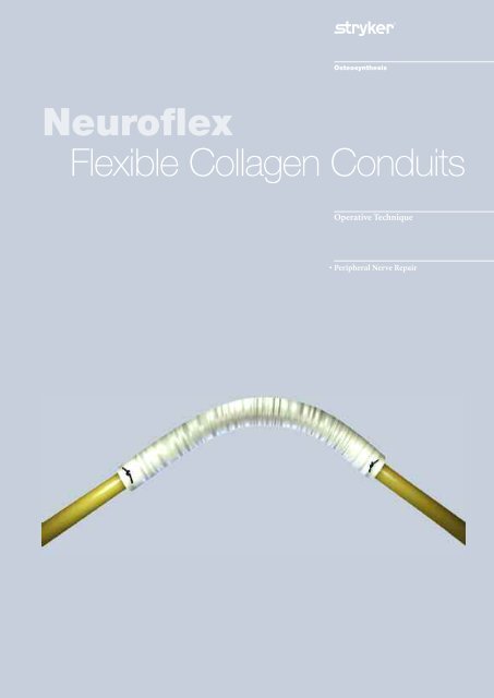
Neuroflex Flexible Collagen Conduits - Stryker
Neuroflex Flexible Collagen Conduits - Stryker
Neuroflex Flexible Collagen Conduits - Stryker

You also want an ePaper? Increase the reach of your titles
YUMPU automatically turns print PDFs into web optimized ePapers that Google loves.
<strong>Neuroflex</strong><br />
<strong>Flexible</strong> <strong>Collagen</strong> <strong>Conduits</strong><br />
Operative Technique<br />
• Peripheral Nerve Repair
Indications and Contraindications<br />
Common Indications<br />
The <strong>Neuroflex</strong> conduit allows repair<br />
without tension of peripheral nerve<br />
discontinuities of less than 3cm. Nerve<br />
gaps may occur in the following types<br />
of discontinuities (gaps) injuries:<br />
1. Crushing Injury (damaged nerve<br />
may need to be resected)<br />
2. Penetrating Injury<br />
Lacerations/Stabbing injuries<br />
Severe Fractures or Dislocations<br />
3. Oncology related excision<br />
4. Repair of Iatrogenic Nerve Injury<br />
5. Failed Primary Repair<br />
Contraindications<br />
1. Acute infections<br />
2. Contaminated wound in the<br />
immediate area surrounding the<br />
peripheral nerve discontinuity<br />
3. Known history of allergic<br />
reactions to collagen and/or<br />
bovine-derived products<br />
See package insert for warnings, precautions, adverse<br />
effects and other essential product information.<br />
Contributing Surgeon<br />
Melvin P. Rosenwasser, MD<br />
Robert E. Carroll, Professor of Hand Surgery<br />
Chief, Orthopaedic Hand & Trauma Service<br />
Director, Trauma Training Center<br />
New York Orthopaedic Hospital<br />
3
Introduction<br />
Repairing Peripheral Nerve Injuries<br />
(PNI) where the gap is less than 3.0cm<br />
with a type 1 collagen conduit offers<br />
several potential advantages:<br />
Eliminates the need to harvest an<br />
autograft in a second surgical<br />
procedure<br />
• Reduces associated morbidity at the<br />
donor site, which may include:<br />
scarring, neuroma formation, and<br />
loss of donor site function.1<br />
• May improve OR efficiency.<br />
• Removing the need to harvest<br />
autograft from the lower extremity<br />
(sural nerve), may allow regional<br />
anesthesia and use at an ambulatory<br />
surgery center.<br />
Multiple nerve conduits are readily<br />
available at the time of surgery, and<br />
can be size-matched to fit the nerve<br />
repair site/s. This may be a benefit<br />
to surgeons addressing multiple<br />
nerves or segmental nerve injuries.<br />
Approximation of nerve fascicles is<br />
not required.<br />
• Studies suggest regenerating axons are<br />
able to align themselves as a result of<br />
various neurotropic and neurotrophic<br />
factors. 2,3,4 This allows growth factors<br />
to influence the proximal growth<br />
cone as a severed nerve ending grows<br />
across a confined gap.<br />
Allows tensionless repair of the nerve<br />
• In many situations, a primary<br />
tensionless repair of the nerve is not<br />
possible, even with extensive<br />
mobilization. Extensive mobilization<br />
may also negatively influence the<br />
epineurial vasculature. The <strong>Neuroflex</strong><br />
nerve conduit allows for a tensionless<br />
repair.<br />
• In many cases, the hand may be<br />
positioned in a more anatomic<br />
position following nerve repair where<br />
the gap is bridged by a conduit than<br />
when the surgeon re-approximates<br />
the nerve endings. This is often<br />
helpful to lessen swelling and<br />
stiffness.<br />
Type 1 <strong>Collagen</strong> is ideally suited to<br />
peripheral nerve repair. 5<br />
• Selective conduit permeability allows<br />
nutrients to diffuse, yet acts as a<br />
barrier to larger fibroblast cells.<br />
• Type 1 collagen may be better<br />
accepted by soft tissue than PGA<br />
based conduits. 2,6,7<br />
• Usually degrades via normal<br />
metabolic pathways within 3-6<br />
months following implantation.<br />
• Hypo-immunogenic. 8<br />
4<br />
<strong>Neuroflex</strong>, the type 1 collagen<br />
conduit with additional flexibility<br />
• In addition to these benefits,<br />
<strong>Neuroflex</strong> adds the patented feature<br />
of being kink-resistant. 9 When flexed,<br />
the type 1 collagen conduit will bend<br />
up to approximately 60 degrees<br />
without forming an occlusion,<br />
potentially impacting nerve repair.<br />
Standard type 1 collagen conduits will<br />
occlude at approximately 20 degrees. 10<br />
• The corrugated sides of the conduit<br />
allow additional flexibility without<br />
changing the established benefits of<br />
standard type 1 collagen conduit<br />
properties, including permeability<br />
and resorption time.
Operative Technique<br />
Step 1: Prepare Nerve<br />
Expose nerve at the appropriate<br />
incision site according to standard<br />
procedures.<br />
Prepare the nerve bed. Examine the<br />
local tissues (fat/muscle), resecting scar<br />
tissue as needed.<br />
The proximal and distal segments of<br />
the injured nerves are debrided to<br />
normal tissue by visual and tactile cues<br />
(Figure 1).<br />
Note: Axoplasmic fluid often will<br />
weep out of the resected nerve ending,<br />
and may be one indication of<br />
adequate debridement.<br />
Figure 1 Figure 2<br />
Figure 3<br />
5<br />
Figure 4
Operative Technique<br />
Step 2: Select and prepare<br />
appropriate size <strong>Neuroflex</strong><br />
conduit<br />
The diameter of the nerve is measured<br />
and an appropriate size <strong>Neuroflex</strong><br />
conduit is selected. The internal<br />
diameter of the chosen nerve cuff<br />
should be slightly larger than the nerve<br />
diameter (Figures 2,5).<br />
Note: For late repairs when a<br />
neuroma/schwanoma exist, there is<br />
often significant swelling of the nerve.<br />
This may be an important<br />
consideration when sizing the nerve<br />
conduit.<br />
Step 3: Hydrate <strong>Neuroflex</strong><br />
Conduit<br />
Hydrate nerve conduit in sterile<br />
physiological saline solution for 5<br />
minutes. Upon hydration, <strong>Neuroflex</strong><br />
will expand approximately 20-30%<br />
of its dry length.<br />
After hydration, the nerve conduit is<br />
trimmed accordingly to at least a<br />
minimum length of 5mm longer than<br />
the measured nerve gap, so that both<br />
the proximal and distal nerve stumps<br />
can be inserted adequately into each<br />
end of the nerve conduit.<br />
Figure 5 Measuring the <strong>Neuroflex</strong> conduit<br />
for repair of radial sensory nerve.<br />
6
Operative Technique<br />
Step 4: Suture nerve into<br />
<strong>Neuroflex</strong> conduit (Entubulation)<br />
Horizontal Mattress Suture Technique<br />
Using atraumatic (8.0 - 9.0 nylon)<br />
suture, pass the suture through the<br />
<strong>Neuroflex</strong> conduit from the outside to<br />
inside, at least 2mm from the end of<br />
the tube.<br />
Pass the suture longitudinally through<br />
the epineurium of the nerve stump and<br />
back through the epineurium again<br />
(u-shape) (Figure 7). Pass the suture<br />
through the inside of the <strong>Neuroflex</strong><br />
conduit to the outside.<br />
Gently draw the nerve stump into the<br />
<strong>Neuroflex</strong> conduit by pulling the<br />
suture such that the nerve stump is<br />
drawn into the conduit.<br />
The final length of insertion of the<br />
nerve stump into the conduit should<br />
be greater than or equal to the nerve<br />
diameter. A tensionless secure knot is<br />
tied in the suture (Figure 8).<br />
Using a syringe gently flush the lumen<br />
of the nerve cuff with sterile saline<br />
or Lactated Ringer’s solution (USP)<br />
(Figure 9). Repeat the suturing<br />
procedure for the other nerve stump<br />
(Figure 10). Repeat the flushing<br />
procedure and fill the interior of the<br />
nerve cuff with saline or Lactated<br />
Ringer’s solution (USP) (Figure 12).<br />
Typically three sutures are placed<br />
approximately 120 degrees apart in a<br />
‘triangulation’ technique (Figure 13).<br />
Note: Avoid tension of the peripheral<br />
nerve to be repaired during the entire<br />
procedure.<br />
Figure 6 Figure 7<br />
Figure 8 Figure 9<br />
Figure 10<br />
Figure 12 Figure 13<br />
7<br />
Figure 11
Operative Technique<br />
Step 5: Post-operative<br />
Considerations<br />
Closure of the surgical field by layers is<br />
routine. Excessive and uncontrolled<br />
movement of the extremity where<br />
nerve repair was performed must be<br />
avoided to prevent possible migration<br />
of the device and failure of the repair.<br />
Figure 14<br />
8
Case Examples<br />
Figure 15: Radial nerve near elbow<br />
entrapped during plating of a radial neck<br />
fracture. Four months post-op nerve injury referred for<br />
treatment<br />
Figure 19: Final repair of radial nerve with <strong>Neuroflex</strong> conduit<br />
9<br />
Figure 16: Nerve resected to healthy tissue<br />
Figure 17: Measuring the conduit Figure 18: Suturing the <strong>Neuroflex</strong><br />
conduit in place
Ordering Information<br />
<strong>Neuroflex</strong> <strong>Flexible</strong> <strong>Collagen</strong> <strong>Conduits</strong><br />
10<br />
REF Inner Diameter Length<br />
CNCF2025 2.0 mm 2.5 cm<br />
CNCF2525 2.5 mm 2.5 cm<br />
CNCF3025 3.0 mm 2.5 cm<br />
CNCF4025 4.0 mm 2.5 cm<br />
CNCF5025 5.0 mm 2.5 cm<br />
CNCF6025 6.0 mm 2.5 cm
References<br />
1. Taras JS, Nanavati V, Steelman P. Nerve conduits. J Hand Ther. 2005 Apr-Jun; 18(2):191-7. Review.<br />
2. Trumble TE, Parisi D, Archibald S, Allan CH. Synthetic Nerve <strong>Conduits</strong>. pp 121-128 Peripheral Nerve Surgery.<br />
Practical Applications in the Upper Extremity. Slutsky and Hentz. Elsevier 2006.<br />
3. Lundborg G, Dahlin LB, Danielson N, Nachemson AK. Tissue specificity in nerve regeneration. Scand J Plast<br />
Reconstr Surg. 1986; 20:279-83.<br />
4. Mackinnon SE, Dellon AL, Lundborg G, Hudson AR, Hunter DA. A study of neurotropism in a primate model. J<br />
Hand Surgery [Am]. 1986; 11:888-94.<br />
5. Li, ST. Peripheral Nerve Repair with <strong>Collagen</strong> <strong>Conduits</strong>. Clinical Materials 9 (1992) 195-200.<br />
6. Weber RA, Breidenbach WC, Brown RE, Jabaley ME, Mass DP. A randomized prospective study of polyglycolic acid<br />
conduits for digital nerve reconstruction in humans. Plast Reconstr Surg 2000 Oct; 106 (5): 1036-45; discussion<br />
1046-8.<br />
7. Bostman O, Pihlajamaki H. Clinical Biocompatibility of biodegradable orthopaedic implants for internal fixation: a<br />
review. Biomaterials 21 (2000) pp 2615-2621.<br />
8. Li ST, Rodkey WG, Yuen D, Hansen P, Steadman JR. Type 1 <strong>Collagen</strong>-Based Template for Meniscus Regeneration.<br />
Tissue Engineering and Biodegradable Equivalents. Scientific and Clinical Applications. 2002 pp 237-266.<br />
9. Li ST, Yuen D: U.S. Patent #6,716,225, Implant Devices for Nerve Repair, 2004.<br />
10. Data on file at <strong>Collagen</strong> Matrix, Inc.<br />
The following products are also available through your<br />
<strong>Stryker</strong> Sales Representative:<br />
HydroSet – Injectable HA Bone Substitute<br />
Inion OTPS – Biodegradable Plates, Screws and Pins<br />
Profyle Modular – Small Fragment System<br />
Twinfix – Cannulated Compression Screw<br />
Hoffmann II Micro – Small Bone External Fixation and Lengthening System<br />
VariAx – Variable Angle Distal Radius Locking Plate System<br />
KnifeLight – Minimally Open Carpal Tunnel Ligament Release<br />
AxSOS – Fixed Angle Locking Plate System<br />
Dynamic Joint Distractor II – Hinged Elbow Fixation System<br />
T2 Proximal Humeral Nail – Intramedulary Nail System<br />
T2 Humeral Nail – Intramedulary Nail System<br />
11
0123<br />
The information presented in this brochure is intended to demonstrate a <strong>Stryker</strong> product. Always refer to the package<br />
insert, product label and/or user instructions before using any <strong>Stryker</strong> product. Surgeons must always rely on their own<br />
clinical judgment when deciding which products and techniques to use with their patients. Products may not be available<br />
in all markets. Product availability is subject to the regulatory or medical practices that govern individual markets.<br />
Please contact your <strong>Stryker</strong> representative if you have questions about the availability of <strong>Stryker</strong> products in your area.<br />
Manufactured by <strong>Collagen</strong> Matrix, Inc., Franklin Lakes, NJ 07417<br />
<strong>Stryker</strong> Corporation or its subsidiary owns the registered trademark: <strong>Stryker</strong><br />
<strong>Collagen</strong> Matrix, Inc. owns the following trademark: <strong>Neuroflex</strong><br />
This product is covered by U.S. Patent: 6,716,225<br />
Literature Number: 90-07563<br />
LOT A1608<br />
Copyright © 2008 <strong>Stryker</strong><br />
325 Corporate Drive<br />
Mahwah, NJ 07430<br />
www.stryker.com


