Page: 92-94; FULL TEXT - Journal of IMAB
Page: 92-94; FULL TEXT - Journal of IMAB
Page: 92-94; FULL TEXT - Journal of IMAB

Create successful ePaper yourself
Turn your PDF publications into a flip-book with our unique Google optimized e-Paper software.
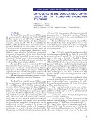
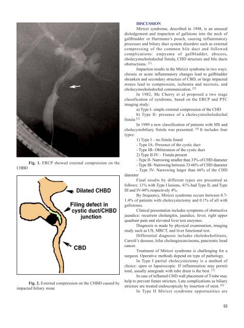
Fig. 1. ERCP showed external compression on the<br />
CHBD<br />
Fig. 2. External compression on the CHBD caused by<br />
impacted biliary stone<br />
DISCUSSION<br />
Mirizzi syndrome, described in 1<strong>94</strong>8, is an unusual<br />
dislodgement and impaction <strong>of</strong> gallstone into the neck <strong>of</strong><br />
gallbladder or Hartmann’s pouch, causing inflammatory<br />
processes and biliary duct system disorders such as external<br />
compressing <strong>of</strong> the common bile duct and followed<br />
complications: empyema <strong>of</strong> gallbladder, abscess,<br />
cholecystocholedochal fistula, CHD structure and bile ducts<br />
obstructions. [1]<br />
Impaction results in the Mirizzi syndrome in two ways:<br />
chronic or acute inflammatory changes lead to gallbladder<br />
shrunken and secondary structure <strong>of</strong> CBD, or large impacted<br />
stones lead to compression, ischemia and necrosis, and<br />
cholecystocholedochal communication. [2]<br />
In 1982, Mc Cherry et al proposed a two stage<br />
classification <strong>of</strong> syndrome, based on the ERCP and PTC<br />
imaging study:<br />
a) Type I- simple external compression <strong>of</strong> the CHD<br />
b) Type II- presence <strong>of</strong> a cholecystocholedochal<br />
fistula [3]<br />
In 1989 a new classification <strong>of</strong> patients with MS and<br />
cholecystobiliary fistula was presented. [4] It includes four<br />
types:<br />
1) Type I – no fistula found<br />
- Type IA- Presence <strong>of</strong> the cystic duct<br />
- Type IB- Obliteration <strong>of</strong> the cystic duct<br />
2) Type II-IV - Fistula present<br />
- Type II- Narrowing smaller than 33% <strong>of</strong> CHD diameter<br />
- Type III- Narrowing between 33-66% <strong>of</strong> CHD diameter<br />
- Type IV- Narrowing larger than 66% <strong>of</strong> the CHD<br />
diameter<br />
Final results by different types are presented as<br />
follows: 11% with Type I lesions, 41% had Type II, and Type<br />
III and IV-44% respectively 4%.<br />
By frequency, Mirizzi syndrome occurs between 0.7-<br />
1.4% <strong>of</strong> patients with cholecystectomy and 0.1% <strong>of</strong> all with<br />
gallstones.<br />
Clinical presentation includes symptoms <strong>of</strong> obstructive<br />
jaundice: recurrent cholangitis, jaundice, fever, right upper<br />
quadrant pain and elevated liver test enzymes.<br />
Diagnosis is made by physical examination, imaging<br />
study such as US, MRCT, and liver functional test.<br />
Differential diagnosis includes choledocholitiasis,<br />
Carroli’s desease, hilar cholangiocarcinoma, pancreatic head<br />
cancer.<br />
Treatment <strong>of</strong> Mirizzi syndrome is challenging for a<br />
surgeon. Operative methods depend on type <strong>of</strong> pathology.<br />
In Type I partial cholecystectomy is a method <strong>of</strong><br />
choice: open or laparoscopic. If inflammation may permit<br />
total, usually antegrade with tube drain is the best. [5]<br />
In case <strong>of</strong> inflamed CHD wall placement <strong>of</strong> T-tube may<br />
help to prevent future stricture. Late complications as biliary<br />
stricture are treated endoscopicaly by insertion <strong>of</strong> stent. [6]<br />
In Type II Mirizzi syndrome opportunities are<br />
93