Page: 92-94; FULL TEXT - Journal of IMAB
Page: 92-94; FULL TEXT - Journal of IMAB
Page: 92-94; FULL TEXT - Journal of IMAB

Create successful ePaper yourself
Turn your PDF publications into a flip-book with our unique Google optimized e-Paper software.
ABSTRACT:<br />
Mirizzi syndrome is a rarely observed complication <strong>of</strong><br />
gallstone disease, causing major biliary problems, if not<br />
diagnosed previously. It was described in 1<strong>94</strong>8 by P. L. Mirizzi<br />
and presents unusual lodged gallstone in either the cystic<br />
duct or most frequently in Hartmann pouch <strong>of</strong> the gallbladder.<br />
Impaction, acute obstruction and wall ischemia are causative<br />
for inflammation and abscess formation. External common<br />
hepatic bile duct compression and obstruction result in<br />
clinical presentation <strong>of</strong> intermittent or constant jaundice. We<br />
report 57-year-old male with extensive mechanical icter, fever,<br />
nausea and vomiting, and upper abdominal pain in epigastria<br />
from five days. Abdominal US evaluation showed 17mm stone<br />
localized in infundibulum and shrunk <strong>of</strong> gallbladder. MRCT<br />
revealed impacted stone, chronic tissue inflammation,<br />
involved common hepatic duct with stricture. Mirizzi<br />
syndrome was diagnosed.<br />
Intraoperatively was found an impacted gallstone in<br />
the Hartmann pouch, extensive fibrosis <strong>of</strong> hepatoduodenal<br />
ligament and abscess cavity formation in the Callot’s triangle<br />
with engagement <strong>of</strong> common hepatic bile duct wall. Antegrade<br />
cholecystectomy was made and T drain was placed. Second<br />
operation and Roux-Y limb anastomosis was performed after<br />
unsuccessful tentative for recanalization <strong>of</strong> distal CBD with<br />
clamping <strong>of</strong> T drain.<br />
Key words: Impacted gallstone, Callot’s triangle<br />
abscess, cystic duct variation, common hepatic duct stricture,<br />
T-drain.<br />
INTRODUCTION<br />
Impaction <strong>of</strong> unique large or multiple small gallstones<br />
between neck <strong>of</strong> gallbladder and confluence <strong>of</strong> cystic duct<br />
and common hepatic duct results pathologic changes in<br />
normal bile flow and local and systematic complications. The<br />
process <strong>of</strong> inflammation, wall ischemia and external<br />
compression lead to erosion <strong>of</strong> the involved tissues and duct<br />
structure <strong>of</strong> common hepatic duct or cholecystocholedochal<br />
fistula formation. Despite modern advances in imaging<br />
<strong>92</strong><br />
<strong>Journal</strong> <strong>of</strong> <strong>IMAB</strong> - Annual Proceeding (Scientific Papers) 2009, book 1<br />
MIRIZZI SYNDROME-RARE CAUSE OF MAJOR<br />
BILIARY COMPLICATIONS. CASE REPORT<br />
Ludmil M. Veltchev 1 , Manol A. Kalniev 2 , Todor A. Todorov 3 ,<br />
1) Fellow, Master’s Program in Hepatobiliary Pancreatic Surgery, Henri Bismuth<br />
Hepatobiliary Institute, 12-14, avenue Paul Vaillant-Couturier, <strong>94</strong>804 Villejuif<br />
Cedex<br />
2) Department <strong>of</strong> Anatomy, Cytology and Histology, University <strong>of</strong> Medicine,<br />
S<strong>of</strong>ia, Bulgaria<br />
3) Department <strong>of</strong> Pathology, University <strong>of</strong> Medicine, S<strong>of</strong>ia, Bulgaria<br />
diagnoses, Mirizzi syndrome presents challenge surgery<br />
treatment situation caused by presentation <strong>of</strong> rare anatomical<br />
variation <strong>of</strong> cystic duct and total change <strong>of</strong> normal anatomy<br />
after long standing inflammation. Good surgical knowledge<br />
for diagnosis and reconstruction is needed.<br />
CASE REPORT<br />
After consultation at emergency room, a 57-year-old<br />
man was admitted in department <strong>of</strong> surgery for resuscitation,<br />
diagnosis and treatment. He presented intensive jaundice<br />
(bilirubin rate: 4.5 mg/dL), fever 38°C, upper right abdominal<br />
pain, predominantly in epigastria and right subcostal region,<br />
nausea and vomiting, and asthenia for last 24 hours.<br />
Abdominal US reveal a 17 mm stone incorporated in<br />
Harmann’s pouch, pericholecystitis, and medially to the<br />
stone, liquid collection aproximally located in Callot’s triangle.<br />
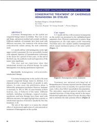
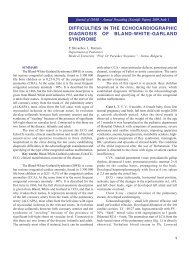
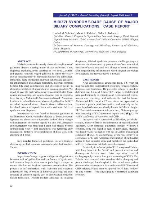
ERCP revealed stone obstructed cystic duct, filiforme passage<br />
with narrowing <strong>of</strong> CHD (common hepatic duct) (Fig.1). No<br />
visible confluence <strong>of</strong> cystic duct and CHD.<br />
Introperativelly scrunched gallbladder, pericholecystatis,<br />
intensive fibrosis and edematous <strong>of</strong> hepatoduodenal<br />
ligament. After bimanual palpation thought Winslow’s<br />
foramen, stone was found in neck <strong>of</strong> gallbladder. Medially<br />
was found “cystic” collection with pus in Callot’s triangle and<br />
evacuated (Fig.2). Microbiological probe was positive in<br />
second day for E. coli. Antegrade mobilization <strong>of</strong> gallbladder<br />
helped to find impacted stone and mobilized the cystic duct<br />
to CHD. No fistula or bile leaks were discovered.<br />
Proximally to inflamed part <strong>of</strong> CHD was placed T-drain,<br />
with long branch to be “stent” and prevent structure and<br />
decompress biliary tree. Intra operative control<br />
cholangiography showed low grade narrowing <strong>of</strong> CHD. The<br />
T-drain was removed after standard dally clamping and<br />
patient discharged from hospital. At first month same patient<br />
presents with subconjuctival icter and control ERCP revealed<br />
CHD stricture. Plastic stent was placed for 30 days. Followup<br />
and control cholangiography confirmed complete<br />
recovery.
Fig. 1. ERCP showed external compression on the<br />
CHBD<br />
Fig. 2. External compression on the CHBD caused by<br />
impacted biliary stone<br />
DISCUSSION<br />
Mirizzi syndrome, described in 1<strong>94</strong>8, is an unusual<br />
dislodgement and impaction <strong>of</strong> gallstone into the neck <strong>of</strong><br />
gallbladder or Hartmann’s pouch, causing inflammatory<br />
processes and biliary duct system disorders such as external<br />
compressing <strong>of</strong> the common bile duct and followed<br />
complications: empyema <strong>of</strong> gallbladder, abscess,<br />
cholecystocholedochal fistula, CHD structure and bile ducts<br />
obstructions. [1]<br />
Impaction results in the Mirizzi syndrome in two ways:<br />
chronic or acute inflammatory changes lead to gallbladder<br />
shrunken and secondary structure <strong>of</strong> CBD, or large impacted<br />
stones lead to compression, ischemia and necrosis, and<br />
cholecystocholedochal communication. [2]<br />
In 1982, Mc Cherry et al proposed a two stage<br />
classification <strong>of</strong> syndrome, based on the ERCP and PTC<br />
imaging study:<br />
a) Type I- simple external compression <strong>of</strong> the CHD<br />
b) Type II- presence <strong>of</strong> a cholecystocholedochal<br />
fistula [3]<br />
In 1989 a new classification <strong>of</strong> patients with MS and<br />
cholecystobiliary fistula was presented. [4] It includes four<br />
types:<br />
1) Type I – no fistula found<br />
- Type IA- Presence <strong>of</strong> the cystic duct<br />
- Type IB- Obliteration <strong>of</strong> the cystic duct<br />
2) Type II-IV - Fistula present<br />
- Type II- Narrowing smaller than 33% <strong>of</strong> CHD diameter<br />
- Type III- Narrowing between 33-66% <strong>of</strong> CHD diameter<br />
- Type IV- Narrowing larger than 66% <strong>of</strong> the CHD<br />
diameter<br />
Final results by different types are presented as<br />
follows: 11% with Type I lesions, 41% had Type II, and Type<br />
III and IV-44% respectively 4%.<br />
By frequency, Mirizzi syndrome occurs between 0.7-<br />
1.4% <strong>of</strong> patients with cholecystectomy and 0.1% <strong>of</strong> all with<br />
gallstones.<br />
Clinical presentation includes symptoms <strong>of</strong> obstructive<br />
jaundice: recurrent cholangitis, jaundice, fever, right upper<br />
quadrant pain and elevated liver test enzymes.<br />
Diagnosis is made by physical examination, imaging<br />
study such as US, MRCT, and liver functional test.<br />
Differential diagnosis includes choledocholitiasis,<br />
Carroli’s desease, hilar cholangiocarcinoma, pancreatic head<br />
cancer.<br />
Treatment <strong>of</strong> Mirizzi syndrome is challenging for a<br />
surgeon. Operative methods depend on type <strong>of</strong> pathology.<br />
In Type I partial cholecystectomy is a method <strong>of</strong><br />
choice: open or laparoscopic. If inflammation may permit<br />
total, usually antegrade with tube drain is the best. [5]<br />
In case <strong>of</strong> inflamed CHD wall placement <strong>of</strong> T-tube may<br />
help to prevent future stricture. Late complications as biliary<br />
stricture are treated endoscopicaly by insertion <strong>of</strong> stent. [6]<br />
In Type II Mirizzi syndrome opportunities are<br />
93
dependent <strong>of</strong> biliary communication and sorrowing inflamed<br />
tissues:<br />
a) Corlette, Bismuth et al. recommende partial<br />
cholecystectomy, oversuturing <strong>of</strong> the gallbladder cuff and<br />
insertion <strong>of</strong> a T-tube through the fistula as an adequate<br />
treatment for Type II. [7]<br />
b) Hepaticojejunostomy with Roux-Y limb is<br />
recommended by many authors as adequate procedure in case<br />
<strong>of</strong> major necrosis and unrepairable defect <strong>of</strong> CHD wall.<br />
Choledochoplasty with neighborhoods tissues and<br />
cholecystoduodenostomy has been described, but not have<br />
introduced as good results. [8-9]<br />
REFERENCES<br />
1. Mirizzi P.L. Syndrome del conducto<br />
hepatico. <strong>Journal</strong> International de Chirurgie<br />
1<strong>94</strong>8; 8: 731-737.<br />
2. Pemberton M., Wells A.D. The<br />
Mirizzi syndrome. Postgrad Med J 1997;<br />
73: 487-490. PMID: 9307740<br />
3. McSherry C.K., Ferstenberg H.,<br />
Virshup M. The Mirizzi syndrome:<br />
suggested classification and surgical therapy.<br />
Surg Gastroenterol 1982;1:219-225.<br />
4. Csendes A., Diaz J.C., Burdiles P.,<br />
Maluenda F., Nava O. Mirizzi syndrome<br />
and cholecystobiliary fistula: a unifying<br />
<strong>94</strong><br />
classification. Br J Surg 1989; 76:1139-1143.<br />
PMID: 2597969<br />
5. Abou-Saif A., Al-Kawas F.H.<br />
Complications <strong>of</strong> gallstone disease: Mirizzi<br />
syndrome, cholecystocholedochal fistula,<br />
and gallstone ileus. Am J Gastroenterol<br />
2002;97: 249-54. PMID: 11866258<br />
6. Binnie N.R., Nixon, S.J., Palmer,<br />
K.R. Mirizzi syndrome managed by<br />
endoscopic stenting and laparoscopic<br />
cholecystectomy. Br J Surg 19<strong>92</strong>;79:647.<br />
PMID: 1643475<br />
7. Corlette M.B., Bismuth H.<br />
Corresponding author:<br />
Ludmil Marinov Veltchev, MD PhD<br />
Mobile: +359 876 259 685<br />
E-mail: drlmarinov@yahoo.com<br />
In conclusion, Mirizzi syndrome is rare pathological<br />
condition that cannot diagnose during physical examination.<br />
It requires imaging study. Management is to determine the<br />
type and best surgical procedure at time <strong>of</strong> laparotomy. In<br />
Type I case, simple cholecystectomy is method <strong>of</strong> choice. If<br />
CHD wall inflammatory changes are found, T-tube placement<br />
is recommended to avoid disruption, leaks and stricture. Type<br />
II-IV patients require complex management. Total isolation <strong>of</strong><br />
inflamed segment with Roux-en-Y hepaticojejunostomy may<br />
have the best long-term outcome.<br />
Biliobiliary fistula. A trap in the surgery <strong>of</strong><br />
cholelithiasis. Arch Surg 1975;110: 377-<br />
83. PMID: 1147754<br />
8. Shah O.J., Dar M.A., Wani M.A.,<br />
Wani N.A. Management <strong>of</strong> Mirizzi<br />
syndrome: a new surgical approach. ANZ J<br />
Surg 2001;71: 423-7. PMID: 11450919<br />
9. Baer H.U., Matthews J.B., Schweizer<br />
W.P., Gertsch P., Blumgart L.H..<br />
Management <strong>of</strong> the Mirizzi syndrome and<br />
the surgical implications <strong>of</strong> cholecystcholedochal<br />
fistula. Br J Surg 1990;77:743-745.<br />
PMID: 2383747