

Mirizzi Syndrome: A Review of the Literature
Mirizzi Syndrome: A Review of the Literature
Mirizzi Syndrome: A Review of the Literature

Create successful ePaper yourself
Turn your PDF publications into a flip-book with our unique Google optimized e-Paper software.
March 2006<br />
INTRODUCTION<br />
KUWAIT MEDICAL JOURNAL<br />
<strong>Review</strong> Article<br />
<strong>Mirizzi</strong> <strong>Syndrome</strong>: A <strong>Review</strong> <strong>of</strong> <strong>the</strong> <strong>Literature</strong><br />
George J Xeroulis, Ward Davies<br />
Department <strong>of</strong> Surgery, University <strong>of</strong> Western Ontario, London, Ontario, Canada<br />
<strong>Mirizzi</strong> syndrome is a rare cause <strong>of</strong> obstructive<br />
jaundice. This entity should be considered in <strong>the</strong><br />
d i ff e rential diagnosis <strong>of</strong> all patients with obstru c t i v e<br />
jaundice. Failure to recognize <strong>the</strong> condition<br />
preoperatively can result in a major bile duct injury,<br />
particularly during laparoscopic surg e r y [ 1 ] . The<br />
syndrome refers to obstruction <strong>of</strong> <strong>the</strong> common<br />
hepatic duct by extrinsic compression usually from<br />
a gallstone impacted in Hartmann’s pouch or <strong>the</strong><br />
cystic duct. Large gallstones that become impacted<br />
in this area produce common hepatic duct<br />
o b s t ruction by two mechanisms: mechanical<br />
obstruction by direct compression <strong>of</strong> <strong>the</strong> common<br />
hepatic duct, or <strong>the</strong>y can cause obstru c t i o n<br />
secondary to repeated bouts <strong>of</strong> local inflammation.<br />
In 1948, A rgentinean surgeon Pablo Luis<br />
<strong>Mirizzi</strong>, first described a syndrome <strong>of</strong> common<br />
hepatic duct obstruction in <strong>the</strong> setting <strong>of</strong><br />
longstanding cholelithiasis and cholecystitis [2] . The<br />
classic description <strong>of</strong> <strong>the</strong> disease includes four<br />
components: (a) a close parallel course <strong>of</strong> <strong>the</strong> cystic<br />
duct and <strong>the</strong> common hepatic duct, (b) an impacted<br />
stone in <strong>the</strong> cystic duct or <strong>the</strong> neck <strong>of</strong> <strong>the</strong><br />
gallbladder, (c) common hepatic duct obstruction<br />
secondary to external compression by <strong>the</strong> cystic<br />
duct stone (and <strong>the</strong> surrounding inflammation),<br />
and (d) jaundice, with or without cholangitis.<br />
<strong>Mirizzi</strong>’s syndrome is a rare complication <strong>of</strong><br />
cholelithiasis, with an estimated incidence <strong>of</strong> 0.05-<br />
2.7% [1,3,4] . It presents as a spectrum <strong>of</strong> disease that<br />
varies from extrinsic compression <strong>of</strong> <strong>the</strong> common<br />
hepatic duct to <strong>the</strong> presence <strong>of</strong> a cholecystobiliary<br />
fistula. Often, this dangerous alteration to anatomy<br />
is not recognized pre o p e r a t i v e l y, and has <strong>the</strong><br />
potential to lead to significant morbidity and<br />
biliary injury, particularly in <strong>the</strong> laparoscopic era.<br />
CLASSIFICATION<br />
There are three classifications which have been<br />
proposed to describe variants <strong>of</strong> <strong>Mirizzi</strong> syndrome,<br />
and to aid in selecting <strong>the</strong> appropriate <strong>the</strong>rapeutic<br />
procedure. The original classification, by McSherry<br />
Kuwait Medical Journal 2006, 38 (1): 3-6<br />
et al [5] , described two types. Type I referred to<br />
compression <strong>of</strong> <strong>the</strong> common hepatic duct by a stone<br />
impacted in <strong>the</strong> cystic duct or Hartmann’s pouch.<br />
Type II referred to erosion <strong>of</strong> <strong>the</strong> calculus from <strong>the</strong><br />
cystic duct into <strong>the</strong> common hepatic duct,<br />
producing a cholecystocholedochal fistula.<br />
Csendes et al [6] created a second classification<br />
taking into account <strong>the</strong> extent <strong>of</strong> fistula. Type I<br />
remained <strong>the</strong> same, external compression <strong>of</strong> <strong>the</strong><br />
common hepatic duct due to a stone impacted at<br />
<strong>the</strong> neck <strong>of</strong> <strong>the</strong> gallbladder or at <strong>the</strong> cystic duct.<br />
Types II to IV lesion referred to <strong>the</strong> presence and<br />
extent <strong>of</strong> a cholecystobiliary (cholecystohepatic or<br />
cholecystocholedochal) fistula, due to erosion <strong>of</strong><br />
<strong>the</strong> anterior or lateral wall <strong>of</strong> <strong>the</strong> common hepatic<br />
duct by impacted stones. The fistula involved less<br />
than one-third <strong>of</strong> <strong>the</strong> circumference <strong>of</strong> <strong>the</strong> common<br />
hepatic duct in type II. Involvement <strong>of</strong> between<br />
one-third and two-thirds <strong>of</strong> <strong>the</strong> circumference <strong>of</strong><br />
<strong>the</strong> common hepatic duct was called a type III<br />
lesion, while destruction <strong>of</strong> <strong>the</strong> entire wall <strong>of</strong> <strong>the</strong><br />
common hepatic duct was called a type IV lesion.<br />
In <strong>the</strong>ir original paper, a total <strong>of</strong> 219 patients were<br />
identified with <strong>Mirizzi</strong>’s syndrome. The incidence<br />
<strong>of</strong> type I lesions was 11 per cent, type II, 41 per cent,<br />
type III, 44 per cent and type IV, four per cent. The<br />
majority had obstructive jaundice.<br />
The third classification, proposed by Nagakawa<br />
and colleagues [7] , expanded upon <strong>the</strong> definition <strong>of</strong><br />
<strong>the</strong> <strong>Mirizzi</strong> syndrome. Type I referred to a stone<br />
impacted in <strong>the</strong> cystic duct or gallbladder neck.<br />
Type II was characterized by a fistula <strong>of</strong> <strong>the</strong><br />
common duct. Type III was defined by hepatic duct<br />
stenosis due to a stone at <strong>the</strong> confluence <strong>of</strong> <strong>the</strong><br />
hepatic and cystic ducts. Type IV was characterized<br />
by hepatic duct stenosis as a complication <strong>of</strong><br />
cholecystitis in <strong>the</strong> absence <strong>of</strong> calculi impacted in<br />
<strong>the</strong> cystic duct or gallbladder neck.<br />
In one series <strong>of</strong> 30 patients, <strong>the</strong> frequency <strong>of</strong><br />
<strong>the</strong>se four types as described by Nagakawa et al<br />
was 14, 2, 6, and 8%, respectively [8] .<br />
Address correspondence to:<br />
George J Xeroulis, Department <strong>of</strong> Surgery, University <strong>of</strong> Western Ontario London, Ontario, Canada. E-mail: wardd@rogers.com
4<br />
<strong>Mirizzi</strong> syndrome is part <strong>of</strong> <strong>the</strong> differential<br />
diagnosis <strong>of</strong> all patients with obstructive jaundice,<br />
and re q u i res a high index <strong>of</strong> suspicion. Most<br />
patients present with jaundice, and right upper<br />
quadrant pain [1] . Elevations in <strong>the</strong> serum concentrations<br />
<strong>of</strong> alkaline phosphatase and bilirubin are<br />
present in over 90 per cent <strong>of</strong> patients [8,9] . The<br />
clinical and laboratory findings are similar to<br />
patients who present with obstructive jaundice<br />
secondary to choledocholithiasis. Once a diagnosis<br />
<strong>of</strong> obstructive jaundice has been made an<br />
abdominal ultrasound is <strong>of</strong>ten <strong>the</strong> first imaging test<br />
preformed. Imaging generally reveals gallstones,<br />
dilated intrahepatic ducts, with a long parallel<br />
cystic duct and a contracted gallbladder [10] . The<br />
presence <strong>of</strong> a stone impacted in <strong>the</strong> gallbladder<br />
neck and an abrupt change to a normal width <strong>of</strong> <strong>the</strong><br />
common duct below <strong>the</strong> level <strong>of</strong> <strong>the</strong> stone are also<br />
very suggestive <strong>of</strong> Mirrizi’s syndrome. The<br />
sensitivity <strong>of</strong> ultrasound in detecting <strong>Mirizzi</strong>’s<br />
s y n d rome is 23-46% [ 3 , 4 ] . In Csendes’ series,<br />
ultrasound revealed dilated ducts in 81% <strong>of</strong><br />
patients and raised suspicion <strong>of</strong> <strong>Mirizzi</strong>’s syndrome<br />
in only 27% <strong>of</strong> cases. CT scanning has a similar<br />
sensitivity to ultrasound, but can be helpful in<br />
diagnosing o<strong>the</strong>r causes <strong>of</strong> obstructive jaundice<br />
such as gallbladder cancer, cholangiocarcinoma, or<br />
metastatic tumor [11] .<br />
CHOLANGIOGRAPHY<br />
Direct cholangiography is usually necessary to<br />
establish <strong>the</strong> correct diagnosis and to delineate <strong>the</strong><br />
hepatic duct anatomy [10] . Pre-operative diagnosis is<br />
<strong>Mirizzi</strong> <strong>Syndrome</strong>: A <strong>Review</strong> <strong>of</strong> <strong>the</strong> <strong>Literature</strong> March 2006<br />
Table 1: Various Classification Systems <strong>of</strong> <strong>Mirizzi</strong>’s <strong>Syndrome</strong><br />
Type I<br />
Type II<br />
McSherry Csendes Nagakawa<br />
Extrinsic compression <strong>of</strong><br />
<strong>the</strong> common hepatic duct<br />
by stones generally impacted<br />
in <strong>the</strong> cystic duct or in<br />
<strong>the</strong> infundibulum <strong>of</strong> <strong>the</strong><br />
gallbladder<br />
Presence <strong>of</strong> cholecystobiliary<br />
fistula<br />
Type I<br />
Type II<br />
Type III<br />
Type IV<br />
Extrinsic compression <strong>of</strong> <strong>the</strong><br />
common hepatic duct by stones<br />
generally impacted in <strong>the</strong> cystic<br />
duct or in <strong>the</strong> infundibulum <strong>of</strong><br />
<strong>the</strong> gallbladder<br />
P resence <strong>of</strong> cholecystobiliary<br />
fistula with diameter one third<br />
<strong>of</strong> circumference <strong>of</strong> <strong>the</strong> common<br />
hepatic duct wall<br />
P resence <strong>of</strong> cholecystobiliary<br />
fistula with diameter two third<br />
<strong>of</strong> circumference <strong>of</strong> <strong>the</strong> common<br />
hepatic duct wall<br />
P resence <strong>of</strong> cholecystobiliary<br />
fistula which involves <strong>the</strong> entire<br />
circumference <strong>of</strong> <strong>the</strong> common<br />
hepatic duct wall<br />
Type I<br />
Type II<br />
Type III<br />
Type IV<br />
Extrinsic compression (stenosis) <strong>of</strong><br />
<strong>the</strong> common hepatic duct by stones<br />
generally impacted in <strong>the</strong> cystic<br />
duct or in <strong>the</strong> infundibulum <strong>of</strong> <strong>the</strong><br />
gallbladder<br />
Fistulization <strong>of</strong> common hepatic<br />
duct from a stone impacted in <strong>the</strong><br />
cystic duct or in <strong>the</strong> infundibulum<br />
<strong>of</strong> <strong>the</strong> gall bladder<br />
Common hepatic duct stone at <strong>the</strong><br />
cystic duct-hepatic duct confluence<br />
Common hepatic duct stenosis<br />
caused by cholecystitis without<br />
stones in <strong>the</strong> cystic duct or<br />
infundibulum <strong>of</strong> <strong>the</strong> gallbladder<br />
essential in avoiding CBD injuries [12,13,14] . If it was<br />
unexpectedly encountered at <strong>the</strong> time <strong>of</strong> surgery, a<br />
cautious approach should be taken. Periductal<br />
inflammation and <strong>the</strong> potential for a cholecystocholedochal<br />
fistula make a trial dissection<br />
particularly challenging and should only be<br />
undertaken by an experienced surgeon. Additional<br />
imaging is <strong>of</strong>ten needed to obtain details <strong>of</strong> <strong>the</strong><br />
biliary anatomy. Intraoperative cholangiogram or<br />
closing and obtaining a postoperative ERCP or<br />
MRCP should be considered. Cholangiography<br />
(intraoperative or ERCP) as well as MRCP will<br />
allow for an accurate assessment <strong>of</strong> anatomy and<br />
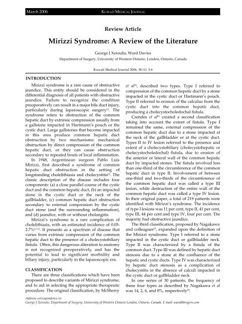
classification <strong>of</strong> <strong>the</strong> type <strong>of</strong> <strong>Mirizzi</strong>’s syndrome (Fig. 1).<br />
The possibility <strong>of</strong> stone retrieval and biliary<br />
stenting during ERCP is an added advantage in<br />
i m p roving surgical outcome, and stenting also<br />
facilitates identification <strong>of</strong> <strong>the</strong> CBD during<br />
operative dissection [ 7 , 1 5 , 1 6 ] . When ERCP i s<br />
unsuccessful or difficult, percutaneous transhepatic<br />
cholangiography (PTC) is a viable alternative.<br />
MRCP and ERCP are equivalent in <strong>the</strong>ir ability<br />
to diagnosis and to delineate details <strong>of</strong> biliary<br />
strictures, and to detect a cholecystocholedochal<br />
fistula [4] . In addition, T2 weighted images can<br />
d i ff e rentiate a neoplastic mass from an<br />
inflammatory one which US or CT scan may not be<br />
capable <strong>of</strong> [17] . Early ERCP is preferred when biliary<br />
sepsis is <strong>the</strong> dominant clinical issue and where a<br />
beneficial endoscopic <strong>the</strong>rapeutic procedure can be<br />
instituted at <strong>the</strong> same time. By contrast, MRCP is<br />
used in <strong>the</strong> non-septic patient to corroborate <strong>the</strong><br />
suspicion <strong>of</strong> malignancy or stones after initial<br />
imaging with US or CT scans [4] .
March 2006<br />
Fig. 1: Endoscopic re t rograde cholangiopancreatography (ERCP) <strong>of</strong><br />
patient with obstructive jaundice and <strong>Mirizzi</strong>’s syndrome. Notice<br />
impacted stone in cystic duct causing obstruction <strong>of</strong> common hepatic<br />
duct. Adapted from UptoDate “<strong>Mirizzi</strong> <strong>Syndrome</strong>” James B McGee.<br />
TREATMENT<br />
Surgery is <strong>the</strong> mainstay <strong>of</strong> <strong>the</strong>rapy <strong>of</strong> <strong>Mirizzi</strong><br />
s y n d rome, <strong>the</strong> dense inflammatory reaction in<br />
Calot’s triangle, as well as <strong>the</strong> frequent aberrant<br />
biliary anatomy, pose a difficult challenge to <strong>the</strong><br />
unsuspecting surgeon when dealing with a <strong>Mirizzi</strong><br />
syndrome. The two principal aims are (a) <strong>the</strong> safe<br />
completion <strong>of</strong> cholecystectomy without injuring<br />
<strong>the</strong> biliary system and (b) <strong>the</strong> appro p r i a t e<br />
management <strong>of</strong> <strong>the</strong> cholecystocholedochal fistula.<br />
Meticulous dissection and vigilance are essential in<br />
order to avoid inadvertent bile duct injury. If <strong>the</strong><br />
diagnosis <strong>of</strong> <strong>Mirizzi</strong> syndrome is made pre o p e r a t i v e , l y<br />
an operative strategy that minimizes <strong>the</strong> risk <strong>of</strong><br />
injury to <strong>the</strong> biliary tract can be carried out.<br />
H o w e v e r, a preoperative diagnosis <strong>of</strong> <strong>Mirizzi</strong><br />
syndrome is seldom made because ERCPand direct<br />
cholangiography are not widely used. ERCP, direct<br />
cholangiography, or magnetic resonance cholangiography<br />
should be performed in patients with<br />
clinical jaundice and signs and symptoms<br />
suggestive <strong>of</strong> biliary obstruction.<br />
A s t a n d a rdized surgical approach has been<br />
recommended based on <strong>the</strong> Csendes classification<br />
<strong>of</strong> <strong>the</strong> variants <strong>of</strong> <strong>Mirizzi</strong> syndrome [6] :<br />
KUWAIT MEDICAL JOURNAL 5<br />
Type I - Cholecystectomy plus common bile<br />
duct exploration with T-tube placement.<br />
Exploration should be performed only if <strong>the</strong> CBD is<br />
easily exposed.<br />
Type II - Suture <strong>of</strong> <strong>the</strong> fistula with absorbable<br />
material or choledochoplasty with <strong>the</strong> re m n a n t<br />
gallbladder.<br />
Type III - Choledochoplasty; suture <strong>of</strong> <strong>the</strong><br />
fistula is not indicated.<br />
Type IV - Bilio-enteric anastomosis is<br />
preferred since <strong>the</strong> entire wall <strong>of</strong> <strong>the</strong> common bile<br />
duct has been destroyed.<br />
The approach may vary with <strong>the</strong> type <strong>of</strong><br />
fistula present; both <strong>the</strong> operative mortality and<br />
postoperative morbidity increase according to <strong>the</strong><br />
severity <strong>of</strong> <strong>the</strong> lesion [6] .<br />
LAPAROSCOPIC SURGERY<br />
The <strong>Mirizzi</strong> syndrome presents a diff i c u l t<br />
challenge for laparoscopic surgery because <strong>the</strong><br />
dense adhesions and edematous inflammatory<br />
tissue cause distortion <strong>of</strong> <strong>the</strong> normal anatomy and<br />
increase <strong>the</strong> risk for biliary injury. While it appears<br />
to be feasible, especially for type I anatomy [18,19] , <strong>the</strong><br />
routine use <strong>of</strong> laparoscopic surgery as <strong>the</strong> primary<br />
treatment <strong>of</strong> <strong>Mirizzi</strong> syndrome is controversial [20,21] .<br />
It has been suggested, that a prudent approach for<br />
type 1 <strong>Mirizzi</strong> syndrome is to perform a trial<br />
laparoscopic dissection, but to have a low threshold<br />
to convert to an open procedure. This approach<br />
should be undertaken only by experienced<br />
laparoscopic surgeons [18,20] .<br />
ENDOSCOPIC THERAPY<br />
Endoscopic treatment with or without<br />
electrohydraulic lithotripsy (EHL) can be effective<br />
as a temporizing measure before surgery and can<br />
be definitive treatment for unsuitable surg i c a l<br />
c a n d i d a t e s [ 9 , 2 2 , 2 3 ] . One report described <strong>the</strong><br />
experience with 14 patients with <strong>Mirizzi</strong> syndrome<br />
treated with EHL [9] . Twelve patients had a single<br />
stone and complete clearance was achieved with<br />
one treatment session; two had multiple stones and<br />
re q u i red an additional treatment session.<br />
Asymptomatic leakage <strong>of</strong> contrast medium from<br />
<strong>the</strong> cystic duct into <strong>the</strong> peritoneal cavity was<br />
observed in one patient after removal <strong>of</strong> a large<br />
impacted cystic duct stone. This patient recovered<br />
with conservative <strong>the</strong>rapy and suffered no adverse<br />
events. In ano<strong>the</strong>r series <strong>of</strong> 25 patients with<br />
cholangiographic evidence <strong>of</strong> <strong>Mirizzi</strong> syndrome, 12<br />
w e re re f e r red for surgery after pre l i m i n a r y<br />
endoscopic <strong>the</strong>rapy and 13 were treated solely with<br />
endoscopy [23] . Stones were completely removed in<br />
three and nine were treated with long-term stents;<br />
complications occurred in four patients [23] .
6<br />
Endoscopic treatment <strong>of</strong> <strong>Mirizzi</strong> syndro m e<br />
should be used as a temporizing measure before<br />
surgery. It can serve as a definitive treatment for<br />
those patients who are unsuitable surg i c a l<br />
candidates when fur<strong>the</strong>r endoscopic attempts can<br />
be made to disimpact and remove <strong>the</strong> stones. Longterm<br />
success appears to be most likely in patients<br />
with type II disease who do not have residual<br />
gallbladder stones [24] .<br />
CONCLUSION<br />
<strong>Mirizzi</strong> syndrome is a rare complication <strong>of</strong><br />
cholelithiasis and requires a high index <strong>of</strong> suspicion<br />
in <strong>the</strong> setting <strong>of</strong> obstructive jaundice. Diagnosis<br />
preoperatively may be elusive with bloodwork, US<br />
and CT alone. Cholangiography (intraoperative<br />
and ERCP) as well as MRCP aids in both <strong>the</strong><br />
diagnosis and identification <strong>of</strong> anatomy and may<br />
p revent serious biliary injury. Surgery is <strong>the</strong><br />
mainstay <strong>of</strong> <strong>the</strong>rapy <strong>of</strong> <strong>Mirizzi</strong> syndrome, and<br />
requires <strong>the</strong> safe completion <strong>of</strong> cholecystectomy<br />
without injuring <strong>the</strong> biliary system and <strong>the</strong><br />
a p p ropriate management <strong>of</strong> <strong>the</strong> cholecystocholedochal<br />
fistula. The aberrant anatomy intrinsic<br />
to this syndrome presents a difficult challenge to<br />
surgeons and <strong>the</strong> laparoscopic approach should be<br />
undertaken with caution and probably left to<br />
specialized minimally invasive centres. Endoscopic<br />
t reatment may be effective as a temporizing<br />
m e a s u re before surgery and can be definitive<br />
treatment for unsuitable surgical candidates.<br />
REFERENCES<br />
1. Waisberg J, Corona A, de Abreu IW, Farah JFM, Lupinacci<br />
RA, G<strong>of</strong>fi FS. Benign Obstruction <strong>of</strong> <strong>the</strong> Common Hepatic<br />
Duct (<strong>Mirizzi</strong> <strong>Syndrome</strong>): diagnosis and operative<br />
management. Arq Gastroenterol 2005; 42:13-18.<br />
2. <strong>Mirizzi</strong>, PL. <strong>Syndrome</strong> del conducto hepatico. J Int de Chir<br />
1948; 8:731-733.<br />
3. Yeh, CN, Jan, YY, Chen, MF. Laparoscopic treatment for<br />
<strong>Mirizzi</strong> syndrome. Surg Endosc 2003; 17:1573-1578.<br />
4. Chan CY, Liau KH, Ho CK, Chew SP. <strong>Mirizzi</strong> syndrome: a<br />
diagnostic and operative challenge. Surgeon 2003;1:273-278.<br />
5. McSherry, CK, Ferstenberg, H, Virshup, M. The <strong>Mirizzi</strong><br />
syndrome: Suggested classification and surgical treatment.<br />
Surg Gastroenterol 1982; 1:219-225.<br />
<strong>Mirizzi</strong> <strong>Syndrome</strong>: A <strong>Review</strong> <strong>of</strong> <strong>the</strong> <strong>Literature</strong> March 2006<br />
6. Csendes, A, Diaz, CJ, Burdiles, P, et al. <strong>Mirizzi</strong> syndrome<br />
and cholecystobiliary fistula: A unifying classification. Br J<br />
Surg 1989; 76:1139-1143.<br />
7. Nagakawa, T, Ohta, T, Kayahara, M, et al. A n e w<br />
classification <strong>of</strong> <strong>Mirizzi</strong> syndrome from diagnostic and<br />
<strong>the</strong>rapeutic viewpoints. Hepatogastro e n t e rology 1997;<br />
44:63-67.<br />
8. I b r a rullah, M, Saxena, R, Sikora, SS, et al. <strong>Mirizzi</strong>’s<br />
syndrome: Identification and management strategy. Aust N<br />
Z J Surg 1993; 63:802-806.<br />
9. B i n m o e l l e r, KF, Thonke, F, Soehendra, N. Endoscopic<br />
treatment <strong>of</strong> <strong>Mirizzi</strong>’s syndrome. Gastrointest Endosc 1993;<br />
39:532-536.<br />
10. Becker, CD, Hassler, H, Terrier, F. Preoperative diagnosis <strong>of</strong><br />
<strong>the</strong> <strong>Mirizzi</strong> syndrome: Limitations <strong>of</strong> sonography and<br />
computed tomography. Am J Roentgenol 1984; 142:591-596.<br />
11. Berland, LL, Lawson, TL, Stanley, RJ. CT appearance <strong>of</strong><br />
<strong>Mirizzi</strong> syndrome. J Comput Assist Tomogr 1984; 8:165-166.<br />
12. Baer, HU, Mat<strong>the</strong>ws, JB, Schweizer, WP, et al. Management<br />
<strong>of</strong> <strong>the</strong> <strong>Mirizzi</strong> syndrome and <strong>the</strong> surgical implications <strong>of</strong><br />
cholecystocholedochal fistula. Br J Surg 1990; 77:743-745.<br />
13. Dewar G, Chung SCS, Li AKC. Operative strategy in<br />
<strong>Mirizzi</strong> syndrome. Surg Gynecol Obstet 1990; 171:157-159.<br />
14. Fan ST, Lau WY, Lee MJR, et al. Cholecysto-hepaticodochal<br />
fistula: <strong>the</strong> value <strong>of</strong> pre-operative recognition. Br J Surg<br />
1985; 72:743-744.<br />
15. Cotton PB. Endoscopic management <strong>of</strong> bile duct stones.<br />
Gut 1984; 25:587-597.<br />
16. Siegel JH, Yatto RP. Biliary endopros<strong>the</strong>sis for <strong>the</strong><br />
management <strong>of</strong> retained bile duct stones. Am J<br />
Gastroenterol 1984; 79:50-54.<br />
17. Choi BW, Kim MJ, Chung JJ, et al. Rdiologic findings <strong>of</strong><br />
<strong>Mirizzi</strong> with emphasis on MRI. Yonsei Med J 2000;<br />
41(1):144-146.<br />
18. Vezakis A, Davides D, Birbas K, et al. Laparo s c o p i c<br />
treatment <strong>of</strong> <strong>Mirizzi</strong> syndrome. Surg Endosc 2000; 10(1): 15-<br />
18.<br />
19. Chowbey PK, Sharma A, Mann V, Khullar R, Baijal M,<br />
Vashistha A. The management <strong>of</strong> <strong>Mirizzi</strong> syndrome in <strong>the</strong><br />
l a p a roscopic era. Surg Laparosc Endosc Percutan Te c h<br />
2000;10:11-14.<br />
20. Targarona EM, Andrade, E, Balague, C, et al. <strong>Mirizzi</strong>’s<br />
syndrome. Diagnostic and <strong>the</strong>rapeutic controversies in <strong>the</strong><br />
laparoscopic era. Surg Endosc 1997; 11:842-845.<br />
21. Sare M, Gurer S, Taskin V, et al. <strong>Mirizzi</strong>’s syndrome: Choice<br />
<strong>of</strong> surgical procedure in <strong>the</strong> laparoscopic era. Surg Laparosc<br />
Endosc 1998; 8:63-67.<br />
22. Binnie NR, Nixon SJ, Palmer KR. <strong>Mirizzi</strong> syndro m e<br />
managed by endoscopic stenting and laparo s c o p i c<br />
cholecystectomy. Br J Surg 1992; 79:647.<br />
23. England RE, Martin, DF. Endoscopic management <strong>of</strong><br />
<strong>Mirizzi</strong>’s syndrome. Gut 1997; 40:272-276.<br />
24. Tsuyuguchi T, Saisho H, Ishihara T, et al. Long-term followup<br />
after treatment <strong>of</strong> <strong>Mirizzi</strong> syndrome by pero r a l<br />
cholangioscopy. Gastrointest Endosc 2000; 52:6390-644.


