Hyaluronans: ls Ctinicat Effectiveness - Crespine Gel
Hyaluronans: ls Ctinicat Effectiveness - Crespine Gel
Hyaluronans: ls Ctinicat Effectiveness - Crespine Gel

You also want an ePaper? Increase the reach of your titles
YUMPU automatically turns print PDFs into web optimized ePapers that Google loves.
<strong>Hyaluronans</strong>: <strong>ls</strong> <strong>Ctinicat</strong> <strong>Effectiveness</strong><br />
Dependent on Motecular Weight?<br />
Peter C. Vitanzo, Jr, MD, and Brian J. Sennett, MD<br />
Abstract<br />
The original rationale for viscosupplementation with<br />
hyaluronans was fluid replacement, suggesting that the<br />
most viscous materia<strong>ls</strong> {eg, those of highest molecular<br />
weight [MW] would provide the most clinical benefits'<br />
Howevei it has become clear that mechanisms of action<br />
for osteoarthritis pain management are not only mechanical<br />
but a<strong>ls</strong>o biological. After intra-articular injection,<br />
hyaluronans exert a range of biological actions within the<br />
joint. Although high- and low- to mid-MW hyaluronans<br />
't'rut not hyaluronans
<strong>Hyaluronans</strong>: <strong>ls</strong> Clinical <strong>Effectiveness</strong> Dependent on Molecular Weight?<br />
in clinical efficacy, as shown by outcoraes such as pain<br />
relief. Data from recent clinical studies carried out in<br />
patients receiving a low- to mid-M.V/ product have suggested<br />
that intra-articular hyaluronan therapy has diseasemodifying<br />
activity and may alter progression of<br />
osteoarthritis.2l-25 Jhess issues are discussed in more detail<br />
later in this reyiew<br />
Pnoposro Mecuaursrrrs 0F AcroN<br />
OF HYÂLURONAN<br />
<strong>Hyaluronans</strong> are responsible for elastoviscosity of synovial<br />
fluid in joints, and elastoviscosity is higher with high-MV/<br />
hyaluronans. This elastoviscosity is shear-dependent,<br />
meaning that the mechanical behavior of hyaluronan<br />
changes with the shear force applied and the speed of flow:<br />
Hyaluronan soiutions behave as viscous fluids when external<br />
forces move them at low speeds but behave as elastic<br />
bodies when subjected to high rates of shear or frequency.<br />
Therefore, hyaluronans are effective as lubricants under<br />
conditions of siow movement but are effective as shock<br />
absorbers when movement is rapid.a<br />
Clearly, this elastoviscous behavior will determine clinical<br />
activity to a large extent, but the pharmacokinetic profile<br />
of hyal*ronans does not support a purely mechanical<br />
role for these substances. During movement, hyaluronan<br />
flows into ihe lymphatic system of the joint capsule, rnovcs<br />
info fhe general circulation, and is ultimately taken up by<br />
the }iver, where it is degraded to water and carbon dioxide.26<br />
Duration of the therapeutic effect of hyaluronan, as<br />
noted in clinical studies, is a<strong>ls</strong>o not consistent with a pure-<br />
1y mechanical role, as treafnent benef,ts can persist for<br />
several months after a course of injections is completed,<br />
whereas the elimination halflife of hyaluronan in the joint<br />
is measured in hours to days.27,28<br />
Of additional interest is the reported increase in the elastoviscous<br />
properlies of synovial fluid after exogenous<br />
administration of hyaluronic acid. Mensitieri and<br />
Ambrosio2e examined synovial fluid from the knee joints<br />
of patients subjected to arthrocentesis or treatment with<br />
hyaluronan compounds of MWs ranging from 500 kDa to<br />
730 kDa. The elastoviscosity of synovial fluid from<br />
patients who received hyaluronan was increased relative to<br />
pretreatment values, as well as to values in patients who<br />
had undergone arlhrocentesis. This increase in elastoviscosity<br />
was present 1 week after administration, well after<br />
the exogenous product would have been expected to have<br />
been cleared from thejoint space, suggesting that the elastoviscosity<br />
of knee synovial fluid benefits from increases<br />
422 The American Journal of Orthopedicso<br />
in both hyaluronic acid concentration and MW<br />
Other potential pharmacologic mechanisms may be<br />
receptor-mediated. These mechanisms include inhibition<br />
of inflammatory mediators, inhibition of phagocytic cell<br />
function, stimulation of cartilage matrix synthesis, and<br />
decreased degradation of carlilage 6stria. 17'30-39<br />
These and other potential mechanisms, and their association<br />
with MW, investigated primarily in vitro, are summarrzed<br />
n Tâble IL17,30-39 Results from in vitro evaluation of<br />
the biological actions of hyaluronaas show that both highand<br />
low- to mid-MW hyaluronans are more or less active<br />
depending on the specific effect examined. For example,<br />
mid-MW hyaluronans (500*999 kDa) appear to inhibit<br />
apoptosis more,35 offer more protection against cartilage<br />
1oss,17 sn6 suppress synovial cell proliferation moreoll'17<br />
whereas high-MW hyaluronans are more effective in<br />
inhibiting prostaglandin EZ and arachidonic acid activities.37'38<br />
Both agents are similar with regard to effects on<br />
leukocyte chemotaxis.3o Although an "ideal" MW rangr<br />
should be related to the biological profile that will optimizàt<br />
clinical benefit for the patient, it is not well understood<br />
which of these actions are most meaningful regarding clinical<br />
benefits, such as pain relief or potential structure (disease)<br />
modification. Potential disease-modifying activities<br />
demonstrated in animal mode<strong>ls</strong> of osteoarthritis (Table I)<br />
may provide a more meaningful picture of the benefit of<br />
low- to mid-MW hyaluronans. Even in this case, however,<br />
the activity may be distinct from the outcome of pain relief.<br />
In a review, Ghosh and Guidolin<strong>ls</strong> concluded that, based on<br />
overall preclinical and clinical data, low- to mid-MW<br />
hyaluronans used clinically for osteoarthritis may indeed<br />
have a more beneficial sffucture- or disease-modifying pro-<br />
Gl-<br />
RruroNsHrps BETwEEN MoLEcULAR WETcHT<br />
OF HYALURONAN AND ETTIcncy<br />
In the United States, hyaluronan products marketed for<br />
treatment of osteoarthriiis of the knee include Hyalgar<br />
(sodium hyaluronate; sanofi-aventis, Bridgewater, NJ),v<br />
Synvisc (hylan G-F 2A; Genzyme Corporation,<br />
Cambridge, Mass), Supartz (sodium hyaluronate; Smith &<br />
Nephew, Memphis, Tenn), Euflexxa (sodium hyaluronate;<br />
Ferring Pharmaceutica<strong>ls</strong>, Suffem, NY), and Orthovisc<br />
(high-MW hyaluronan; DePuy Mitek, Raynham, Mass).<br />
Characteristics of these preparations are summarized in<br />
Table III.<br />
Molecular Weight and Half-Life<br />
Exogenous hyaluronan'begins to leave the joint within 2<br />
hours of injection, though some remains up to 3 days (this<br />
is particularly true of high-MW products). Synvisc remains<br />
up to 3 days,a0 whereas Hyalgan,al Supafiz,az Euflexxa,43<br />
and Orthoviscaa remain less than 24 hours. Although the<br />
high-MW hylan B in Synvisc has a half-life of some<br />
weeks,26 it is biologically inert (not in soiution to engage<br />
receptors). Results from a study involving 1311-rad'olabeling<br />
showed that hyaluronan is eliminated from the human
Asari et altt<br />
Ghosh et all2<br />
Ghosh et all3<br />
Adamla<br />
Ghosh et al<strong>ls</strong><br />
Yoshimi et all6 Rabbit<br />
Kikuchi et al7 Rabbit<br />
Shimizu et alrT Rabbit<br />
Table l. Preclinical Studies of lntra-Articular Hyaluronan {HA}<br />
Preparations in Animal Mode<strong>ls</strong> of Osteoarthritis {OA)*<br />
OA lnduction Method<br />
ACLT, then HA x 5 wk (starting 4 wk PO)<br />
UTMM, then HA or saline x 5 wk<br />
(stading 4 mo PO)<br />
UTMM, then HA or saline x 5 wk<br />
(starting 4 mo PO)<br />
TBLM, then HA or saline x 5 wk<br />
(starting 4 mo PO and after SF aspiration)<br />
PM, then HA or saline immediately<br />
PO Zx/wk x 2-4 wk<br />
Seikagaku (840 kDa or<br />
2300 kDa)<br />
Seikagaku (840 kDa);<br />
Nippon Roussel<br />
(2000 kDa)<br />
Seikagaku (BaO kDa);<br />
Nippon Roussel<br />
(2000 kDa)<br />
Seikagaku (840 kDa<br />
or 2300 kDa)<br />
Female sheeo TBLM, then HA or saline x 5 wk Fidia (50&-730 kDa);<br />
(starting 4 mo PO and after SF aspiration) Pharmacia-Upjohn<br />
{s00M900 kDa)<br />
ACLT, then single-dose HA or saline Hikarl (2020 kDa<br />
or 950 kDa)<br />
Seikagaku (840 kDa<br />
or 1900 kDa)<br />
P. C, Mtanzo and B. J, Sennett<br />
Pathology { significantly<br />
more with 840 kDa than<br />
with 2300 kDa. Synovial<br />
penetration of 840 kDa ><br />
2300 kDa<br />
Loading of operated joints I<br />
and radiologic scores I with<br />
HAs vs saline. Histologic<br />
scores for MTPC worse with<br />
2000 kDa<br />
PG synthesis in MIPC of<br />
operated joints ! with HA vs<br />
placebo, but PG release I<br />
SF HA MW (determined by<br />
multiangle laser light-scattering<br />
photometry) 1 at 5 wk<br />
PO with both HAs, but<br />
mean t with 840 kDa ><br />
2300 kDa<br />
SF storage modulus and<br />
viscosity at 5 wk PO t vs<br />
saline with both HAs,<br />
lmprovement with 500-730 kDa<br />
> 3000-6900 kDa<br />
Both HAs protected against<br />
histologic cartilage loss.<br />
Nonsignificant trend toward<br />
greater efficacy with 2020<br />
kDa<br />
Cartilage histology<br />
suggested grealer<br />
protection with 840 kDa<br />
than with 1900 kDa or saline<br />
Proiective effect in cartilage<br />
and synovium: higher MW<br />
> MW 500-730 kDa.<br />
Suppression of synovial cell<br />
proliferation: 840 kDa ><br />
larger HAs.<br />
"lnvestigations shown are those in which HAs with differing l\4ws were compared directly. Compounds were âdministered intra-afiicularly in all studies. MW indicales<br />
molecular weighi; ACLI anterior cruciate ligament transeclion; PO, postoperatively; UTMM, unilateral total medial meniscectomy; MTPC, medial tibial plâteau cartilages;<br />
PG, proteoglycan; TBLM, total bilaleral lateral meniscectomy; SF, synoviâl fluid; PM, partial meniscectomy.<br />
knee joint in 3 distinct phases (half-lives of 1.5 hours, 1.5<br />
days, and 4 weeks) fitting a 3-erponent function.a5 In this<br />
study, conducted with a high-MW-nonanimal stabilized<br />
hyaluronic acid product (NASHA), it was suggested that<br />
the rapid initial phase was linked to elimination of low-<br />
MW fragments and the second phase to elimination of<br />
high-MW labeled hyaluronan. During the slow third phase,<br />
the daily decline in radioactivity was comparable to that in<br />
the urine, indicating a slow release of hyaluronan or degradation<br />
products from the ge1 in &e knee and subsequent<br />
excretion by the kidneys. The possibility of uptake of<br />
NASHA and its products by the synovial layer and<br />
ACLT, then HA x 5 wk (starting 4 wk PO) Fidiâ (500 kDa);<br />
Seikagaku (840 kDa);<br />
Pharmacia {3600 kDa);<br />
Biomairix {6000 kDa)<br />
popliteal lyrnph nodes was a<strong>ls</strong>o postulated.as Results from<br />
another study showed increased clearance of hyaluronan<br />
with osteoarthritis but no increase in normal joints.a0 The<br />
elimination half-life of hyaluronan increased to 23.5 hours<br />
as osteoarthritis developed but fell ta l'1.4 hours by the<br />
time the disease was fully established.<br />
lflolecutar Weight and Tissue Penetration<br />
The discrepancy between retention in thejoint and duration<br />
of clinical effecl may be attributable, at least in part, to<br />
enhanced penetration of low- to mid-MV/ hyaluronans into<br />
the extracellular matrices of the synoyial tissues and per-<br />
September 2006 423
Hyaiuronans: <strong>ls</strong> Clinical <strong>Effectiveness</strong> Dependent on Molecular Weight?<br />
!1. Molecular Weight and Effect" on BiologicalActivity of Hyaluronan<br />
Biological Activity Low {6000)<br />
haps the cartilage. There is some evidence-from a canine<br />
osteoarthritis model evaluating increased expression of<br />
prostaglandin E2, thickening of synovial lining layers, and<br />
vacuolar changes in lining cel<strong>ls</strong>, together with fluorescein<br />
staining of hyaluronan-that low- to mid-M\Y hyaluronans<br />
(ie, 840 kDa) penetrale diseased tissues more effectively<br />
than high-MW hyaluronans (ie, 230û kDa). Pathologic<br />
changes suggested more accessibility of synovial tssues to<br />
the 840-kDa product.ll However, there remains no direct<br />
evidence that this phenomenon correlates with or explains<br />
a clinical benefit.<br />
lrtolecular l#eight and Elastoviscosity<br />
Increasing the mean MW of hyaluronan clearly increases<br />
the elastoviscous properties of the solution.a Although this<br />
result would be advantageous if the mechanism of pain<br />
relief were only mechanical, it seems counterintuitive<br />
when biological mechanisms are involved. It does not fo1low<br />
that côllulâr activation of the precisely regulated<br />
hyaluronans and other associated pathways would occur in<br />
response to a higher concentration and higher than normal<br />
M'W of hyaluronan in the knee. Within the cartilage matrix,<br />
struçtural propeflies of the exffacelluiar rnatrix ars deter-<br />
424 The American Journal of Orthopedics@<br />
Genzyme Corporation<br />
B0% hylan A (6000),<br />
rnined by aggregates of endogenous hyaluronan and proteoglycans<br />
that include aggrecan and link proteins.4T<br />
Hyaluronan MW seems to be determined by the binding of<br />
elongating nascent hyaluronan to a receptor. Saturation of<br />
these receptors may stall elongation of the nascent hyaluro- V<br />
***r*r/<br />
{€,q! {{}*ll<br />
gK{*l<br />
XûJ<strong>ls</strong>d<br />
DePuy Mitek<br />
1 000-2900<br />
HA<br />
d<br />
i:CELL<br />
MOLICI Lt3<br />
fening Pharmaceuticd<br />
2400-3600<br />
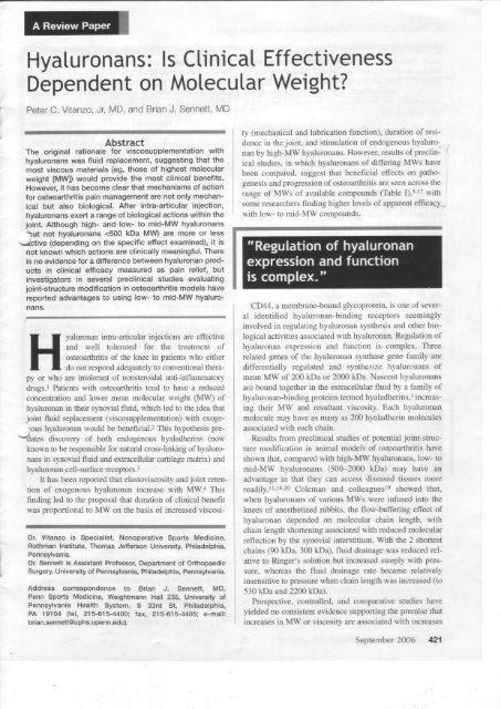
Figure. In vitro synthesis of hyaluronan by synovial fibroblasts<br />
is inlluenced by eoncentration and molecular weight (MW) of<br />
êxogenous hyaluronan {HA). The signal for synthesis is provided<br />
by HA in the MW range of 500 lo 4000 kDa in a concentration-dependent<br />
manner. Figure adapted with permission from<br />
Smith MM, Ghosh P. The synthesis ol hyaluronic acid by<br />
human synovial fibroblasts is influenced by the nature of the<br />
hyaluronate in the extracellular environment. Rheumatol lnt.<br />
1987:7:113-122<br />
\<br />
tAS 1&ô c f*t{ s,Xlô8 l&t<br />
1 I<br />
J
nan chains, resulting in an intracellular response that<br />
affects both hyaluronan and proteogiycan synthesis.<br />
Although an efficacy advantage has beea claimed for<br />
products of a MW similar to that of normal, healthy synovial<br />
fluid, analysis of the clinical evidence has not shown<br />
a correlation between MW and degree of improvement or<br />
duration of efficacy.as<br />
Molecular Weight and Receptor Binding<br />
It is thought that hyaluronan concentration is monitored by<br />
synovial fibroblasts and that homeostasis is maintained<br />
through specific cell-surface receptors. These receptors<br />
have a<strong>ls</strong>o been shown in vitro to be activated by exogenous<br />
hyaluronan, with maximal responses being elicired by<br />
exogenous hyaluronans of certain size ranges.36 In one<br />
study, investigators found the most marked response by<br />
synovial fibroblasts from an osteoarthritic joint exposed io<br />
hyaluronan of MW >5t0 kDa, whereas smaller molecules<br />
had little or no effect.36 The investigators a<strong>ls</strong>o found that a<br />
Jirigh-MW hyaluronan (4700 kDa) was less effecrive in<br />
stimulating synthesis than a compound of MW 3800 kDa.<br />
They thought this finding indicates that synovial fibroblasts<br />
do not increase synthesis of endogenous hyaluronans in the<br />
presence of functionally acceptable (ie, very high MW or<br />
high concentration) hyaluronans.<br />
Study results have shown that hyaluronan binding at the<br />
cell surface is a complex interplay of multivalent binding<br />
events affected by the size of the multivalent hyaluronan<br />
ligand, the density of cell-surface CD44, and the CD44<br />
activation state.4e The most significant feature dislinguishing<br />
CD44 from other hyaluronaa-binding proteins is that,<br />
with CD44, binding to hyaluronan takes place at the cell<br />
surface, where multiple, closely arrayed CD44 receptor<br />
molecules interact with the highly multivalent repeating<br />
disaccharide chain of hyaluronan. The actual afËnity of a<br />
single CD44*hyaluronan binding domain for hyaluronan is<br />
likely to be very low. Thus, binding of a CD44-positive cell<br />
to a soluble hyaluronan molecule must involve multiple<br />
s/vveuç receptor*ligaad interactions, dependent on the relative<br />
valency of the native hyaluronan molecule. In high-<br />
MW hyaluronan polymers, each molecule interacts with<br />
more than one receptor*binding site, and this increases the<br />
likelihood that the molecule will remain bound to the<br />
receptor, thereby increasing the receptor aftnity (avidity)<br />
of the molecule. Beyond a certain limit, howevet, very<br />
large hyaluronan molecules will be less efficient in engaging<br />
multiple receptors because of steric trindrance-hence<br />
the bimodal nature of many biological activities. This suggests<br />
that the maximal response would be produced by<br />
hyaluronans within a specific size range (neither too big<br />
nor too small). The existence of isoforms of the hyaluronan<br />
receptors may a<strong>ls</strong>o contribute to differential binding.<br />
However, these ideas remain speculative at this time, and the<br />
exact mechanism of receptor binding is not known. Either<br />
the response may be proportional to the number of receptors<br />
bound, or it may be an "all-or-nothing" phenomenon that<br />
depends on the simultaneous binding of 2 or more recep-<br />
P. C. Vitanzo and B. J. Sennett<br />
tors.18 In addition, biological activation is a<strong>ls</strong>o balanced with<br />
"bioavailabiiity," the ability to penetrate iato diseased tissues.<br />
Again, larger hyaluronan molecules may be less ef[cient<br />
in penetrating into the synovium (see Figure).tt<br />
ClrNlcal Ernclcy<br />
There have been several reviews1,5O-sz and meta-analyses53'54<br />
on the clinical efficacy of intra-articular hyaluronans<br />
in the treatment of pain associated with osteoarthritis-including,<br />
most recently, publication by the Cochrane<br />
Collaboration of the most comprehensive meta-analysis of<br />
this class.5s However, there have been no reports of any<br />
well-controlled, randomized clinical studies directly comparing<br />
hyaluronans across a wide range of MWs<br />
(500-6000 kDa). Several clinical studies have compared 2<br />
or more hyaluronans of differing MWs. Wobig and col-<br />
1eagues56 reported on a study fhat was said to compare<br />
hylan G-F 20 (MVi =>60û0 kDa) with sodium hyaluronate<br />
(MV/ = 800 kDa) and find the high-MW hyaluronan superior.<br />
However, it was subsequently noted thaT the original<br />
study was a 4-arm trial that a<strong>ls</strong>o included another sodium<br />
hyaluronate (MW = 2300 kDa) and a control group of a<br />
degraded, nonelastoviscous hylan G-F 20 preparation.sT In<br />
the total analysis, it was shown that none of the hyaluroîans<br />
was statistically different from the control group. A<br />
retrospective clinical practice reporl compared hylan G-F<br />
20 with sodium hyaluronate (MW = 615 kDa) and found<br />
that, for both products, a1l patients repofied a statistical<br />
improvemenl in pain after treatment.s8 The report further<br />
suggested fhat, compared with sodium hyaluronate, hylan<br />
G-F 20 had a superior response in many parameters.<br />
However, it was subsequently reported thar significant limits<br />
in the scope and design of the study called its conclusions<br />
into question.se<br />
Three recent prospective studies from clinical practice<br />
a<strong>ls</strong>o compared the clinical efficacy of hylan G-F 20 (MW<br />
-6000 kDa) with that of sodium hyaluronate (MW<br />
500-73û kDa): Brown and colleagues60 (N = 54) reported<br />
no evidence for a benefit of either product over the other,<br />
and Garcia6l (N = 51) and Richardson and colleagues62 (N<br />
= 90) reported in 2 separate studies that the short-term efficacy<br />
of hylan G-F 20 (MW = 6000 kDa) and sodium<br />
hyaluronate (MV/ 5û0*730 kDa) could not be distinguished.<br />
However, a1l 3 reports documeated a product-specific<br />
pseudoseptic reactioa to hylan G-F 20-a<strong>ls</strong>o termed<br />
severe acute inflammatory reaction {SAIR). None of these<br />
studies reported any SAIRs to the sodium hyaluronate<br />
injections. Incidence of SAIRs in these reports ranged kom<br />
5.3Vo ta -24.77a of the patients injected. Similarly,<br />
Hamburger and colleagues63 reported that in clinical practice<br />
study af 171 patients, 5 weekly injections of sodium<br />
hyaluronate O,{W 500*730 kDa) had a mean duration of<br />
benefit of 447 days, whereas 3 weekly injections ofhylan<br />
G-F 20 had a mean duration of 370 days (P = .018 in favor<br />
of sodium hyaluronate). F*rthermore, incidence of SAIRs<br />
was 16.3Vo with hylan G-F 20 versus 07o for sodium<br />
hyaluronate (P
<strong>Hyaluronans</strong>: <strong>ls</strong> Clinical Ëffectiveness Dependent on Molecular Weight?<br />
Kar<strong>ls</strong>son and colleagues6a recently reported on a multicenîer,<br />
randomized, saline-controlled study comparing the<br />
clinical efficacy of hylan G-F 20 (MW * 6û00 kDa) or<br />
sodium hyaluronate (MW - 890 kDa) with that of an intraarticular<br />
saline control. Patienis with knee osteoarthritis<br />
treated with injection of hyaluronan or saline showed clinical<br />
improvement during the frst 26 weeks of trealment,<br />
though nei*rer hyaluronan preparalion demonstrated any<br />
significant difference from t}le saline control. Collecrively,<br />
the limited clinical trial evidence suggests {hat there is no<br />
defînitive difference in efficacy (sympiomatic relief)<br />
between hyaluronans ranging in MV/ from 5Û0 kDa to<br />
6000 kD4 though there seem to be clear differences in<br />
safety profiles and other clinically relevant benefi<strong>ls</strong> (eg,<br />
disease-modifuing) in several studies.<br />
Kirshner and Marshall compared the safety and effectiveness<br />
of sodium hyaluronate (MW 24æ-3600-kDa) prepared<br />
by biological fermentation wilh those of avian-derived<br />
hyaluronan ftylan G-F 20 (MW * 6û00 kDa). The effectiveness<br />
was not inferior aad lhere was a significantly higher<br />
incidence of post-injection effusions in hylan G-F 20.65<br />
This conclusion was reiterated in the American College<br />
of Rbeumatology 2000 treatment guidelines,o6 in which it<br />
was recommended that intra-articular hyaluronan therapy<br />
be considered a possible therapy for knee osteoarthritis:<br />
"To date, differences in clinical efficacy between these<br />
preparations fHyalgan and Synvisc] as a function of<br />
molecular weight have not been demonstrated." In agreement,<br />
the Cochrane meta-analysis concluded that no evidence<br />
would lead one ts conclude that hyaiuronan products<br />
differ in pain-relieving efficacy, though it was<br />
acknowledged that few well-designed, head-to-head studies<br />
have been conducted.5s<br />
426 The American Journal of Orthopedics@<br />
there have been several case reports of patients having a<br />
SAIR to hylan G-F 2A ard subsequently being treated with<br />
sodium hyaluronate with good clinical results and without<br />
further sequelae,Tl adding further support for a hylan G-F<br />
20*specific reaction.<br />
Antibodies to hylan have been noted in the sera of some<br />
patients.Tl In addition, the immunologic response may<br />
progress to more serious conditions such as granulomaious<br />
reactions. Cases of hylan G-F 20-related granulomatous<br />
reactions have been reported in patients who had knee<br />
osteoarthritis treaied with intra-articular injections of this<br />
agent.71 The reactions take the form of chronic inflarnmation<br />
of the perisynovial area and surrounding tissues and<br />
are shown by histologic evaluation to include histiocytic<br />
and foreign body giant cel<strong>ls</strong>. A recent report a<strong>ls</strong>o characterized<br />
this response as a pseudosatcama.T2 A recent animal<br />
study comparing the biocompatibility of sodium<br />
hyaluronate and saline with hylan G-F 20 has confirmed<br />
these clinical observations.T3 After intradermal injection in<br />
guinea pigs and intramuscular injection in rabbits, hylan G- V<br />
F 20 induced definitive macroscopic changes in guinea<br />
pigs by day 14 and in rabbits by day 28. Severe granulomatous<br />
inflammation in guinea pigs and acute inflammation<br />
with minimal infiltration of macrophages and foreign<br />
body giant cel1s in rabbits were seen on histologic assessment.<br />
Furtbermore, speciflc antibodies against hylan G-F<br />
20 were demonstrated in guinea pigs by passive cutaneous<br />
anaphylaxis, and substantial deposits of immunoglobulin G<br />
on hylan G-F 20 were evident by immunohistochemistry.T3<br />
Couct-ustoNs<br />
Data on intra-articular hyaluronan therapy for osteoarthritis<br />
pain-which have been accumulating for almost 2<br />
ReurroNsHrp BETwEEN MoLEcULAR WEtcHT<br />
AND TOLERABILITY<br />
decades-have shown rhat the original (1980s) hyporhesis<br />
of a link between hyaluroaan MW and duration of efficacy<br />
is not bome out by precliaical and clinical evidence. It is<br />
As a class, the hyaluronans have a well-documented toler- instead likely that the mechanism of action of hyaluronan<br />
ability profile, with no known systemic effects and few is based on several probable biological activities mediated<br />
contraindications or drug interactions.6T The most cornmon through receptor-based pharmacologic mechanisms that go V<br />
side effect aoted ia most clinical studies has boen injection- beyond the physical, mechanical, and elastoviscous propersite<br />
pain. Although any intra-articular injection can elicit an ties of these products.<br />
inflammatory response, a clinically distinct reaction known Current evidence sugges<strong>ls</strong> that there is an optimal MW<br />
as pseudosepsis or SAIRs has emerged after hyaluronan range for induction of various biological activities. In pri-<br />
injeclions. Although it is not clear whether pseudoseptic marily in vitro analyses, some of these activities are<br />
reactions can occur with any hyaluronan, al1 published induced more effectively by high-MW hyaluronans, where-<br />
reports to date have been linked to hylan G-F 20 (Synvisc). as others are induced by low- to mid-MW hyaluronans. In<br />
Whereas the manufacturing practices for all hyaluronan contrast, preclinical studies evaluating joint-structure out-<br />
products have similar quantitative specifications for the comes in animal mode<strong>ls</strong> of osteoarlhritis indicate that low-<br />
amount of allowable proteins within the products (generalto mid-MW hyaluronans may have more potential for disly<br />
P, C. Vitanzo and B. J. Sennett<br />
disease progressiol, which has been described for low- to<br />
mid-MW sodium byaluronate (500-730 kDa).22'2s'7+'ts<br />
When risk-benef,t assessment is used in making treatment<br />
decisions, lack of evidence for a superior clinical benefit<br />
from use of high-MW hyaluronans versus low- to mid-MW<br />
hyaluronans should be considered together with the finding<br />
that cross-linked polymer products may be associated with<br />
increased risk of adverse events in the knee, specifically<br />
SAIRs and granulomas.<br />
AurnoR AcxNowleDGMENTs AND<br />
Drsclosunr STATEMENT<br />
This study was supporled by a grar* from Sanofi-<br />
Synthelabo. Dr. Vitanzo wishes to note that he is a consultant.<br />
and lecturer for sanofi-aveatis and for Smith &<br />
Nephew. Dr. Sennett wishes tû note that he has received a<br />
grant from sanofi -aventis.<br />
\'<br />
V<br />
RrrERrNcrs<br />
, 1. Altnran RD. Status ot hyaluronan supplementation therapy in osteoarthritis,<br />
Cun Rheumatol Rep.2da35:7-14.<br />
2. Batazs EA, Denlinger JL. Clinical uses of hyaluronat'. Ciba Found Symp.<br />
1989;143:265-275.<br />
3. Frasêr JR, Laurent TC, Lâurent UB. Hyaluronan: ks nature, distribution, functions<br />
and turnover. J lntern Md. 1997:'242:27-33.<br />
4. Balazs ÊA, Denlinçr JL. Viscosupp'emontâtion: a new concept in the treatment<br />
of osteoarthriÊ. J Rharmatol Suppl 1993;39:3-9.<br />
5. Shimâu C, Yoshioka M, Coutts RD, et al. Longterm eftects of hyalufonan on<br />
experimental osteoarthritis in the fabbil knee. Osfeoa,'ihrlfis Cartilage.<br />
i ooe.A l -o<br />
6. Yoshioka M, Shimizu C, Haruood FL, Coutts RD, Arfliel D. The effects of<br />
hyaluronan during the development of osteoarthritis. Ostæafthritis Câttilâge.<br />
1 997;5:251 -260.<br />
7, Kikuchi I Yamada H, Shimmei lvl. Effect of high molecular weight hyalurcnan<br />
on cartilage degenerâtion in â râbbit model o{ osteoarlhrii<strong>ls</strong>. Osteoa/thritis<br />
Çaftilage. 1 996;4:99-1 1 0^<br />
8. Armstrong S, Fead R, Ghosh P The effec<strong>ls</strong> oT intmarticular hyaluronan on cartilage<br />
and subchondal bone changes in an ovine model ol early osteoarthritis.<br />
J Rheumato| 1 994;21 :680-688.<br />
9. Schiavinato A, Lini Ë, Guidolin D, et al. Intraarticular sodium hyaluronate injections<br />
in the Pond-Nuki experimental model oT osteoadhritis in dogs. ll.<br />
Morphological findings. C/ln Orthop. 1989i241 :286-299.<br />
10. Wenz W Craf J, Brocai DR, et al" Wirksamkeit von intraarTikular applizierter<br />
Hyalurcnsauro auf Fruhformen der Femoropatellararthrose*Fine experimentelle<br />
Untersuchung an Hunden. Afthop lhre Grervgeb. 1998;136:298-<br />
303.<br />
11. Asari A, Miyauchi S, Matsuzaka S, Ito I Kominami a, Uchiyâma Y. Molecular<br />
weighl-dependent effects o{ hyaiuronate on the arthritic Enovium, Arch Hislol<br />
) ct40l. 1998:61:125 135.<br />
tz. G'nosh q Read R, Numata Y Smlth S, Annstrong S, Wi<strong>ls</strong>on D. The effects oi<br />
ir'ltra-a{icular administration of hyaluronan rn â model oT early osteoarthritis in<br />
sheep. ll, Cartilage composition and proteoglycan metabolism. SernrhArlhrirs<br />
Rhotm. 1 993;22(suppl 1 ):31 -42.<br />
13. Ohosh R Read R, Numatâ Y Smith S, Armstrong $, Wi<strong>ls</strong>on D. The eTfects of<br />
intra-articular administration of hyalu.onan in a môdel of early osteoafthritis in<br />
sheep. l. Gait analysis, radiological and moqrhological studies. Sernih Athrfa<br />
Rheum. 1 993;22(suppl 1 ):1 8-30.<br />
'14.<br />
Adam N. Studies on Hyaluronan and Glycosaminoglycans of Normal and<br />
Pathological Synovial Fluid lmaster's thesisl, Sydney, Australia: University of<br />
Sydney; 1999.<br />
15. Ghosh e Kipic B, Swain M, Smith MM, Cake M, Read R. lmprovement in &eological<br />
parameters of synovial fluids aspirated from OA joints of sheep administered<br />
intra-afticular hyaluronan (MW * 0.5-0.73 x 10ô Da) but not a higher<br />
MW preparation. In press.<br />
1 6, Yoshimi 1l Kikuchi T, Obara I Yamaguchi T, Sâkakibafa Y lto T' Effects of highmolecular-weight<br />
sodium hyaluronale on experimental osteoarthritis induced<br />
by the resêcUon of rabbit anterior cruciate ligament. Ain Orthop.<br />
1 994;298:296-304.<br />
17. Shimizu C, Kubo I Hirasawâ Y Coutts RD, Arniel D. Histomorphomeiric ând<br />
biochemical effêct oi various hyaluronans on early osteoarthritis.<br />
J Rheumatol. 1 998;2ô:1 81 3-1 81 9.<br />
'18.<br />
Ghosh P Guidolin D, Potential mechanism oT action of intra-articular hyaluronan<br />
therapy in osteoarthritis: are the effects molecular weight dependent?<br />
Semin Arlhitis Rhcum. 2ffi?'32'.1A37.<br />
19, Coleman PJ, Scott D, Mason fiM, Levick JR. Role of hyaluronan chain length<br />
in buffering interstitial tlow across synovium in rabbits. J physlot 20. Scott D, Coleman PJ, Mason RM, Levick JR. Concentration depe.dence of<br />
interslitiai flow buffering by hyaluronan in synovial ioints. M/brovasc Res,<br />
2000;59:345-353.<br />
21. Jubb R, Piva S, Beinat l, Dacre J, Gishen P Structure modifying study of<br />
hyaluronan (500-730 kDa, Hyalgan) on osieoa.thritis of lhe knee. Arflrl<strong>ls</strong><br />
Rheum. 2OO1 ;44tsuppl): 1 55.<br />
22. Pasquali Ronchetti l, GueTfâ D, Taparelli f, el al. Morphological analysis of knee<br />
slnovial membrane biopsiæ Irom a randornizod controlled clinical study comparing<br />
the effecis of sodium hyaluronâe (Hyalgan) and methylprednisolone<br />
acetate (Depomedrol) in osteoa$hdtis. Rheumatobgy. 2001 ;40:1 58-1 69.<br />
23. Guidolin DD, Ronchetti lq Linl E, Guena D , Fnzziero L Morphological analysis<br />
of articular cartilage biopsies Trom a randomized, clinicâl study comparing The<br />
efects of 500-730 kDa sodium hyaluronate (Hyalgan) and methylprednisoione<br />
acelâte on primary osteoefthritis of the k^ee. Osteaafthitis Cartilqge.<br />
2OO1 :9:371-381 ,<br />
24. Frizziero L, Govoni [, Bacchini P. Intra-aniculâr hyaluronac acid in the treatment<br />
of ost€oârlhrjtis of the knee:<br />
2000;526(pt<br />
?):425-434.<br />
'clinical and mophological study. Clih Ëip<br />
Rheumatol. 1 998:1 6:441 -449.<br />
25. Ustral V Ayral X, Patarnello f, et al. Arthroscopic evaluation of potential slructure<br />
modi{ying activity of hyaluronân fiyalgan) in osteoarthritis of the knee.<br />
Asboafthitis Caftilage. 1 997;5:1 53-1 60.<br />
26. Weiss C, Band P Musculoskeletal applications of hyaluronan and hylan.<br />
Potential uses in the foot and anlde. Clin Podiatr Med Surg. 1995',12:497 -517 .<br />
27. Wobig M, DickhLrt A, Maier R, Vetter G. \Iscosupplementaiion with hylan G-F<br />
20: a 26-week controlled trial of efficacy and safety in the osteoarth*tic knee.<br />
Cli n Tha 1 998:2O:41 0 -423.<br />
28. Altman RD, Moskowitz R. Intraarticulaf sodium hyaluronâte (Hyalgan) in the<br />
treatment of patients with osteoarlhritis of the knee: a rândomized clinical trial.<br />
Hyalgan Study Group lpublished conection appea.s in J Rheumatol.<br />
1 999;26:1 21 61. J Rheumatol. 1998:25:2203-2212.<br />
29. Mensiiieri ML, Ambrosio L. Viscoetaslic evaluation for differeni knee<br />
osieoarthritis therapies. J Materia! Sci. 1 995;6:1 30-137.<br />
30. Iamoto K Nochi H, Tadâ M, et al. High-molæular-weight hyaluronic acids<br />
inhibit chemotaxis and phagocytosis bul not lysosomal enzyrne release<br />
induced by receptor-mediated stimulations in guinea pig phagocytes.<br />
Microbiol lmmunol. 1 994;38:73-80.<br />
31. Peluso Gl Perbellini A, Tajana F. The efiect oT high and low rnoleoJar weight<br />
hyaluronic acid on mitogen-induced lymphocyte proliferaiion. Cun Ther Res,<br />
199O;47:437-443.<br />
32. Presti D, Scott JE. Hyaluronan-mediated protective efiecl against cell damage<br />
caused by enzymâtically produced hydroxyl (OH) radicâ<strong>ls</strong> is dependent on<br />
hyaluronan molecular mass. Cdl Biochem Funct. 1994;12:281 -288.<br />
33. Tobetto K, Nakai K, Akatsuka M, Yâsui T, Ando I Hirano S. Inhibitory efects<br />
oi hyaluronan on neulrophil-mediated canilage degradation. Connæt Tasue<br />
Êes. 1993;29:181-190.<br />
34. Yasui 1 Akatsuka M, Tobetto K, Hayaishi M, Ando I The effect of hyaluronan<br />
on intedeukin-] âlpha-induced prostaglandin E2 production in humân<br />
osteoarthritic synovial cel<strong>ls</strong>. Agenfs Actibns. 1 992;37: 1 55-1 56.<br />
35. Lisignoli G, Grâssi F, Zini N, et al. Anti-Fas-induced apoptosis in chondrccytes<br />
reduced by hy"aluronan: evidence ïor CD44 and CD54 (intercellular adhesion<br />
molecule 1 ) invo,vement. Arlh ritis Rheum. 2001 ;44: 1 8@- 1 807.<br />
36. Smith MM, Ghosh P The synthesis of hyaluronic acid by hufiân synovial<br />
fibroblasts is iniuenced by the nature of the h),€luronate in the extracellular<br />
environment, Rheumatol lnt. 1987',7 :1 13-122.<br />
37. Aka<strong>ls</strong>uka M, Yamamolo Y Iobetto K, Yasui I Ando T. ln vitro eTfects of<br />
hyaluronan on prostaglandin E2 induction by interleukjn-1 in rabbit articular<br />
chondrocytes. Agen<strong>ls</strong> Actrons. 1 993;38:1 ?2-1 25,<br />
38. Tobetto K, Yasui T, Ando ï ei al. Inhibitory effects oT hyaluronan on<br />
ll4Olarachidonic acid release Trom labeled human synoùal ibroblasts. Jpn J<br />
Pharmacol. 1 992:60:79-84.<br />
39. Gotoh S, Onaya J, Abe M, et al. Elfects of the molecular weight of hyaluronic<br />
acid and its action mechanisms on êxperimentaljoint pain in rats. Ann Rheurn<br />
Dis. 1993i521817-822.<br />
40. Synvisc lpackage insert]. Ridgefield, NJ: Genzyme Biosurgery; 2002.<br />
41. Hyalgan lpackage insertl. New Yor{, NY Sanofi Pharmaceutica<strong>ls</strong> lnc; 2000.<br />
42. Supar? lpackage insertj. Tolqo, Japan: Seikagaku Corporation; 2001.<br />
43. Nimrod A, Greenman B, l(anner D, Landsberg M. BioTechnology General<br />
Corp. High molecular weight sodium hyaluronate. US Patent No, 4,784,99A.<br />
Nov 15, 1988.<br />
44. Orthovisc lpackage irsert]. Woburn, Mass: Anika Therapeutics lnc; 2004.<br />
45. Undqvist U, Tolmachev V Kairomo K, Ast!'om G, Jonsson [, Lundqvist H.<br />
Elimination of stabilised hyaiuronan lom the knee joint in heâlthy men. Clin<br />
Pharmacokinet. 2002;41 :603-61 3.<br />
46. Lindenhayn k, Heilmann HH, Niederhausen T, Waither HU, Pohlenz K.<br />
Êlimination oT tritiumjabelM hyâluronic âcid fom normal ând osteoafthritic<br />
rabbit knee ;oints. Ëur J Clin Chem Clin Biochem. 1997;35:355-363.<br />
47. Knudson CB, Knudson W Hyaluronan-binding proteins in developr,lenl, tissue<br />
homeostasis, and disease. FASEB J. 1993;7:1233-1241.<br />
48. Aviad AD, l'loupt JB. The rnolecular weight of therapeutic hyaiuronan (sodium<br />
hyaluronate): how signiflcani is it? J Rheumatol, 1994121:297 -3O1 .<br />
49. Lesley J, Hascall VC, Tammi M, Hyman B. Hyaluronan binding by cell sudace<br />
CDM. J Biol Chem. 2æAi27 5:26987 -2697 5.<br />
50. Maheu E, Ayral X, Dougados M. A hyaluronan preparation (500-730 kDa) in<br />
the treatrnenl of osteoarth,itis: a review of clinical irja<strong>ls</strong> with Hydgan. l$ J Ain<br />
Pract. 2002;56:804-81 3.<br />
51. Kelly MA, Goldberg Vlvl, Healy WL, Pagnano MW Hamburger Ml.<br />
Ostêoarthritis and beyond: a consensus on the past, present, aild future ol<br />
hyaluronans in o{hopedics. Orthopedics. 2003;26:1 064 1 079,<br />
September 2A06 427
<strong>Hyaluronans</strong>: <strong>ls</strong> Clinical <strong>Effectiveness</strong> Dependent on Molecular Weight?<br />
52. Êspallargues M, Pons JM. Eficacy and safety of viscosupplementation with<br />
hylan G-F 20 for the treatment of knee osteoarthriiis: a systematic roview. Int<br />
J ?â*nolAssess Health Care. 2003;.1 9:41 -56.<br />
53. Wang CI Un J, Chang CJ, Un Yl Hou $M. Therapeutic effects of hyaluronic<br />
acid on osteoarthdtis of the knee. A meta-analysis of randomized controlled<br />
lnds. J Bar,e Jaint Sury Arn.2004;86:538-545.<br />
54. Lo GH, Lavalley M, McAlindon 1 fuison DT. lntra-articular hyaluronic acid in<br />
treatment ot knee osteoa.thritis: a meta-anâysis. JAMA 2003;290:3115-<br />
3121.<br />
55. Bellamy N, Campbell J, Robinson V Gee T, .Boume R, Wel<strong>ls</strong> G.<br />
\Iscosupplernentation for tie tr€atment of ostooarthritis of the knee.<br />
Cnchwle '€,tabase Syst Reu æ05;Apr 18iCDOt5321.<br />
56. Wobig M, Bach G, Beks q et al. The fole of elastoviscosity in the effcaey of<br />
viscosup$enientalist for osteoarthritis of the knee: a compâison of hylan G-<br />
F 2O and a lower-molecular-ra;eight hyaluronan. Clin Ther. 1999;21:1549-<br />
1562.<br />
57, Allard S, O'Regan M. The role of elastoviscosity in the eTficacy of viscosupplementation<br />
irr osieoarthritis of the knee: a comparison o1 hy{an ô-F 20 and a<br />
lower-molecuf ar-weighl hyaluronan. Clin Thed 2AAO;22:792-7 95.<br />
58. Pdtchard CH, Sripadâ R Bankes e Srnith D, Schneider D. A retrospective<br />
comparison oT the effcacy and tolerability of sodium hyaluronate and hylan G-<br />
F 20 in the treatment ot osteoarthritis of the knee. J Musculoskelet Res.<br />
2æ2i6]'197-245.<br />
59. Moskowitz R. Comrnentary onPritchard et al comparati\e relospective study<br />
ot hylan G-F 20 and ssdium hyaluro!1ale. J Mrcculoskdetalfres. 2004;7:v-vii.<br />
60. Brown DJ, Wood EV Hannah HM, Rao VS, Tۉnby.D. Prospeclive comparison<br />
of sodium hyaluronate and hy{an G-F 20 in a clinical practice: comment<br />
on the concise communicaTion by Manens Iletterl. e16;i11t p1r**.<br />
2004;50:1697-1698.<br />
61. Garcia B. A comparative study: pssdoseptic reactions to Synvisc and not<br />
Hyalgan injections. Presented at Annual Me€ting ofthe Arnerican Academy<br />
of Orthopaedic Sugêons; Mârch 1A-14, 2m4: San Francisco, Calif.<br />
62. Richardson M, Larsen KM, Moore KD, Droege l- Hæld DC. Êandomized<br />
safety ând eff,cacy compafison of hylan G-F 20 and sodium hyaluronate in<br />
knee osteoarth.itis: a safety distinction ldetter]. J Bone Joint Surg Am. June<br />
30, 2m4. Availâble at http://vwwv.ejbjs.or0/cgileletters/84Æl1 619. Accessed<br />
March 10.2005.<br />
63. Hamburger Ml, Brandt L, Bennett R, et â1. Retrospective comparative anal!€is<br />
of the safety and e{ficacy of infaa{icular hplun3nan therapies in clinical prac-<br />
428 The American Journal of Orthopedicso<br />
tice. Ost€oarlh/ûs Çattilage. !@{;12:S146.<br />
64. lGr'<strong>ls</strong>son J, Sjogen LS, Lrhmânder LS. Cornparison ol iwo hyaluronan drugs<br />
and daæbo in @ients with knee osteoaûfitis. A controllod, mndomized, doubfe-blind,<br />
parâllef-design multicgltre study. RTetmatobgy. 2ffi2;41:j240-1248.<br />
65. Kirschner M. Marshall, D.A. double-blind randomized controlled trial compâring<br />
alternate torms of high molecular weight hyaluronan for the trsatment<br />
of osteoarthritis of the knee. OsteoArthritis and Cartilage.<br />
2006;14:154*162.<br />
ô6. ACB Subcommiitee on Osteoarthritis Guidelines. Recommendations for the<br />
medical manâgement of osteoarthritis o{ tfre hip and knee: 2æ0 updâ1e.<br />
Attllri(s Rheum. 2000:43:1 905-1 91 5.<br />
67. Hamburger Ml, Lâkhanpal S, Mooaf PA, Osler D. Intra-aftjçular hyaluronans:<br />
a review of product-specific safety profiles. Semin' Arthitis Rheum.<br />
2003;32:296€09.<br />
68. Bucher W, Otto I Hamburger Ml. Difterentlation of hyaluronate products by<br />
qualiiative diffsence in their immunog*ricfu in rabbits; possible mechanism<br />
tor prcduci-specific severe adverse reactions? Arthritis Rheum.<br />
2N2:46:,2543-2544.<br />
69. Bâlazs Ë, Leschiner A, Larsen N, Band B inenTors; Eornatrix lnc, assigneo.<br />
Hylan preparaiion and melhod of recovery ihereof from animâl tissues. US<br />
patent 5,099,013. 1992.<br />
20. Sct*avinato A, Finæso M, Cortivo fi, Abatangelo G. Comparison of the effects<br />
of intra-aniculâr injections of hyalurcnan and its chemically cross-linked dedvative<br />
(hylan G-F20) in normal rabbit knee joints. Clin F:p Rheumatal.<br />
2N2;20:445454.<br />
7.1. Goldberg VM, Couus RD. Pseudoseptic ræctions to hylan viscosupplementation:<br />
diagnæis and ?eâtment. C[n Adwp 2A04;419134-137.<br />
72. Jones KB, Patd PB Deyoung BR, Budaralter JA. Viscosupplemêntation<br />
pssjdotumor. A case rço*. J Sorn Joint Surg 41'n. 2005;87:1113- 1119.<br />
73. Sasaki M, Miyazaki Y Takahashi T. Hylân G-F 20 induces delayed foreign body.<br />
innammiion in guirH pigs and rabbiæ. Toxlcot Pathot. 2û{;91:321-OZS. - V<br />
74. Jubb RW; Pr./a S, Beinât L, Dâcre J, Gishen P A one-year, randomised, placebo<br />
{saline) conTolled clinicâl triâl of 500-730 kDa sodium hyalurcnate (Hyalgan)<br />
on the radiological change in osteoarthritis of the knee. lnt J Clin Pract.<br />
2ffi3;57:.467-474.<br />
75.FweroL- Pasquali Ronchetti l. Intra-articulartreatment of osteoarlhritis-of the<br />
knee: an arthroscopic and clinical compâiso{] betvveen sodium hyaluronate<br />
{500-730 kDa) and methylprednisone acêtâto. J Orthop Traumatol<br />
2002;3:89-96.<br />
v