General Introduction to In Situ Hybridization
General Introduction to In Situ Hybridization
General Introduction to In Situ Hybridization
Create successful ePaper yourself
Turn your PDF publications into a flip-book with our unique Google optimized e-Paper software.
5<br />
74<br />
Procedures for <strong>In</strong> <strong>Situ</strong> <strong>Hybridization</strong> <strong>to</strong> Chromosomes, Cells, and Tissue Sections<br />
ISH <strong>to</strong> whole chromosomes<br />
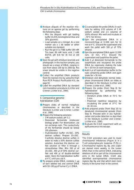
6 Analyze aliquots of the reaction mixtures<br />
on an agarose gel by performing<br />
the following steps:<br />
Mix the aliquots with gel loading<br />
buffer (0.25% bromophenol blue and<br />
30% glycerol).<br />
Load samples on a minigel. As a size<br />
marker use a 1 kb ladder or other<br />
suitable size standard.<br />
Run the gel in 1x TBE buffer (89 mM<br />
Tris base, 89 mM boric acid, 2 mM<br />
EDTA; pH 8.0) for 30 min at 100<br />
volts.<br />
7 Stain the gel with ethidium bromide and<br />
pho<strong>to</strong>graph. <strong>In</strong> the reaction samples, you<br />
should see a smear of DNA, ranging in<br />
size from about 200 bp <strong>to</strong> 2000 bp. No<br />
smear should be visible in the negative<br />
control.<br />
8 Collect the amplified DNA products<br />
from the reaction mix by using the High<br />
Pure PCR Product Purification Kit, see<br />
page 50.<br />
9 Label the amplified DNA by standard<br />
nick translation procedures (Lichter and<br />
Cremer; Lichter et al., 1994).<br />
II. Comparative genomic<br />
hybridization (CGH)<br />
1 Prepare slides of normal metaphase<br />
chromosomes as described in the<br />
literature (Lichter and Cremer; Lichter<br />
et al., 1990).<br />
2 Prepare the following solutions:<br />
3 M sodium acetate, pH 5.2.<br />
Deionized formamide (molecular<br />
biology grade): For deionization, use<br />
ion exchange resin. The conductivity<br />
of the formamide should be below<br />
100 µSiemens.<br />
<strong>Hybridization</strong> buffer (4 x SSC, 20%<br />
dextran sulfate): Prepare 20 x SSC.<br />
Carefully dissolve dextran sulfate in<br />
water <strong>to</strong> make a 50% dextran sulfate<br />
solution. Au<strong>to</strong>clave the dextran sulfate<br />
solution or filter it through a<br />
nitrocellulose filter. Mix 200 µl of<br />
20 x SSC, 400 µl of 50% dextran sulfate<br />
and 400 µl of double-distilled<br />
water. S<strong>to</strong>re the hybridization buffer<br />
at 4°C until you use it.<br />
3 To prepare probe DNA, combine 1 µg<br />
of each labeled test and control DNA<br />
with 50–100 µg of human Cot-1 DNA.<br />
4 Co-precipitate the probe DNAs in each<br />
tube by adding 1/20 volume of 3 M<br />
sodium acetate and 2.5 volumes of<br />
100% ethanol. Mix well and incubate at<br />
–70°C for 30 min.<br />
5 Spin the precipitated DNA in a<br />
microcentrifuge at 12,000 rpm for 10<br />
min at 4°C. Discard the supernatant and<br />
wash the pellet with 500 µl of 70%<br />
ethanol.<br />
6 Spin the precipitated DNA again (12,000<br />
rpm, 10 min, 4°C). Discard the<br />
supernatant and lyophilize the pellet.<br />
7 Add 6 µl deionized formamide <strong>to</strong> the<br />
lyophilizate and resuspend the probe<br />
DNA by vigorously shaking the tube<br />
for > 30 min at room temperature.<br />
8 Add 6 µl of hybridization buffer <strong>to</strong> the<br />
tube containing probe DNA and again<br />
shake for > 30 min.<br />
9 Denature and dehydrate normal metaphase<br />
chromosomal DNA on slides as<br />
described in the literature (Lichter and<br />
Cremer; Lichter et al., 1990).<br />
10 Prepare the probe (from Step 8) for<br />
hybridization by performing the<br />
following steps:<br />
Denature probe DNA at 75°C for<br />
5 min.<br />
Preanneal repetitive sequences by<br />
incubating the probe at 37°C for<br />
20–30 min.<br />
11 Add prepared probe (from Step 10) <strong>to</strong><br />
denatured chromosomal spreads (from<br />
Step 9). Proceed with in situ hybridization<br />
and probe detection as described<br />
in the literature (Lichter and Cremer;<br />
Lichter et al., 1990).<br />
12 Evaluate the CGH experiment with an<br />
epifluorescence microscope.<br />
Results<br />
The CGH procedure was used <strong>to</strong> reveal<br />
chromosomal imbalances (Figure 3) in a<br />
T-cell prolymphocytic leukemia (T-PLL).<br />
Chromosomal regions 6q, 8p, and 11qter<br />
are stained more weakly in the tumor<br />
DNA than in the control DNA, indicating<br />
deletions of all or part of the chromosomal<br />
arms in the tumor DNA. CGH also<br />
identified overrepresented chromosomal<br />
regions (6p, 8q, and 14q) which stain more<br />
heavily in the tumor than in the control.<br />
CONTENTS<br />
INDEX


