The Genus Serratia
The Genus Serratia
The Genus Serratia

You also want an ePaper? Increase the reach of your titles
YUMPU automatically turns print PDFs into web optimized ePapers that Google loves.
Prokaryotes (2006) 6:219–244<br />
DOI: 10.1007/0-387-30746-x_11<br />
CHAPTER 3.3.11<br />
eTh<br />
<strong>The</strong> <strong>Genus</strong> <strong>Serratia</strong><br />
FRANCINE GRIMONT AND PATRICK A. D. GRIMONT<br />
<strong>The</strong> genus <strong>Serratia</strong> a member of the Enterobacteriaceae (see<br />
Introduction to the Family Enterobacteriaceae from the<br />
second edition.), is comprised of a group of bacteria that are<br />
related both phenotypically and by DNA sequence. <strong>The</strong> type<br />
species of the genus is <strong>Serratia</strong> marcescens. Some species and<br />
biotypes of <strong>Serratia</strong> produce a nondiffusible red pigment,<br />
prodigiosin, or 2-methyl-3-amyl-6-methoxyprodigiosene<br />
(Williams and Qadri, 1980). <strong>The</strong> multiplication of redpigmented<br />
<strong>Serratia</strong> was incriminated in the appearance of<br />
bloodlike spots (e.g., on bread, consecrated wafers [sacramental<br />
Hosts], and polenta) with rather disastrous sociological<br />
consequences. In this context, several scholars have<br />
traced the history of the genus <strong>Serratia</strong> back to antiquity<br />
(Gaughran, 1969; Harrison, 1924; Reid, 1936). However, several<br />
bacterial species outside the genus <strong>Serratia</strong> produce prodigiosin<br />
or prodigiosin-like pigments (Williams and Qadri,<br />
1980) or many other kinds of red pigments, and the identity<br />
of microorganisms involved in these striking phenomena can<br />
only be surmised.<br />
Bizio (1823) named the red-pigmented microorganism he<br />
observed on polenta <strong>Serratia</strong> marcescens. Ehrenberg (1848)<br />
named a motile bacterium isolated from red spots on food<br />
“Monas prodigiosa.” No cultures of these organisms were preserved,<br />
but the name <strong>Serratia</strong> marcescens was preferred over<br />
the name “Erythrobacillus pyosepticus”—a culture of which<br />
was preserved as ATCC 275 (Fortineau, 1904)—by Breed and<br />
Breed (1924, 1927) and by the editors of Bergey’s Manual of<br />
Determinative Bacteriology (Bergey et al., 1923). <strong>The</strong> name<br />
<strong>Serratia</strong> marcescens is now universally accepted, and a neotype<br />
strain has been designated (Martinec and Kocur, 1961a).<br />
At the start of this century, more than 76 nomenspecies<br />
had been described with red or pink pigmentation (Hefferan,<br />
1904), and 23 <strong>Serratia</strong> species were listed in the first edition<br />
of Bergey’s Manual (Bergey et al., 1923). This number progressively<br />
decreased to five in the fifth edition of Bergey’s<br />
Manual (Breed et al., 1957), and later to one species: S.<br />
marcescens (Ewing et al., 1959; Martinec and Kocur, 1960,<br />
1961a, 1961b, 1961c, 1961d). <strong>The</strong> only <strong>Serratia</strong> species recognized<br />
in the eighth edition of Bergey’s Manual was S. marcescens<br />
(Sakazaki, 1974). <strong>The</strong>n, new objective approaches,<br />
such as numerical taxonomy and DNA relatedness applied<br />
to strains recovered from diverse habitats, delineated an<br />
increasing number of species in the genus <strong>Serratia</strong>. Five and<br />
seven species, respectively, were mentioned in the first edition<br />
of <strong>The</strong> Prokaryotes (Grimont and Grimont, 1981) and<br />
in Bergey’s Manual of Systematic Bacteriology (Grimont and<br />
Grimont, 1984). Ten species are presently known to belong<br />
in the genus <strong>Serratia</strong>. <strong>The</strong>se species (and synonyms) are:<br />
1. <strong>Serratia</strong> marcescens Bizio 1823: “Typical” S. marcescens<br />
(Colwell and Mandel, 1965; Ewing et al., 1959; Martinec and<br />
This chapter was taken unchanged from the second edition.<br />
sun<br />
Ge<br />
Kocur, 1961a); <strong>Serratia</strong> pattern 1 (Fulton et al., 1959); <strong>Serratia</strong><br />
biotype 1 (Bascomb et al., 1971); phenon A (Grimont and<br />
Dulong de Rosnay, 1972; Grimont et al., 1977b); S. marcescens<br />
DNA hybridization group (Steigerwalt et al., 1976). <strong>The</strong><br />
neotype strain is ATCC 13880, CDC 813-60, Grimont 504,<br />
and CCM 303 (Martinec and Kocur, 1961a). <strong>The</strong> name<br />
appeared in the Approved Lists of Bacterial Names (Skerman<br />
et al., 1980). <strong>The</strong> type strains of “Bacillus indicus” (Eisenberg,<br />
1886), “Erythrobacillus pyosepticus” (Fortineau, 1904),<br />
“Bacillus sphingidis” (White, 1923a), and “<strong>Serratia</strong> anolium”<br />
(Duran-Reynals and Clausen, 1937) are all referable to the<br />
taxonomic entity now known as S. marcescens. Strains<br />
labeled “S. marcescens subsp. kiliensis” according to Ewing<br />
et al. 1962 were Voges-Proskauer-negative variants of S.<br />
marcescens.<br />
2. <strong>Serratia</strong> liquefaciens (Grimes and Hennerty 1931)<br />
Bascomb et al. 1971: “Aerobacter liquefaciens” Grimes and<br />
Hennerty 1931; “Aerobacter lipolyticus” Grimes 1961;<br />
Enterobacter liquefaciens (Grimes and Hennerty, 1931)<br />
Ewing 1963; phenon Clab (Grimont et al., 1977b); <strong>Serratia</strong><br />
liquefaciens sensu stricto (Grimont et al., 1982a, 1982b). <strong>The</strong><br />
type strain is ATCC 27592, CDC 1284-57, and Grimont 866.<br />
<strong>The</strong> name appeared in the Approved Lists of Bacterial Names<br />
(Skerman et al., 1980). It should be mentioned that the strain<br />
considered to be the type strain of “Aerobacter liquefaciens,”<br />
or of “Aerobacter lipolyticus” by Grimes (1961), was ATCC<br />
14460 (now the type strain of S. grimesii). Since this strain<br />
was considered atypical, another strain (ATCC 27592) was<br />
given as the type strain in the Approved Lists.<br />
3. <strong>Serratia</strong> proteamaculans (Paine and Stansfield 1919)<br />
Grimont et al. 1978b: S. proteamaculans sensu stricto<br />
(Grimont et al., 1982a, 1982b). <strong>The</strong> type strain is ATCC<br />
19323, Grimont 3630, ICPB XP176, and NCPPB 245. <strong>The</strong><br />
name appeared in the Approved Lists of Bacterial Names<br />
(Skerman et al., 1980). S. proteamaculans and S. liquefaciens<br />
were thought to be synonymous on the basis of DNA<br />
relatedness (Grimont et al., 1978b). However, subsequent<br />
observation of significant thermal instability of DNA<br />
hybrid fragments supported the separation of both species<br />
(Grimont et al., 1982a).<br />
Biogroup RQ was named S. proteamaculans subspecies<br />
quinovora (Grimont et al., 1982a, 1982b) with strain Grimont<br />
4364, CIP 8195, and ATCC 33765 as the type strain.<br />
4. <strong>Serratia</strong> grimesii Grimont et al. 1982a, 1982b: phenon<br />
Cld (Grimont et al., 1977b); S. liquefaciens hybridization<br />
group (Steigerwalt et al., 1976). <strong>The</strong> type strain is ATCC<br />
14460 and Grimont 503. This strain had been considered as<br />
the type strain of S. liquefaciens prior to the publication of<br />
the Approved Lists.<br />
5. <strong>Serratia</strong> plymuthica (Lehmann and Neumann 1896)<br />
Breed et al. 1948: “Bacterium plymuthicum” Lehmann and<br />
ai<br />
tar<br />
reS
220 F. Grimont and P.A.D. Grimont CHAPTER 3.3.11<br />
Neumann 1896; excluded from the genus <strong>Serratia</strong> (Ewing et<br />
al., 1959); “atypical” S. marcescens (Colwell and Mandel,<br />
1965); “S. marcescens var. kiliensis” according to Martinec<br />
and Kocur (1961d) (not Ewing et al., 1962); <strong>Serratia</strong> III (Mandel<br />
and Rownd, 1964); <strong>Serratia</strong> pattern 2 (Fulton et al., 1959);<br />
atypical S. rubidaea (Ewing et al., 1972, 1973); and phenon<br />
C2 (Grimont et al., 1977b). <strong>The</strong> type strain is Grimont 510,<br />
CCM 640, and ATCC 183. <strong>The</strong> type strains of “Bacterium<br />
kiliense” reference is not an exact matchLehmann and<br />
Neumann 1986 and “<strong>Serratia</strong> esseyana” Combe 1933 are S.<br />
plymuthica.<br />
6. <strong>Serratia</strong> rubidaeaStapp 1940 (Ewing et al., 1973): “Bacterium<br />
rubidaeum” Stapp 1940; “Prodigiosus” VIII (Hefferan<br />
1904); S. marinorubra ZoBell and Upham 1944; <strong>Serratia</strong><br />
biotype 2 (Bascomb et al., 1971); phenon B (Grimont and<br />
Dulong de Rosnay, 1972; Grimont et al., 1977b); S. rubidaea<br />
DNA hybridization group (Steigerwalt et al., 1976). <strong>The</strong><br />
name appeared in the Approved Lists of Bacterial Names<br />
(Skerman et al., 1980). <strong>The</strong> type strain (neotype) is ATCC<br />
27593, CDC 2199–72, and Grimont 864. <strong>The</strong> former type<br />
strain (holotype) of S. marinorubra was NCTC 10912, ATCC<br />
27614, and Grimont 288. However, the Approved Lists of<br />
Bacterial Names (Skerman et al., 1980) listed S. marinorubra<br />
with ATCC 27593 as the type strain. Thus, both names (S.<br />
rubidaea and S. marinorubra) were made objective synonyms<br />
and the name S. rubidaea has priority. Three subspecies can<br />
be delineated: S. rubidaea subsp. rubidaea S. rubidaea subsp.<br />
burdigalensis, and S. rubidaea subsp. colindalensis (Grimont<br />
et al., manuscript in preparation).<br />
7. <strong>Serratia</strong> odorifera Grimont et al. 1978a: Strains similar<br />
to the unclustered <strong>Serratia</strong> strain 38 (Grimont and Dulong<br />
de Rosnay, 1972; Grimont et al., 1977b). <strong>The</strong> type strain is<br />
ATCC 33077, CDC 1979-77, Grimont 1073, ICPB 3995, and<br />
NCTC 11214. <strong>The</strong> name appeared in the Approved Lists of<br />
Bacterial Names (Skerman et al., 1980).<br />
8. <strong>Serratia</strong> ficaria Grimont et al. 1979c: the type strain is<br />
ATCC 33105, Grimont 4024, CIP 79.23, and ICPB 4050.<br />
9. <strong>Serratia</strong> entomophila Grimont et al. 1988: the type strain<br />
is ATCC 43705, Jackson A1, and CIP 102919.<br />
10. <strong>Serratia</strong> fonticola Gavini et al. 1979: Lysine-positive<br />
Citrobacter-like (Steigerwalt et al., 1976). <strong>The</strong> type strain is<br />
ATCC 29844, CUETM 77.165, Grimont 4011, and CIP 78.64.<br />
<strong>The</strong> name appeared in the Approved Lists of Bacterial Names<br />
(Skerman et al., 1980).<br />
<strong>The</strong> inclusion of the genus <strong>Serratia</strong> in the tribe Klebsielleae<br />
is no longer tenable. Studies on DNA relatedness, immunological<br />
cross-reaction between isofunctional enzymes, and<br />
the physical properties, regulation, and amino acid sequences<br />
of enzymes have all shown the genus <strong>Serratia</strong> to be consistently<br />
different from the group composed of the genera<br />
Escherichia, Shigella, Salmonella, Citrobacter, Klebsiella, and<br />
Enterobacter (reviewed by Grimont and Grimont, 1978a).<br />
Until the late 1950s, <strong>Serratia</strong> spp. were rarely isolated from<br />
human patients. Later on S. marcescens became more and<br />
more frequently involved in nosocomial infections and nonpigmented<br />
S. marcescens strains are now a serious threat<br />
in surgical and intensive care units (Daschner, 1980; von<br />
Graevenitz, 1980; Yu, 1979).<br />
Ecology<br />
For a long time, the confused status of <strong>Serratia</strong><br />
taxonomy prevented any precise knowledge of<br />
the habitat of <strong>Serratia</strong> species. After the aforementioned<br />
<strong>Serratia</strong> species had been defined,<br />
however, it became apparent that they occupied<br />
different habitats. Table 1 shows the distribution<br />
of the major species of the <strong>Serratia</strong> strains isolated<br />
from small mammals and their territories,<br />
and from water, plants, and hospitalized human<br />
patients. Table 2 shows the distribution of the<br />
major biotypes of the S. marcescens strains isolated<br />
from these same habitats.<br />
<strong>Serratia</strong> in Water and Soil<br />
Water is probably the principal habitat of S.<br />
plymuthica. Nomenspecies resembling S.<br />
plymuthica (Breunig’s Kiel bacillus, “Bacillus<br />
miniaceus,” “<strong>Serratia</strong> miquelii,” and “S. esseyana”)<br />
have also been isolated from water. Seawater<br />
isolates belong to the same species as<br />
terrestrial water isolates. “<strong>Serratia</strong> marinorubra”<br />
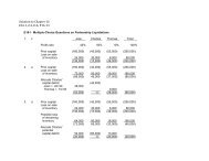
Table 1. Percent distribution among <strong>Serratia</strong> species of 1,543 strains isolated from different habitats.<br />
Species<br />
Small mammals a<br />
(92 isolates)<br />
a Rodents and shrews.<br />
b From plants and soil around the traps where small mammals were captured.<br />
c All plants except figs and coconuts.<br />
Animal territories b<br />
(51 isolates)<br />
Percentage of isolates in habitat<br />
Water<br />
(155 isolates)<br />
Plants c<br />
(137 isolates)<br />
Hospitalized patients<br />
(1,108 isolates)<br />
S. marcescens 10 2 75 10 97<br />
S. plymuthica 4 2 1 5 0<br />
S. liquefaciens 43 55 11 38 2<br />
S. proteamaculans 39 29 8 32 0<br />
S. grimesii 3 10 5 7 0.5<br />
S. rubidaea 0 0 0 1 0.2<br />
S. odorifera 0 0 0 4 0.1<br />
S. ficaria 0 2 0 4 0<br />
Total percentage 100 100 100 100 100
CHAPTER 3.3.11 <strong>The</strong> <strong>Genus</strong> <strong>Serratia</strong> 221<br />
Table 2. Percent distribution among <strong>Serratia</strong> marcescens biogroups of 1,210 strains isolated from four different habitats.<br />
Biogroup<br />
a Isolated at the Pellegrin Hospital (Bordeaux, France) from 1968 through 1975.<br />
b In addition, 72 strains of biotype A2b were isolated from infants (mostly from feces) in the neonatology ward.<br />
was not a true marine species, as it showed<br />
no sodium requirement (P. Baumann, personal<br />
communication). Many strains of S. fonticola<br />
have been isolated from well waters and springs<br />
(Gavini et al., 1979). In a systematic search for<br />
<strong>Serratia</strong> in river water, 150 strains were isolated<br />
and were distributed in the following species:<br />
S. marcescens (75%), S. liquefaciens (11%), S.<br />
proteamaculans (8%), S. grimesii (5%), and S.<br />
plymuthica (1%) (F. Agbalika, F. Grimont, and<br />
P.A.D. Grimont, unpublished observations). <strong>The</strong><br />
selective medium used (caprylate-thallous agar)<br />
did not allow isolation of S. fonticola.<br />
Strains producing the non-diffusible red pigment<br />
prodigiosin seem to be toxic to protozoa<br />
(Groscop and Brent, 1964), and this may be an<br />
ecological advantage in water and soil. However,<br />
it seems that pigmented bacteria are more often<br />
isolated from unpolluted water (from springs or<br />
wells) than from polluted water (river water<br />
downstream from cities).<br />
In soil, S. marcescens might play a role in the<br />
biological cycle of metals by mineralizing organic<br />
iron and dissolving gold and copper (Parès,<br />
1964). A mineralization role has also been attributed<br />
to cold-tolerant <strong>Serratia</strong> associated with<br />
low-moor peat (Janota-Bassalik, 1963).<br />
<strong>Serratia</strong> on Plants<br />
<strong>Serratia</strong> proteamaculans was once isolated from<br />
a leaf spot disease of the tropical plant Protea<br />
cynaroides (the King Protea) (Paine and Stansfield,<br />
1919). However, the experimental lesions<br />
caused by S. proteamaculans on detached leaves<br />
of Protea suggest a hypersensitivity reaction<br />
(Paine and Berridge, 1921). Similar lesions were<br />
obtained with other species of <strong>Serratia</strong> (Grimont<br />
et al., 1978b). Inoculation of S. marcescens ICPB<br />
2875 and S. marinorubra ICPB 2881 on tobacco<br />
and bean leaves also produced a typical hypersensitivity<br />
reaction (Lakso and Starr, 1970). A<br />
root disease complex of alfalfa (Medicago<br />
sativa) involving a Fusarium sp., a Pseudomonas<br />
Percentage of isolates in habitat<br />
Water (107 isolates) Plants and rodents (37 isolates) Hospitalized patients (1,066 isolates) a<br />
A1 12 19 0.1<br />
A2/6 23 5 7 b<br />
A3 21 38 7<br />
A4 34 38 26<br />
A5/8 7 0 47<br />
TCT 3 0 13<br />
Total 100 100 100<br />
sp., and a bacterium named “Erwinia amylovora<br />
var. alfalfae” was observed by Shinde and<br />
Lukezic (1974). <strong>The</strong>se isolates of “Erwinia<br />
amylovora var. alfalfae” were later identified as<br />
<strong>Serratia</strong> marcescens biotype A4a (Grimont et al.,<br />
1981).<br />
Characteristic <strong>Serratia</strong> populations were found<br />
in figs. S. ficaria was recovered from most figs<br />
collected in California, Tunisia, France (Grimont<br />
et al., 1979c), Sicily (Giammanco and Amato,<br />
1982; Grimont and Deval, 1982), and Greece<br />
(P.A.D. Grimont, unpublished observations).<br />
This species could be associated with S. marcescens<br />
(biogroups A1, A2, or A3) or other genera.<br />
Figs of the Calimyrna variety (Smyrna variety<br />
adapted to California) deserve special mention.<br />
Calimyrna figs contain only pistillate flowers and<br />
in order to become edible, they need to be pollinated<br />
specifically by the fig wasp Blastophaga<br />
psenes. <strong>The</strong> fig wasp has a life cycle limited to the<br />
caprifig, an inedible fruit produced by the caprifig<br />
tree. When a young female fig wasp makes her<br />
way out of a caprifig, she covers herself with<br />
pollen and a specific fungal and microbial flora<br />
(Phaff and Miller, 1961). <strong>The</strong> fig wasp will then<br />
enter an unpollenated caprifig of a new crop,<br />
pollinate it, oviposit in a pistillate flower, and die;<br />
or it may be carried by the wind to a Calimyrna<br />
fig—a cul-de-sac in the wasp life cycle—where<br />
the fig wasp will deposit pollen and the microbial/fungal<br />
flora in its desperate attempt to oviposit.<br />
Since 1927 (Caldis, 1927; Phaff and Miller,<br />
1961; Smith and Hansen, 1931), a red-pigmented<br />
and a nonpigmented <strong>Serratia</strong> have been repeatedly<br />
isolated from caprifigs, pollinated Calimyrna<br />
figs, and fig wasps. <strong>The</strong> pigmented and<br />
nonpigmented isolates were identified as <strong>Serratia</strong><br />
marcescens biotype A1b and S. ficaria, respectively<br />
(Grimont et al., 1979c; Grimont et al.,<br />
1981). <strong>The</strong> fact that the Calimyrna fig is internally<br />
sterile until the fig wasp enters it provides<br />
an ecological niche for these <strong>Serratia</strong> strains. <strong>The</strong><br />
multiplication of <strong>Serratia</strong> in caprifigs or Calimyrna<br />
figs is limited, and these organisms do not
222 F. Grimont and P.A.D. Grimont CHAPTER 3.3.11<br />
cause fig spoilage. Washing of a syconium cavity<br />
can yield 10 to 300 colony-forming-units (CFU)<br />
of S. ficaria.<br />
<strong>Serratia</strong> rubidaea has been repeatedly isolated<br />
from coconuts bought in France (originating<br />
mostly from Ivory Coast) and in California (Grimont<br />
et al., 1981). <strong>The</strong> three subspecies (former<br />
biotypes B1 to B3) have been associated with<br />
coconuts, and when present, S. rubidaea<br />
numbered 3× 10 4 to 6× 10 6 CFU per gram of<br />
coconut milk or flesh (Bollet et al., 1989). It is<br />
believed that coconut palms have been disseminated<br />
worldwide from an Indo-Pacific origin.<br />
It would thus be interesting to compare the<br />
bacterial flora of coconuts collected in diverse<br />
countries.<br />
In the course of an ecological survey and using<br />
the selective caprylate-thallous agar medium,<br />
<strong>Serratia</strong> species were frequently found associated<br />
with plants (Grimont et al., 1981). With respects<br />
to the presence of <strong>Serratia</strong>, plants (other than figs<br />
and coconuts) were classified into three prevalence<br />
groups. In the first group, which is composed<br />
of vegetables, mushrooms, mosses, and<br />
decaying plant material (i.e., wet plants), about<br />
54% of the samples carried <strong>Serratia</strong> in counts<br />
varying from 2,000 to over 20,000 CFU per gram<br />
of plant (highest counts in mushrooms and<br />
leaves of radish, lettuce, cauliflower, and brussels<br />
sprout). In the second group, which is composed<br />
of grasses, <strong>Serratia</strong> prevalence was about 24%. In<br />
the third group composed of trees and shrubs,<br />
8% of the samples (leaves) contained <strong>Serratia</strong><br />
(10 to 300 CFU per gram).<br />
<strong>Serratia</strong> strains isolated from plants other than<br />
figs and coconuts were distributed among eight<br />
<strong>Serratia</strong> species, although S. liquefaciens and S.<br />
proteamaculans were predominant (Table 1).<br />
<strong>The</strong> selective medium used (caprylate-thallous<br />
agar) was unable to support the growth of S.<br />
fonticola. S. entomophila was not isolated<br />
although this species can grow on the selective<br />
medium. Pigmented S. marcescens biotypes were<br />
rarely isolated from plants. Nonpigmented S.<br />
marcescens biogroups A3 and A4 were isolated<br />
from plants, but biogroups A5/8 and TCT were<br />
not (Table 2).<br />
Vegetables used in salads might bring <strong>Serratia</strong><br />
strains to hospitals and contaminate the patient’s<br />
digestive tract. S. marcescens, S. liquefaciens, and<br />
S. rubidaea were found in 29%, 28%, and 11%<br />
(respectively) of vegetable salads served in a<br />
hospital in Pittsburgh (Wright et al., 1976). Similar<br />
results were found in a Paris hospital<br />
(Loiseau-Marolleau and Laforest, 1976). However,<br />
it still needs to be proven that biotypes/<br />
serotypes found in patients are the same as those<br />
found in salads. In the first described case of S.<br />
ficaria infection, the patient was very fond of figs<br />
(Gill et al., 1981).<br />
<strong>Serratia</strong> in Insects<br />
<strong>The</strong>re is an extensive literature on <strong>Serratia</strong> associated<br />
with insects. This topic has been reviewed<br />
in detail (Bucher, 1963a; Grimont and Grimont,<br />
1978a; Steinhaus, 1959). <strong>The</strong> insects involved<br />
belong to numerous species and genera of the<br />
orders Orthoptera (crickets and grasshoppers),<br />
Isoptera (termites), Coleoptera (beetles and<br />
weevils), Lepidoptera (moths), Hymenoptera<br />
(bees and wasps), and Diptera (flies). Taxonomic<br />
uncertainties make a retrospective evaluation of<br />
the role of <strong>Serratia</strong> spp. in insect infections difficult.<br />
Red-pigmented <strong>Serratia</strong> were easily recognized<br />
(although not determined as to species<br />
with any certainty), whereas nonpigmented<br />
strains were often referred to genera other than<br />
<strong>Serratia</strong>. For example, S. proteamaculans or S.<br />
liquefaciens strains were named “Bacillus noctuarum”<br />
(White, 1923b), “Bacillus melolonthae liquefaciens”<br />
(Paillot, 1916), “Paracolobactrum<br />
rhyncoli” (Pesson et al., 1955), and “Cloaca” B<br />
type 71–12A (Bucher and Stephens, 1959); nonpigmented<br />
S. marcescens strains were named<br />
“Bacillus sphingidis” (White, 1923a) and “Bacillus<br />
apisepticus” (Burnside, 1928).<br />
<strong>The</strong> distribution among the various <strong>Serratia</strong><br />
species of 48 collection strains isolated from<br />
insects (Grimont et al., 1979b) showed a predominance<br />
of S. marcescens and S. liquefaciens.<br />
<strong>The</strong> absence of S. plymuthica (a pigmented species)<br />
in this collection is unexplained. Steinhaus<br />
(1941) reported the presence of S. plymuthica in<br />
the gut of healthy crickets (Neombius fasciatus),<br />
but the taxonomic schemes in vogue at that time<br />
did not allow a definite identification of S.<br />
plymuthica. <strong>The</strong> rarity of S. rubidaea (also a pigmented<br />
species) in insects might be explained by<br />
its inability to produce chitinase—a virulence<br />
factor for insect-associated <strong>Serratia</strong> species<br />
(Lysenko, 1976).<br />
<strong>The</strong> red-pigmented <strong>Serratia</strong> isolates associated<br />
with the fig wasp Blastophaga psenes and formerly<br />
identified as S. plymuthica (Phaff and<br />
Miller, 1961) were reidentified as S. marcescens<br />
biotype A1b (Grimont et al., 1981). It is noteworthy<br />
that S. ficaria was isolated from both the fig<br />
wasp Blastophaga psenes and from a black ant,<br />
i.e., Hymenoptera (Grimont et al., 1979c).<br />
S. liquefaciens and S. marcescens were found<br />
in sugar-beet root-maggot development stages<br />
(Tetanops myopaeformis), suggesting an insectmicrobe<br />
symbiosis, as well as a nutritional interdependence<br />
(Iverson et al. 1984). A relationship<br />
appeared to exist between adult fly emergence<br />
and enzymatic chitin degradation of the puparium<br />
by the bacterial symbionts.<br />
S. marcescens biotype A4b was repeatedly isolated<br />
from diseased honeybee (Apis mellifera)<br />
larvae in Sudan (El Sanoussi et al., 1987).
CHAPTER 3.3.11 <strong>The</strong> <strong>Genus</strong> <strong>Serratia</strong> 223<br />
<strong>Serratia</strong> marcescens, S. proteamaculans and<br />
S. liquefaciens are considered potential insect<br />
pathogens (Bucher, 1960). <strong>The</strong>y cause a lethal<br />
septicemia after penetration into the hemocoel.<br />
More than 70 species of insects were found to be<br />
susceptible to inoculation with <strong>Serratia</strong> (Bucher,<br />
1963a). <strong>The</strong> lethal dose (LD 50) of inoculated <strong>Serratia</strong><br />
(by intrahemocoelic injection) was calculated<br />
for several insects 10–50 <strong>Serratia</strong> cells per<br />
grasshopper (Bucher, 1959), 5.1 cells per adult<br />
bollweevil (Slatten and Larson, 1967), 7.5 and<br />
14.5 cells per third and fourth instar larva<br />
(respectively) of Lymantria dispar (Podgwaite<br />
and Cosenza, 1976), and 40 cells per Galleria<br />
mellonella larva (Stephens, 1959). <strong>The</strong> LD 50 of<br />
ingested S. marcescens is much higher. <strong>The</strong><br />
hemolymph of insects—normally bactericidal for<br />
nonpathogens—cannot prevent multiplication of<br />
potential pathogens (Stephens, 1963). Lecithinase,<br />
proteinase, and chitinase play a role in the<br />
virulence of <strong>Serratia</strong> for insects, and purified<br />
<strong>Serratia</strong> proteinase or chitinase is very toxic<br />
when injected into the hemocoel (Kaska, 1976;<br />
Lysenko, 1976). <strong>Serratia</strong> strains in the insect<br />
digestive tract probably originate from plants.<br />
<strong>The</strong> multiplication of <strong>Serratia</strong> strains in the insect<br />
digestive tract has not been quantitatively studied.<br />
Antibacterial substances in ingested leaves<br />
might interfere with bacterial multiplication, but<br />
<strong>Serratia</strong> strains were found resistant to these<br />
(Kushner and Harvey, 1962). How potential<br />
pathogens such as <strong>Serratia</strong> can enter the<br />
hemolymph from the gut is generally unknown.<br />
However, spontaneous gut rupture, which happens<br />
in about 10% of grasshoppers, may allow<br />
<strong>Serratia</strong> strains to invade the hemocoel (Bucher,<br />
1959). Direct injection occurs when Itoplectis<br />
conquisitor contaminated with <strong>Serratia</strong> stings<br />
host pupae to oviposit into their bodies (Bucher,<br />
1963b). <strong>Serratia</strong> epizootics are common among<br />
reared insects (Bucher, 1963a). However, until<br />
recently, no genuine epizootic of <strong>Serratia</strong> infection<br />
among insects had been observed in the field.<br />
Strains of S. entomophila and S. proteamaculans<br />
can be pathogenic for the grass grub Costelytra<br />
zealandica, which is a major pasture pest in<br />
New Zealand (Stucki and Jackson, 1984; Trought<br />
et al., 1982). Larvae of Costelytra zealandica feed<br />
on grass, clover, and other plant roots. Typically,<br />
their populations grow to a peak (about 600 larvae/m<br />
2 ) in 4 to 6 years after the pasture is sown<br />
and then collapse (to about 50 larvae/m 2 ). Grass<br />
grub population collapse was found associated<br />
with the presence of a disease called amber disease<br />
(Trought et al., 1982). S. entomophila and S.<br />
proteamaculans were isolated from naturally<br />
infected larvae and shown experimentally to produce<br />
the disease when transmitted orally to<br />
healthy larvae (Grimont et al., 1988; Stucki and<br />
Jackson, 1984). <strong>The</strong> bacteria are ingested from<br />
the soil. Infected larvae stop feeding within a few<br />
days, become translucent and then amber colored<br />
and lose weight until death occurs 4 to 6<br />
weeks later. <strong>The</strong> bacteria colonize the gut and<br />
cause disease symptoms without invading the<br />
hemocoele. Field trials have shown that control<br />
of the grass grub was feasible by application of<br />
S. entomophila suspensions on pastures (Jackson<br />
and Pearson 1986). Reductions of 30 to 59% in<br />
the larval populations were obtained, with 47%<br />
of the remaining larvae being infected. Such bacterial<br />
treatment resulted in a 30% increase in<br />
grass production (dry matter).<br />
<strong>Serratia</strong> in Vertebrates<br />
<strong>Serratia</strong> has been associated with chronic<br />
infections of cold-blooded vertebrates: nodular<br />
infection of Anolis equestris, the Cuban lizard<br />
(Duran-Reynals and Clausen, 1937); subcutaneous<br />
abscess of iguanid lizards (Boam et al., 1970);<br />
arthritis in the lizard Tupinambis tequixin (Ackerman<br />
et al., 1971); and ulcerative disease in the<br />
painted turtle Chrysemys picta (Jackson and Fulton,<br />
1976). <strong>Serratia</strong> strains have also been recovered<br />
from the healthy, small, green pet turtle<br />
Pseudemys scripta elegans (McCoy and Seidler,<br />
1973) and from geckos and turtles in Vietnam<br />
(Capponi et al., 1956).<br />
Poultry may be contaminated with <strong>Serratia</strong>. A<br />
deadly <strong>Serratia</strong> epizootic among chick embryos<br />
was observed in a Japanese hatchery (Izawa et<br />
al., 1971). <strong>The</strong> hens carried S. marcescens in their<br />
digestive tract, but were themselves unaffected.<br />
Contamination of chicken carcasses with S. liquefaciens<br />
(Lahellec et al., 1975) and spoilage of<br />
eggs by red-pigmented <strong>Serratia</strong> (Alford et al.,<br />
1950) have been reported. A disseminated suppurative<br />
infection in a blue and gold macaw (Ara<br />
ararauna) affected with a chronic lymphoid atrophy<br />
has also been recorded (Quesenberry and<br />
Short, 1983).<br />
<strong>Serratia</strong> strains (mostly red-pigmented) are<br />
responsible for 0.2–1.5% of cases of mastitis in<br />
cows (Barnum et al., 1958; Roussel et al., 1969;<br />
Wilson, 1963). Raw milk, therefore, may occasionally<br />
contain <strong>Serratia</strong> spp., and S. liquefaciens<br />
and S. grimesii are common in dairy products<br />
(Grimes and Hennerty, 1931). <strong>Serratia</strong> strains<br />
have been involved in septicemia in foals (Deom<br />
and Mortelmans, 1953), goats (Wijewanta and<br />
Fernando, 1970), and pigs (Brisou and Cadeillan,<br />
1959); they have also been implicated in conjunctivitis<br />
of the horse (Carter, 1973) and abortion in<br />
cows (Smith and Reynolds, 1970). <strong>The</strong> isolation<br />
of <strong>Serratia</strong> from the anal sac of the red fox Vulpes<br />
vulpes (Gosden and Ware, 1976) has been<br />
reported.<br />
<strong>Serratia</strong> strains have rarely been systematically<br />
searched for in the gut of animals. S. fonticola
224 F. Grimont and P.A.D. Grimont CHAPTER 3.3.11<br />
was isolated in fecal samples from seven sparrows<br />
and unidentified birds. <strong>The</strong> study involved<br />
90 wild European birds (Müller et al., 1986).<br />
About 40% of trapped wild rodents and<br />
shrews carried <strong>Serratia</strong> strains in their gut without<br />
any visible sign of infection upon autopsy.<br />
<strong>The</strong> following animal species were found to carry<br />
<strong>Serratia</strong> spp.: 75/180, Apodemus sylvaticus; 7/23,<br />
Microtus arvalis; 3/11, Clethrionomys glareolus;<br />
1 /2, Micromys minutus; (rodents); 7/9, Sorex; and<br />
1 /3, Crocidura (shrews) (P. Giraud, F. Grimont,<br />
P.A.D. Grimont, unpublished observations). <strong>The</strong><br />
S. liquefaciens complex (S. liquefaciens, S. proteamaculans<br />
and S. grimesii) represented 73 to<br />
94% of all <strong>Serratia</strong> isolated from the gut of small<br />
mammals and from the soil and plants around<br />
traps (Table 1).<br />
<strong>Serratia</strong> in Humans<br />
<strong>The</strong> healthy human being does not often become<br />
infected by <strong>Serratia</strong>, whereas the hospitalized<br />
patient is frequently colonized or infected. At<br />
present, S. marcescens is the only known nosocomial<br />
species of <strong>Serratia</strong> (Table 1). S. liquefaciens<br />
and S. rubidaea are occasionally isolated from<br />
clinical specimens, but their pathogenic role is<br />
not established. <strong>The</strong> isolation of other <strong>Serratia</strong><br />
species is anecdotal (Farmer et al., 1985; Gill et<br />
al., 1981).<br />
Clinically, <strong>Serratia</strong> infections do not differ<br />
from infections by other opportunistic pathogens<br />
(von Graevenitz, 1977): respiratory tract infection<br />
and colonization of intubated patients (e.g.,<br />
Cabrera, 1969; von Graevenitz, 1980); urinary<br />
tract infection and colonization of patients with<br />
indwelling catheters (e.g., Maki et al., 1973); surgical<br />
wound infection or superinfection (e.g.,<br />
Cabrera, 1969); and septicemia in patients with<br />
intravenous catheterization or complicating a<br />
local infection (osteomyelitis, ocular or skin<br />
infections) (e.g., Altemeier et al., 1969). Meningitis,<br />
brain abscesses, and intraabdominal infections<br />
are more exceptional.<br />
<strong>The</strong> relationship between a <strong>Serratia</strong> strain and<br />
a patient may be in the form of an ephemeral<br />
association (in gut or throat, on hands or skin),<br />
a long-term colonization (in gut or urinary tract<br />
or on the skin), or a localized or generalized<br />
infection. <strong>The</strong> form of relationship might depend<br />
on the species or strain of <strong>Serratia</strong>, the entry<br />
route (ingestion, injection, catheter), an ecologic<br />
advantage (antibiotic treatment), or the patient’s<br />
physiologic status. Patient factors have been<br />
reviewed by von Graevenitz (1977). <strong>The</strong> localization<br />
of a hospital-acquired infection is often<br />
determined by the kind of instrumentation or<br />
intervention done (i.e., the entry route). <strong>The</strong><br />
same strain may cause a urinary infection in a<br />
urology ward, a bronchial colonization in an<br />
intensive care unit, and a wound infection in a<br />
surgery unit. Five epidemiological situation models<br />
can be described (adapted from Farmer et al.,<br />
1976):<br />
Model 1: “Endogenous,” nonepidemic infections.<br />
Sporadic cases of infection are observed<br />
that are associated with different <strong>Serratia</strong> strains.<br />
<strong>The</strong> strains are often susceptible to several antibiotics.<br />
<strong>The</strong> presence of a <strong>Serratia</strong> strain in feces<br />
is not a sufficient proof of the endogenous origin<br />
of the infection (<strong>Serratia</strong> are probably ingested<br />
daily with food). <strong>The</strong>re is no prevention mechanism<br />
in this epidemiological model.<br />
Model 2: Common source epidemics. A single<br />
strain (species, biotype, serotype) is found to<br />
colonize or infect several patients. Any type of<br />
<strong>Serratia</strong> can be involved, including pigmented<br />
biotypes of S. marcescens or environmental species<br />
(e.g., S. liquefaciens, S. grimesii, S. rubidaea).<br />
<strong>The</strong> strain is often susceptible to several antibiotics.<br />
An investigation can reveal a common<br />
infection source such as a breathing machine<br />
(the nebulizer and tubings should be sampled),<br />
a batch of perfusion or irrigation fluid, or an<br />
antiseptic solution. Identification of the source<br />
usually allows efficient control of the epidemic.<br />
Model 3: Patient-to-patient spread. <strong>The</strong> <strong>Serratia</strong><br />
strain involved is typically a multiresistant<br />
member of a nonpigmented biogroup of S.<br />
marcescens (biogroups A3, A4, A5/8, or TCT).<br />
No common source is found although several<br />
secondary sources are possible (sink or sponge<br />
in patients’ rooms, high rate of fecal carriage).<br />
Disinfection of inanimate sources often has no<br />
effect on the endemic state. In fact, the reservoir<br />
is usually the infected patient, and spread<br />
among patients occurs by transient carriage on<br />
the hands of nursing or medical staff. Handling<br />
of urinary catheters, wound drains, or tracheal<br />
tubes of infected (or colonized) patients contaminates<br />
the hands of personnel (Maki et al.,<br />
1973; Traub, 1972b). Hasty hand washing in an<br />
overbusy ward or in the course of an emergency<br />
(for example, (in an intensive care unit) allows<br />
the transmission of the strain to uninfected<br />
patients. <strong>The</strong>se patients are often immunocompromised,<br />
treated preventively with broad<br />
spectrum antibiotics and subjected to diverse<br />
instrumentation. <strong>The</strong> situation is typically<br />
endemic with epidemic peaks in periods of time<br />
when the ward is crowded. Transfer of infected<br />
or colonized patients from one ward to another<br />
or from one hospital to another often results in<br />
the spread of the outbreak to other wards or<br />
hospitals. Prevention of this epidemiological<br />
model is difficult (and in some countries, hopeless).<br />
Proper handwashing should be enforced.<br />
Some proposed solutions deal with ward/hospital<br />
management (e.g., smaller wards, higher<br />
nurse/patient ratio, separation of infected from
CHAPTER 3.3.11 <strong>The</strong> <strong>Genus</strong> <strong>Serratia</strong> 225<br />
noninfected patients, and separation of their<br />
respective nurses).<br />
Model 4: Colonization of the newborn intestinal<br />
tract. Typically, an investigation of a case of<br />
<strong>Serratia</strong> infection leads to the discovery that<br />
most newborns are colonized by a red-pigmented,<br />
drug-susceptible strain of S. marcescens.<br />
A common source can be identified (“sterile”<br />
water or oily antiseptic solution used to clean the<br />
baby’s skin). Contamination may occur on the<br />
first day of life. Multiplication of the strain in<br />
soiled diapers may show a red discoloration (reddiaper<br />
syndrome). Control of this situation is<br />
sometimes difficult due to the size of the reservoir.<br />
Newly sterilized solutions are quickly contaminated<br />
again. Enforcement of handwashing<br />
and frequent sterilization (daily or more) of<br />
incriminated solutions may be helpful.<br />
Model 5: Pseudoepidemics. A drug-sensitive<br />
strain of any <strong>Serratia</strong> species (e.g., S. liquefaciens)<br />
is unreproducibly isolated from several patients<br />
who show no sign of infection. Investigation of<br />
the plastic material used to “sterilely” collect<br />
blood occasionally allows the isolation of the<br />
environmental strain that contaminated the<br />
system (plastic tubing, EDTA, or citrate<br />
solution).<br />
<strong>The</strong> above situations are sometimes mixed or<br />
less clear. References to reports fitting with the<br />
above models can be found in Farmer et al., 1976;<br />
Schaberg et al., 1976; von Graevenitz, 1977, 1980;<br />
and Daschner, 1980.<br />
Properties Relevant to Pathogenicity<br />
in Humans<br />
<strong>Serratia</strong> marcescens is generally an opportunistic<br />
pathogen causing infections in immunocompromised<br />
patients. Among the possible pathogenicity<br />
factors found in <strong>Serratia</strong> strains are the<br />
formation of fimbriae, the production of potent<br />
siderophores, the presence of cell wall antigens,<br />
the ability to resist to the bactericidal action of<br />
serum, and the production of proteases.<br />
In practice, each strain of the genus <strong>Serratia</strong><br />
produces one to three different kinds of fimbrial<br />
hemagglutinin (HA) (Old et al., 1983). Five<br />
types of fimbriae have been observed in<br />
serratiae:<br />
Type 1 fimbriae: thick, channelled fimbriae of<br />
external diameter 8 nm, associated with a mannose-sensitive<br />
hemagglutinin (MS-HA) reacting<br />
strongly with untanned fowl or guinea pig erythrocytes.<br />
<strong>The</strong> production of MS-HA is increased<br />
by serial, static broth cultures in air at either 20,<br />
30, or 37°C. Production of MS-HA was found to<br />
be correlated with the ability of S. marcescens<br />
cells to attach to human buccal epithelial cells<br />
(Ismail and Som, 1982) or to the human urinary<br />
bladder surface (Yamamoto et al., 1985). This<br />
type of HA was found to be produced by all (Old<br />
et al., 1983) or almost all (Franczek et al., 1986)<br />
S. marcescens strains, whether environmental or<br />
clinical, and in some strains of other <strong>Serratia</strong> species,<br />
except S. plymuthica and S. fonticola.<br />
Type 3 fimbriae: thin, non-channelled fimbriae<br />
of external diameter 4–5 nm associated with a<br />
mannose-resistant hemagglutinin reacting with<br />
tannic acid-treated, but not fresh, oxen erythrocytes<br />
(MR/K-HA) (Old et al., 1983). MR/K-HA<br />
was found to be produced by almost all strains<br />
of all <strong>Serratia</strong> species studied by Old et al. 1983.<br />
However, Franczek et al. 1986 found MR/K-HA<br />
was more frequently produced by clinical than<br />
by environmental strains of S. marcescens. <strong>The</strong><br />
MR/K-HA of all <strong>Serratia</strong> species, except S.<br />
rubidaea, were immunologically related. MR/K-<br />
HA from S. rubidaea was immunologically<br />
related to a Klebsiella MR/K-HA (Old et al.,<br />
1983).<br />
Thin fimbriae associated with a mannose-resistant<br />
hemagglutinin reacting with fowl, guinea<br />
pig, and horse erythrocytes (type FGH MR/P-<br />
HA). This HA was produced by strains from all<br />
species except S. plymuthica, S. odorifera, and S.<br />
fonticola (Old et al., 1983). <strong>The</strong> corresponding<br />
fimbriae are immunologically related in the different<br />
species.<br />
Thin fimbriae associated with a mannoseresistant<br />
hemagglutinin reacting with fowl erythrocytes<br />
only (type F MR/P-HA). This HA was<br />
produced by some S. rubidaea strains and was<br />
immunologically unrelated to other hemagglutinins<br />
(Old et al., 1983).<br />
Thick, channelled fimbriae of external diameter<br />
9–10 nm: associated with a mannose-resistant<br />
hemagglutinin reacting with fowl erythrocytes<br />
only (type F MR/P-HA). This HA was produced<br />
by some S. fonticola strains (Old et al., 1983).<br />
Nearly all clinical or environmental S. marcescens<br />
strains produce potent siderophore(s)<br />
capable of scavenging iron from ethylenediamine<br />
di-O-hydroxyphenylacetic acid, a chelator<br />
with an association constant for ferric iron of<br />
10 33.9 (Franczek et al., 1986). <strong>Serratia</strong> strains (S.<br />
marcescens and S. liquefaciens were tested) generally<br />
produce enterobactin (Reissbrodt and<br />
Rabsch, 1988) but only rarely produce aerobactin<br />
(Martinez et al., 1987). A novel iron (III)<br />
transport system named SFU, was evidenced in<br />
a S. marcescens strain. In this system, no siderophore<br />
production is involved (Zimmermann et<br />
al., 1989).<br />
At least two patterns of hemolysis were found<br />
to be produced by S. marcescens on horse blood:<br />
a clear-cut narrow zone of hemolysis under<br />
the colony, evoking the action of a cell-bound<br />
hemolysin, and a fuzzier, diffusing zone of<br />
hemolysis evoking the production of a soluble<br />
hemolysin (Grimont and Grimont, 1978b). <strong>The</strong>
226 F. Grimont and P.A.D. Grimont CHAPTER 3.3.11<br />
latter hemolysis pattern was produced only by S.<br />
marcescens biogroup A4.<br />
<strong>The</strong> cell-bound hemolysin has been studied<br />
and direct contact between erythrocytes and S.<br />
marcescens cells has been demonstrated (Braun<br />
et al., 1985). <strong>The</strong> lysis of erythrocytes requires<br />
actively metabolizing bacteria and does not need<br />
calcium ions. A 7.3-kilobase-pair chromosomal<br />
fragment encoding the hemolytic activity has<br />
been cloned in Escherichia coli and sequenced<br />
(Poole et al., 1988). Two open reading frames<br />
designated shlA and shlB have been observed.<br />
<strong>The</strong> hemolysin protein (molecular weight<br />
165,056) is encoded by shlA, and the product of<br />
shlB somehow activates shlA. Protein shlA integrates<br />
into the erythrocyte membrane and causes<br />
osmotic lysis through channel formation (Schiebel<br />
and Braun, 1989). When an E. coli strain<br />
carrying the <strong>Serratia</strong> hemolysin gene was compared<br />
to the isogenic strain without such a gene<br />
in an experimental rat model (10 7 CFU/ml<br />
injected via urethra into the bladder), the<br />
strain carrying the hemolysin gene colonized the<br />
urinary tract more and led to a stronger inflammatory<br />
response compared to the hemolysinnegative<br />
strain (Marre et al., 1989).<br />
A capsulated <strong>Serratia</strong> strain injected in the<br />
peritoneal cavity of the mouse multiplied and<br />
killed the mouse whereas an uncapsulated variant<br />
was avirulent (Ohshima et al., 1984).<br />
Traub (1983) has shown that antibodies<br />
directed against O antigens of challenge strains<br />
afford passive protection in mice. <strong>The</strong> predominance<br />
of the O6 and O14 serotypes of S. marcescens<br />
in infections, the surface localization of<br />
LPS and its masking of other antigens suggests<br />
that LPS is a prime candidate for exploitation as<br />
a protective antigen in a vaccine against S.<br />
marcescens (Jessop, 1985).<br />
Traub and Fukushima (1979a) classified S.<br />
marcescens strains into three categories with<br />
respect to their serum susceptibility. In solutions<br />
with 80% (vol/vol) of fresh serum, 88% of strains<br />
were found to be “delayed serum-sensitive” i.e.,<br />
they were killed after a few hours exposure; 6%<br />
of strains were “promptly serum-sensitive” i.e.,<br />
they were killed within a matter of minutes; and<br />
6% of strains resisted an overnight exposure to<br />
serum. <strong>The</strong> delayed serum-sensitive strains were<br />
shown to be killed via the activation of the alternative<br />
pathway of the human complement system<br />
since killing was unaffected by inhibition of<br />
the classical pathway (Traub and Kleber, 1976).<br />
Conversely, promptly serum-sensitive strains<br />
were killed in a delayed fashion when the classical<br />
pathway was inhibited (Traub and Kleber,<br />
1976). Depletion of fresh human serum of C3<br />
with hydrazine hydrate completely abolished the<br />
bacterial activity against both promptly serumsensitive<br />
and delayed serum-sensitive strains<br />
(Traub and Fukushima, 1979a). Complement is<br />
of prime importance with respect to efficient<br />
opsonization and subsequent phagocytic killing<br />
by human peripheral blood granulocytes (Traub,<br />
1982). In an intraperitoneal mouse model, treatment<br />
of animals with cyclophosphamide (generating<br />
a leukopenia) or zymosan (depletion of<br />
complement) did not increase susceptibility to S.<br />
marcescens. A combination of both treatments<br />
was necessary to significantly raise susceptibility<br />
to S. marcescens (Traub et al., 1983).<br />
Crude culture filtrates have been shown to<br />
produce dermal hemorrhage after intradermal<br />
inoculation (Liu, 1961), corneal damage (Kreger<br />
and Griffin, 1975), and endophthalmitis after<br />
intravitreal injection (Salceda et al., 1973). <strong>The</strong>se<br />
are due to one or more extracellular proteases<br />
produced by S. marcescens. Up to four proteases<br />
were purified (Lyerly and Kreger, 1979; Matsumoto<br />
et al., 1984)—two metalloproteases of 56<br />
and 60 KDa and two thiol proteases of 73 KDa.<br />
<strong>The</strong>se proteases play a prominent role in<br />
the pathogenesis of experimental pneumonia<br />
(Lyerly and Kreger, 1983) and keratitis (Lyerly<br />
et al., 1981). Most studies focused on the 56-KDa<br />
protease. Protease(s) were shown to cause: 1)<br />
liquefactive necrosis of the cornea (Kamata et<br />
al., 1985); 2) inactivation of five major proteinase<br />
inhibitors including α1-proteinase inhibitor<br />
(Virca et al., 1982), α 2-macroglobulin, Cl inhibitor,<br />
α 2-antiplasmin, and antithrombin III (Molla<br />
et al., 1989), which participate in regulating various<br />
cascade mechanisms (fibrinolysis and clotting<br />
cascade, complement system, inflammatory<br />
response); 3) degradation of immunoglobulins G<br />
and A, fibronectin, and other serum proteins<br />
(Molla et al., 1986; Traub and Bauer, 1985); 4) a<br />
potent toxic effect of fibroblasts that was mediated<br />
by internalization of a proteinase-α2-macroglobulin<br />
complex via the α 2-macroglobulin<br />
receptor, followed by regeneration of the proteinase<br />
activity in the cell (Maeda et al., 1987);<br />
5) activation of the Hageman factor-kallikrein<br />
system generating kinin, which leads to<br />
enhanced vascular permeability (Matsumoto et<br />
al., 1984); and 6) cleavage of human C3 and C5<br />
to yield leukotactic fragments (Ward et al.,<br />
1973).<br />
Although resistance plasmids probably play<br />
no role in the virulence of S. marcescens in experimental<br />
models (Traub et al., 1983), multiple<br />
drug resistance may affect the course and prognosis<br />
of infections.<br />
Non-pigmented strains of S. marcescens are<br />
generally more resistant to antibiotics than<br />
pigmented strains because they often harbor<br />
resistance plasmids. Environmental strains of S.<br />
marcescens or strains isolated before the antibiotic<br />
era are resistant to colistin, cephalothin,<br />
ampicillin (low level of resistance), tetracycline,
CHAPTER 3.3.11 <strong>The</strong> <strong>Genus</strong> <strong>Serratia</strong> 227<br />
and nitrofurantoin and are usually susceptible to<br />
carbenicillin, third-generation cephalosporins,<br />
chloramphenicol, streptomycin, kanamycin, gentamicin,<br />
tobramycin, amikacin, trimethoprim/<br />
sulfamethoxazole, fosfomycin, nalidixic acid, and<br />
other quinolones (P.A.D. Grimont, unpublished<br />
observations). Clinical strains harboring plasmids<br />
often show additional resistance to various<br />
antibiotics (Farrar, 1980; Hedges, 1980), with a<br />
single plasmid being able to confer resistance to<br />
one to eleven antibiotics (Hedges, 1980; Olexy et<br />
al., 1982). Mutants resistant to nalidixic acid are<br />
often encountered in urology wards, and these<br />
mutants are often resistant to other quinolones.<br />
<strong>The</strong> antibiotics most often active against nosocomial<br />
strains of S. marcescens are amikacin, moxalactam,<br />
and cefotaxime. However, some strains<br />
may produce enzymes inactivating amikacin<br />
(Farrar, 1980) or may overproduce a class C βlactamase<br />
(low-level resistance to cefotaxime<br />
and some other third-generation cephalosporins),<br />
which may combine with decreased<br />
permeability to β-lactams (high resistance to<br />
cefotaxime and some other third-generation<br />
cephalosporins) (Hechler et al., 1989).<br />
Compounds Produced by <strong>Serratia</strong><br />
Pigments<br />
Prodigiosin, a nondiffusible red pigment, is a secondary<br />
metabolite formed by the enzymatic<br />
condensation of 2-methyl-3-amylpyrrole (MAP)<br />
and 4-methoxy-2,2′-bipyrrole-5-carboxyaldehyde<br />
(MBC), leading to a tripyrrole derivative, 2methyl-3-amyl-6-methoxyprodigiosene(Williams<br />
and Qadri, 1980). Little is known about the<br />
biosynthetic pathways of MAP or MBC, except<br />
that a proline molecule is incorporated intact<br />
into one of the pyrrole groups of MBC (Williams<br />
and Qadri, 1980). Pigment synthesis require air,<br />
probably molecular oxygen. In the genus <strong>Serratia</strong>,<br />
prodigiosin is only produced by strains of S.<br />
marcescens, S. plymuthica, and S. rubidaea. In S.<br />
marcescens, prodigiosin is produced by biogroups<br />
A1 and A2/6 and never by biogroups A3,<br />
A4, A5/8, or TCT (Grimont, P. A D., 1977). Nonpigmented<br />
strains of biogroups A1 or A2/6 are<br />
often blocked in the synthesis of either MAP or<br />
MBC (Grimont, P. A. D., 1977; Williams and<br />
Qadri, 1980). Strains in the nonpigmented biogroups<br />
are likely to lack the condensing enzyme<br />
(Ding and Williams, 1983). Some genes encoding<br />
prodigiosin biosynthesis were cloned by Dauenhauer<br />
et al. 1984 and expressed in Escherichia<br />
coli. <strong>The</strong> clones obtained had acquired the ability<br />
to condense MAP with MBC or to produce MAP<br />
in addition to the condensing enzyme.<br />
Strains belonging to the ubiquitous biotype A4<br />
can synthesize a pink diffusible pigment called<br />
pyrimine (Grimont and Grimont, 1984; Williams<br />
and Qadri, 1980). Pyrimine, or ferrorosamine A,<br />
is a ferrous complex of L-2(2-pyridyl)-D′-pyrroline-5-carboxylic<br />
acid, a secondary metabolite<br />
also known to be produced by Erwinia rhapontici<br />
(Feistner et al., 1983).<br />
A yellow diffusible pigment, 2-hydroxy-5carboxymethylmuconic<br />
acid semialdehyde, is<br />
produced from the meta cleavage of 3,4dihydroxyphenylacetic<br />
acid (3,4-DHP) by the<br />
enzyme 3,4-DHP 2,3-dioxygenase (Trias et al.,<br />
1988). This enzyme is induced by tyrosine in all<br />
S. marcescens strains. However, presently only S.<br />
marcescens strains of biotype A8a, which have<br />
lost the ability to grow on aromatic compounds,<br />
can produce the yellow pigment. An uncolored<br />
muconic acid, β-cis-cis-carboxymuconic acid,<br />
can be produced by the ortho cleavage of 3,4dihydroxybenzoate.<br />
Biosurfactants<br />
Both pigmented and some nonpigmented strains<br />
of <strong>Serratia</strong> marcescens produce a biosurfactant<br />
that can act as a wetting agent. This wetting agent<br />
is produced in large amounts at 30°C but not at<br />
37°C during the stationary phase of growth.<br />
Three aminolipids, designated W1 to W3 and<br />
having the wetting activity, were separated by<br />
thin-layer chromatography (Matsuyama et al.,<br />
1986). W1 was identified as serratamolide, a<br />
cyclodepsipeptide earlier discovered by Wasserman<br />
et al. 1961.<br />
Fatty Acids<br />
<strong>The</strong> major fatty acids components in whole-cell<br />
methanolysates of <strong>Serratia</strong> species are 3-hydroxytetradecanoic,<br />
n-hexadecanoic, hexadecanoic,<br />
and octadecanoic (not separated from octadecadienoic)<br />
acids. <strong>The</strong>se contribute 50–80% of the<br />
component fatty acids in each strain. Significant<br />
quantities are also contributed by ntetradecanoic<br />
acid (Bergan et al., 1983).<br />
Flavors<br />
Alkyl-methoxypyrazines are responsible for the<br />
potatolike odor produced by all strains of S.<br />
odorifera and S. ficaria and some strains of S.<br />
rubidaea. <strong>The</strong> major alkyl-methoxypyrazine produced<br />
by these <strong>Serratia</strong> strains is 3-isopropyl-2methoxy-5-methylpyrazine.<br />
Minor compounds<br />
include 3-sec-butyl-2-methoxy-5(6)-methylpyrazine<br />
and 3-isobutyl-2-methoxy-6-methylpyrazine<br />
(Gallois and Grimont, 1985). Production of<br />
these three molecules by other bacterial species<br />
has never been observed. <strong>The</strong> food industry<br />
uses pyrazines to improve the flavor of some<br />
products.
228 F. Grimont and P.A.D. Grimont CHAPTER 3.3.11<br />
Enzymes<br />
<strong>The</strong> most probable potential use of chitinase is<br />
in the treatment of the chitin-containing wastes<br />
produced by the seafood packing industry, biocontrol<br />
agents against plant pathogenic fungi,<br />
production of adhesives and wound dressings,<br />
heavy metal recovery from waters, delayedrelease<br />
agrochemicals or drugs, and dialysis<br />
membranes (Joshi et al., 1989). S. marcescens<br />
produces five proteins (molecular masses of 57,<br />
52, 48, 36, and 21 kDa) with chitinolytic activity<br />
(Fuchs et al., 1986) whereas S. liquefaciens produces<br />
three proteins (molecular masses of 56, 51,<br />
and 36 kDa) with such activity (Joshi et al., 1989).<br />
<strong>The</strong> sequences of two S. marcescens chitinase<br />
genes, chiA encoding the 57 kD enzyme and<br />
chiB encoding the 52 kD enzyme, have been<br />
published (Jones et al., 1986; Harpster and<br />
Dunsmuir, 1989).<br />
Biotechnology<br />
<strong>Serratia</strong> strains have been used in whole-cell<br />
bioconversion processes. Resting-cell suspensions<br />
of S. marcescens are able to convert<br />
vanillin to vanillic acid. At high substrate concentration<br />
(0.3%), 75% vanillin is converted<br />
(Perestello et al., 1989). 2,5-Diketogluconic acid,<br />
a key intermediate in the synthesis of ascorbic<br />
acid, can be produced from glucose at 20°C by<br />
S. marcescens (without requirement for a cofactor)<br />
and by S. liquefaciens and S. grimesii when<br />
supplied with the cofactor, pyrroloquinoline<br />
quinone (Bouvet et al., 1989).<br />
Some genetic tools are available to construct<br />
S. marcescens strains of biotechnological interest.<br />
<strong>The</strong>se tools include general transduction (Matsumoto<br />
et al., 1973), transformation (Reid et al.,<br />
1982), and use of nuclease-deficient, antibioticsensitive,<br />
and restrictionless mutants (reference<br />
is not an exact match Takagi and Kisumi, 1985).<br />
Recombinant strains obtained were histidaseless<br />
regulatory mutants producing L-histidine<br />
(Kisumi et al., 1977), L-arginine-producing<br />
mutants (Kisumi et al., 1978), an isoleucine-producing<br />
strain (Komatsubara et al., 1980), and a<br />
threonine-hyperproducing strain (Komatsubara<br />
et al., 1983).<br />
Isolation<br />
Underlying Principles<br />
Pigmented species and biotypes of <strong>Serratia</strong> often<br />
exhibit pink or red colonies on nutrient agar.<br />
Use of low-phosphate agar without glucose,<br />
such as peptone-glycerol agar (Difco peptone,<br />
5 g; glycerol, 10 ml; Difco agar, 20 g; distilled<br />
water, 1 liter), is best in order to demonstrate<br />
pigmentation (Williams and Hearn, 1967).<br />
Although the genus <strong>Serratia</strong> includes all the redpigmented<br />
enterobacteria, identification of a<br />
pink or red colony should be confirmed by biochemical<br />
tests because a few nonenterobacteria<br />
may also produce prodigiosin (see “Introduction”).<br />
On nutrient agar, nonpigmented species<br />
or biotypes of <strong>Serratia</strong> give opaque-whitish,<br />
mucoid, or transparent smooth colonies. None<br />
of these traits is specific for the isolation of<br />
<strong>Serratia</strong> from a mixture of enteric bacteria.<br />
Colonies of S. odorifera and S. ficaria and<br />
occasionally S. rubidaea give off a specific,<br />
potato-like odor. Detection of this odor may<br />
suggest the presence of at least one colony of<br />
that species on the plate.<br />
<strong>The</strong> salt tolerance and relatively low minimal<br />
growth temperature of all <strong>Serratia</strong> species may<br />
help devise a means of enriching them. A nutrient<br />
broth containing 4% NaCl, incubated at 15–<br />
20°C, will allow multiplication of <strong>Serratia</strong> but not<br />
of Enterobacter cloacae or Aeromonas hydrophila<br />
(P. A. D. Grimont, unpublished observations).<br />
This procedure has been used by us, but<br />
no systematic study of its effectiveness has been<br />
conducted.<br />
Strains of the genus <strong>Serratia</strong> do not normally<br />
require addition of growth factors to a minimal<br />
medium. Thus, an enrichment medium for <strong>Serratia</strong><br />
can be a minimal medium with a carbon<br />
source that is commonly used by <strong>Serratia</strong> spp. but<br />
not by non-<strong>Serratia</strong> spp.<br />
Production of extracellular gelatinase, lecithinase,<br />
and DNase are essential characteristics of<br />
the genus <strong>Serratia</strong> (Ewing, 1986; Grimont et al.,<br />
1977b), S. fonticola excepted. Agar media that<br />
show the presence of one or a combination of<br />
extracellular enzymes can be used to differentiate<br />
<strong>Serratia</strong> colonies.<br />
Finally, <strong>Serratia</strong> spp. are resistant to several<br />
compounds, including colistimethate, cephalothin<br />
(Greenup and Blazevic, 1971), and thallium<br />
salts (Starr et al., 1976), and one of these<br />
compounds can be used to make enrichment or<br />
differential media selective for <strong>Serratia</strong>. However,<br />
some S. plymuthica or S. rubidaea strains<br />
may be more susceptible to colistimethate, cephalothin,<br />
or ampicillin than is S. marcescens.<br />
No medium has yet been devised to selectively<br />
isolate one particular <strong>Serratia</strong> species (other than<br />
S. marcescens), although specific carbon sources<br />
or conditions are listed in taxonomic papers<br />
(Grimont et al., 1977b). Occasionally, a selective<br />
medium may be devised according to a specific<br />
pattern of antibiotic resistance displayed by a<br />
nosocomial strain of S. marcescens (Denis and<br />
Blanchard, 1975), but such media cannot be of<br />
general utility.
CHAPTER 3.3.11 <strong>The</strong> <strong>Genus</strong> <strong>Serratia</strong> 229<br />
Selective Media Based on DNase<br />
Production and Antibiotic Resistance<br />
Farmer et al. 1973 proposed the following deoxyribonuclease-toluidine<br />
blue-cephalothin (DTC)<br />
agar.<br />
DTC Agar for Isolating <strong>Serratia</strong> (Farmer et al., 1973)<br />
Deoxyribonuclease test agar (BBL) 21 g<br />
Agar (Difco) 2.5 g<br />
Toluidine blue O 0.05 g<br />
Distilled water 500 ml<br />
Mix on a mechanical stirrer until the dye dissolves, autoclave<br />
with a Teflon stirring bar at 121°C for 15 min, cool<br />
to 50°C, add 5 ml (500 mg) of cephalothin (Keflin<br />
injectible, Eli Lilly), stir and dispense in sterile petri dishes.<br />
Typically, <strong>Serratia</strong> spp. grow on this blue DTC<br />
medium producing a red halo extending several<br />
millimeters around the colonies. “False negatives”<br />
(no growth or no halo) are very rare<br />
among S. marcescens isolates, but several strains<br />
of S. rubidaea and S. plymuthica failed to give the<br />
proper reaction (Starr et al., 1976).<br />
A medium with DNase test agar, toluidine<br />
blue, cephalothin (30 µg/ml and colistimethate<br />
(30µ/ml) has been proposed by Cate (1972); and<br />
a medium with DNase test agar, toluidine blue,<br />
egg yolk, and cephalothin (100µ/ml) has been<br />
proposed by Goldin et al. 1969. <strong>The</strong> latter combines<br />
DNase and lecithinase detection.<br />
Berkowitz and Lee (1973) devised the following<br />
medium for the isolation of S. marcescens:<br />
DNase Medium for Isolating <strong>Serratia</strong> marcescens<br />
(Berkowitz and Lee, 1973)<br />
Deoxyribonuclease test agar<br />
with methyl green (Difco) 42 g<br />
L-Arabinose 10 g<br />
Phenol red 0.05 g<br />
Methyl green, 1% 4 ml<br />
Distilled water up to 1 liter<br />
After autoclaving, add ampicillin (5 µg/ml), colistimethate<br />
(5 µg/ml), cephalothin (10 µg/ml), and amphotericin<br />
B (2.5 µg/ml).<br />
<strong>Serratia</strong> colonies hydrolyze DNA, and the<br />
green component of the medium’s dark color<br />
disappears around the colonies. S. marcescens (or<br />
S. entomophila), unable to ferment L-arabinose,<br />
will give colonies surrounded by a red halo,<br />
whereas other <strong>Serratia</strong> species will give a yellow<br />
halo. <strong>The</strong> other <strong>Serratia</strong> species, however, may be<br />
inhibited by the antibiotic mixture. Wright et al.<br />
1976 used this medium to isolate and enumerate<br />
S. marcescens from vegetable salads.<br />
Selective Media Based on Carbon-Source<br />
Utilization<br />
A minimal medium with meso-erythritol as<br />
the sole carbon source was devised by Slotnick<br />
and Dougherty (1972). A modification of this<br />
medium that included the antiseptic “Irgasan”<br />
(4′,2′,4′-trichloro-2-hydroxydiphenylether), has<br />
been proposed (Lynch and Kenealy, 1976).<br />
Unfortunately, <strong>Serratia</strong> liquefaciens, S. plymuthica,<br />
S. entomophila, S. proteamaculans biotypes<br />
Clc and RB, S. odorifera biotype 1, and the nosocomial<br />
biotypes A5, A8a, A8b, A8c and TCT of<br />
S. marcescens cannot grow with erythritol as sole<br />
carbon source (Grimont et al., 1977b; Grimont<br />
et al., 1978a; Starr et al., 1976). <strong>The</strong>refore, these<br />
media should not be used in epidemiological or<br />
ecological surveys unless the study is knowingly<br />
limited to erythritol-positive serratiae.<br />
A minimal medium with meso-inositol as sole<br />
carbon source has been proposed for the selective<br />
isolation of Klebsiella pneumoniae and <strong>Serratia</strong><br />
spp. (Legakis et al., 1976). However, only a<br />
few genera and species have been studied by<br />
these authors, and it has been shown (Grimont<br />
et al., 1977b) that Enterobacter aerogenes, E. cloacae,<br />
Erwinia herbicola, and pectinolytic Erwinia<br />
spp. can also grow on a minimal medium with<br />
inositol as sole carbon source.<br />
A caprylate-thallous (CT) agar medium has<br />
been devised for the selective isolation of all <strong>Serratia</strong><br />
species and biotypes. This CT agar, derived<br />
from M70 minimal medium (Véron, 1975), is<br />
made up as follows (Starr et al., 1976):<br />
CT Agar for Selective Isolation of <strong>Serratia</strong><br />
(Starr et al., 1976)<br />
First, a trace element solution (Véron, 1975) is prepared:<br />
H3PO4<br />
1.96 g<br />
FeSO4·7H2O 0.0556 g<br />
ZnSO4·4H2O 0.0287 g<br />
MnSO4·4H2O 0.0223 g<br />
CuSO4·5H2O 0.025 g<br />
Co(NO3)2·6H2O 0.003 g<br />
H3BO3<br />
0.0062 g<br />
Distilled water 1 liter<br />
Store at 4°C (keeps well for at least 1 year). <strong>The</strong>n<br />
the following solutions (solution A and solution B) are<br />
prepared:<br />
Solution A:<br />
CaCl2·2H2O 0.0147 g<br />
MgSO4·7H2O 0.123 g<br />
KH2PO4<br />
0.680 g<br />
K2HPO4<br />
2.610 g<br />
Trace element solution (see above) 10 ml<br />
Caprylic acid<br />
Yeast extract (Difco)<br />
1.1 ml<br />
(5% wt/vol solution) 2 ml<br />
Thallous sulfate 0.25 g<br />
Distilled water up to 500 ml<br />
<strong>The</strong> pH is adjusted to 7.2 with NaOH. Autoclave at 110°C<br />
(or 120°) for 20 min.<br />
Solution B:<br />
NaCl 7 g<br />
(NH4)2SO4 1 g<br />
Agar (Difco) 15 g<br />
Distilled water 500 g
230 F. Grimont and P.A.D. Grimont CHAPTER 3.3.11<br />
<strong>The</strong> pH is adjusted to 7.2. Autoclave at the same time as<br />
solution A. After autoclaving, solutions A and B are<br />
mixed aseptically, and the resulting medium is poured in<br />
thick layers into sterile, plastic petri dishes (25–30 ml for<br />
petri dishes 9–10 cm in diameter). <strong>The</strong> medium is no<br />
longer effective if it is remelted, but plates of CT agar<br />
keep well for several weeks at 4°C if contamination and<br />
desiccation are prevented.<br />
CT agar is very useful for the isolation of <strong>Serratia</strong><br />
spp. (except S. fonticola) from feces, sputum,<br />
and any other polymicrobial clinical sample.<br />
<strong>Serratia</strong> colonies are apparent within 3 days, and<br />
further incubation allows colonies grow (2–5<br />
mm). Occasionally, Providencia spp., Acinetobacter<br />
spp., or Pseudomonas spp. may develop<br />
colonies on this medium. Other bacteria give<br />
only pinpoint colonies. When samples are from<br />
the natural environment or food, the results are<br />
less clear, especially if the sample contains nutrients.<br />
Several colonies must then be checked for<br />
DNase. Preenrichment in nutrient broth with<br />
4% NaCl, incubated overnight at 20°C, can be<br />
used; about 0.1 ml of the enrichment culture is<br />
streaked onto CT agar. Broth that has supported<br />
overnight bacterial growth usually does not<br />
contain enough nutrients to adversely affect<br />
the selective isolation of <strong>Serratia</strong> spp. (P. A. D.<br />
Grimont, unpublished observations).<br />
Identification<br />
<strong>The</strong> usual methods for the identification of<br />
serratiae and other Enterobacteriaceae can be<br />
found in Ewing (1986). However, following the<br />
pioneering example of Stanier et al. (1966) with<br />
the pseudomonads, developments in enterobacterial<br />
taxonomy (Bouvet et al., 1985; Grimont<br />
and Ageron, 1989; Grimont et al., 1977b; Grimont<br />
et al., 1988) have demonstrated the usefulness<br />
of carbon source utilization tests. Also, it<br />
should be noted that utilization tests are preferable<br />
to fermentation tests since strains able to<br />
utilize a polyalcohol sometimes fail to produce<br />
enough acid products to give a positive reaction<br />
in fermentation tests. Although all species can be<br />
identified with carbon source utilization tests,<br />
some conventional tests can also be used:<br />
tetrathionate-reduction and β-xylosidase tests<br />
are useful in clinical identification (Le Minor et<br />
al., 1970; Brisou et al., 1972). <strong>The</strong> methodology<br />
of carbon source utilization tests and other procedures<br />
that are not in general use, but are essential<br />
to <strong>Serratia</strong> identification, are detailed herein.<br />
Conditions of Incubation<br />
Best results are obtained when <strong>Serratia</strong> cultures<br />
are incubated at 30°C. At 37°C, pigmentation<br />
often fails to appear, and the Voges-Proskauer<br />
test is often negative. S. plymuthica may even fail<br />
to grow at 37°C. Otherwise, there is little difference<br />
between the results of tests held at 30°C<br />
and those held at 37°C as far as S. marcescens is<br />
concerned.<br />
Carbon Source Utilization Tests<br />
Two methods can be used for carbon utilization<br />
tests: a minimal agar medium or API strips (API<br />
System, La Balme-les-Grottes, France).<br />
<strong>The</strong> minimal agar medium employs the same<br />
M70 medium (Véron, 1975) used in the caprylate-thallous<br />
selective agar (see the recipe), but<br />
omits the yeast extract, thallous sulfate, and<br />
caprylate from solution A. Neutralized carbon<br />
sources (the purest brand available) are added<br />
to solution A to give a 0.1% final concentration<br />
(weight of the anion/vol) in the complete<br />
medium (0.2% in the case of carbohydrates).<br />
Sterilization of the carbon source in aqueous<br />
solution is achieved by filtration throught membrane<br />
filters or by heating at 80°C for 20 min.<br />
Plates are examined for growth after 4 days<br />
(early reading) and 14 days (late reading).<br />
<strong>The</strong> API strip method uses API 50 CH (containing<br />
49 carbohydrates), API 50 AO (49<br />
organic acids), and API 50 AA (49 aminoacids)<br />
galleries and a minimal medium containing<br />
growth factors provided by the manufacturer.<br />
<strong>The</strong>se were described by Gavini et al. (1980).<br />
Since about one-third of the carbon sources are<br />
useless in the study of the Enterobacteriaceae,<br />
we use special galleries containing 99 selected<br />
carbon sources (including new compounds not<br />
included in the above-mentioned galleries). <strong>The</strong><br />
inoculated galleries are examined for growth<br />
after 2 days (early reading) and 4 days (late reading).<br />
<strong>The</strong>se galleries have been used for the<br />
description of several bacterial groups (Bouvet<br />
et al., 1985; Grimont et al., 1988; Grimont and<br />
Ageron, 1989).<br />
In our experience, carbon source utilization<br />
tests (CSUT) gave reproducible and clear-cut<br />
results. Utilization of carbohydrates and polyalcohols<br />
often give more useful results than fermentation<br />
tests (FT). More than 95% of CSUT<br />
and FT using D-xylose, L-arabinose, trehalose,<br />
sorbitol, sucrose, and rhamnose gave the same<br />
results (P. A. D. Grimont, 1977). However,<br />
results with CSUT and FT are not parallel when<br />
adonitol, inositol, cellobiose, and lactose are<br />
studied. Whereas almost all <strong>Serratia</strong> strains can<br />
grow on inositol, acid from inositol was produced<br />
by only 76% of S. marcescens, 36% of S.<br />
rubidaea, 92% of S. liquefaciens-S. proteamaculans<br />
and 35% of S. plymuthica strains within 2<br />
days. CSUT with adonitol is a very good test for<br />
separating S. marcescens from S. liquefaciens<br />
(99% vs. 0% positive strains, respectively), but
CHAPTER 3.3.11 <strong>The</strong> <strong>Genus</strong> <strong>Serratia</strong> 231<br />
FT with adonitol is a poor test for separating<br />
these two species (32% vs. 0% positive strains in<br />
2 days, respectively).<br />
Glucose Oxidation Test<br />
<strong>The</strong> Lysenko test (Lysenko, 1961) was extensively<br />
modified by Bouvet et al. 1989. A 1-liter<br />
portion of glucose oxidation medium is composed<br />
of basal medium containing 8 g of nutrient<br />
broth (Difco), 0.02 g of bromcresol purple, and<br />
0.62 g of MgSO 4·7H 2O (2.5 mM). To 9.2 ml of<br />
autoclaved (121°C, 15 min) basal medium is<br />
added 0.4 ml of a filter-sterilized 1 M D-glucose<br />
solution (final concentration, 40 mM). This<br />
medium is supplemented with 0.4 ml of a fresh,<br />
sterile 25 mM iodoacetate solution (final concentration,<br />
1 mM). <strong>The</strong> glucose oxidation medium is<br />
distributed (0.5 ml portions) into glass tubes (11<br />
by 75 mm), which are plugged with sterile cotton<br />
wool. Bacteria grown overnight at 20 or 30°C<br />
on tryptocasein soy agar (Diagnostics Pasteur,<br />
Marnes-la-Coquette, France) supplemented with<br />
0.2% (wt/vol) D-glucose are collected with a<br />
platinum loop, suspended in a sterile 2.5 mM<br />
MgSO 4 solution and adjusted to an absorbance<br />
(at 600 nm) of about 4. Glucose oxidation<br />
medium is then inoculated with 50µ1 of bacteria<br />
suspension and vigorously shaken at 270 strokes<br />
per minute overnight at 20 or 30°C. <strong>The</strong> test is<br />
positive if a yellow color develop (acid production).<br />
Otherwise, the medium remains purple.<br />
When negative, the glucose oxidation test is<br />
repeated in the presence of 10µM pyrroloquinoline<br />
quinone (PQQ). <strong>The</strong> control strains used are<br />
the type strains of S. marcescens (positive without<br />
requirement for PQQ) and S. liquefaciens<br />
(positive only when PQQ is supplied) (Bouvet<br />
et al., 1989).<br />
Gluconate and 2-Ketogluconate<br />
Dehydrogenase Tests<br />
<strong>The</strong> gluconate dehydrogenase test is performed<br />
as follows (Bouvet et al., 1989): Bacteria are<br />
grown overnight at 20°C on tryptocasein soy<br />
agar supplemented with 0.2% D-gluconate, then<br />
collected with a platinum loop and suspended in<br />
sterile distilled water to an absorbance (at 600<br />
nm) of about 4. <strong>The</strong> reaction medium contains<br />
0.2 M acetate buffer (pH5), 1% (wt/vol) Triton<br />
X-100, 2.5 mM MgSO 4, 75 mM gluconate, and<br />
(added immediately before use) 1 mM iodoacetate.<br />
A control medium contains the same ingredients<br />
except gluconate. <strong>The</strong> reaction medium<br />
and the control medium are dispensed (100µl<br />
portions) into 96-well microtiter plates (Dynatech<br />
AG, Denkendorf, Germany). Bacterial suspensions<br />
(10µl) are added to the reaction and the<br />
control media, and the microtiter plates are incubated<br />
at 20°C for 20 min. <strong>The</strong>n, 10-µl portions of<br />
a 0.1 M potassium ferricyanide solution (kept in<br />
the dark at room temperature for no longer than<br />
1 week) are added to the wells, and the plates are<br />
gently shaken and incubated at 20°C for 40 min.<br />
<strong>The</strong>n 50 µ1 of a reagent containing Fe2(SO 4) 3,0.6<br />
g; sodium dodecyl sulfate, 0.36 g; 85% phosphoric<br />
acid, 11.4 ml; distilled water to 100 ml is added<br />
to each well. <strong>The</strong> plates are examined for the<br />
development of a green- to-blue color (due to<br />
Prussian blue) within 15 min at room temperature.<br />
<strong>The</strong> color in the uninoculated control<br />
medium remains yellow. <strong>The</strong> suggested control<br />
strains used are Escherichia coli K-12 (negative)<br />
and the type strain of <strong>Serratia</strong> marcescens<br />
(positive).<br />
<strong>The</strong> 2-ketogluconate dehydrogenase test is<br />
the same as above, except that the control and<br />
reaction media are adjusted to pH 4.0, and 2ketogluconate<br />
is used in place of gluconate in<br />
the reaction medium. Suggested control strains<br />
used are Escherichia coli K-12 (negative) and the<br />
type strain of <strong>Serratia</strong> marcescens (positive at<br />
20°C, not at 30°C) (Bouvet et al., 1989).<br />
Tetrathionate Reductase Test<br />
<strong>The</strong> tetrathionate reductase test was found to be<br />
very useful for the identification of <strong>Serratia</strong> spp.<br />
<strong>The</strong> liquid medium of Le Minor et al. (1970) is<br />
described below:<br />
Tetrathionate Reductase Test for Identifying <strong>Serratia</strong><br />
spp. (Le Minor et al., 1970)<br />
K2S4O6<br />
5 g<br />
Bromthymol blue (0.2%<br />
aqueous solution) 25 ml<br />
Peptone water (peptone [Difco], 10 g;<br />
NaCl, 5 g; distilled water, 1 liter) Up to 1 liter<br />
Adjust pH to 7.4. Filter-sterilize and dispense 1 ml into<br />
12-x-120-mm tubes. Within 1 or 2 days, reduction of<br />
tetrathionate will acidify the medium, indicated by a yellow<br />
color. Negative tubes remain green or turn blue.<br />
Voges-Proskauer Test<br />
Results of the Voges-Proskauer test are variable<br />
with some <strong>Serratia</strong> spp., probably because the<br />
end products of the butanediol pathway are<br />
metabolized (Grimont et al., 1977b). Many<br />
strains of S. plymuthica and S. odorifera give negative<br />
results with O’Meara’s method (O’Meara,<br />
1931) and positive results with Richard’s procedure<br />
(Richard, 1972). In Richard’s procedure,<br />
0.5-ml aliquots of Clark and Lubs medium<br />
(BBL) are inoculated in 16-mm-wide test tubes.<br />
After overnight incubation at 30°C, 0.5 ml of αnaphthol<br />
(6 g in 100 ml absolute ethyl alcohol)<br />
and 0.5 ml of 4 M NaOH are added. Positive<br />
tubes will turn red.
232 F. Grimont and P.A.D. Grimont CHAPTER 3.3.11<br />
Gas Production<br />
Gas production differentiates <strong>Serratia</strong> marcescens<br />
from S. liquefaciens—S. proteamaculans<br />
better when glucose agar is used than when a<br />
liquid medium with a Durham inverted tube is<br />
used (Grimont et al., 1977b). Glucose agar consists<br />
of nutrient agar (meat extract [Liebig], 3 g;<br />
yeast extract [Difco], 10 g; agar [BBL], 15 g; distilled<br />
water to 1 liter; pH 7.4) supplemented with<br />
1% (wt/vol) glucose. Dispense into tubes (160 ×<br />
10 mm). Autoclave 20 min at 120°C, cool at 50°C,<br />
and inoculate before the agar sets. Tubes are<br />
examined for bubbles of gas for up to 3 days.<br />
β-Xylosidase<br />
<strong>The</strong> method of Brisou et al. (1972) for demonstrating<br />
β-xylosidase can conveniently be<br />
adapted for use with sterile microculture plates.<br />
Aqueous (1% wt/vol) p-nitrophenyl-β-xyloside<br />
is dispensed into the wells in 0.05-ml amounts,<br />
followed by 0.05 ml of a fresh bacterial suspension<br />
in 0.25 M phosphate buffer, pH 7. Plates are<br />
examined for a yellow color after 24 h.<br />
Identification of <strong>Serratia</strong> at the <strong>Genus</strong> Level<br />
Members of the genus <strong>Serratia</strong> share the characteristics<br />
defining the family Enterobacteriaceae.<br />
Only occasionally can a nitrate-negative strain<br />
be isolated. <strong>The</strong> properties that best define the<br />
genus <strong>Serratia</strong> are listed in Table 3. Although<br />
lipase activity on tributyrin or corn oil is listed,<br />
S. odorifera strains are only weakly lipolytic. A<br />
weak urease activity, lack of motility, or presence<br />
of a capsule are occasionally observed. <strong>Serratia</strong>e<br />
are clearly differentiated from Klebsiella spp.,<br />
Enterobacter aerogenes, and E. cloacae by production<br />
of gelatinase, lipase, DNase, and by<br />
growth on caprate or caprylate as sole carbon<br />
source. Some soft-rot Erwinia spp. produce<br />
extracellular proteinase and DNase, but these<br />
strains are pectinolytic and lack glucose and gluconate<br />
dehydrogenases (Grimont et al., 1977b).<br />
Identification of <strong>Serratia</strong> Species<br />
<strong>The</strong> characteristics best allowing identification of<br />
each <strong>Serratia</strong> species are given in Tables 4 and 5.<br />
Carbon source utilization tests are invaluable for<br />
unambiguous identification. Indole production<br />
by S. odorifera is not reproducibly observed<br />
when peptone water is used. A defined medium<br />
containing trytophane (e.g., “urée-indole”<br />
medium, Diagnostics Pasteur, Marnes-la-<br />
Coquette, France; or the indole test in API 20E<br />
strips) gives consistently positive results with this<br />
species. With the use of classical tests (Ewing,<br />
1986), S. plymuthica may be confused either with<br />
Table 3. Characteristics defining the genus <strong>Serratia</strong>. a<br />
Positive characteristics of all species of the genus <strong>Serratia</strong> b<br />
Motile rods<br />
Growth at 20°C in 1 day<br />
Growth at pH 9<br />
Growth with 4% NaCl<br />
Growth on minimal medium without addition of growth<br />
factor<br />
Acid from maltose<br />
Acid from mannitol<br />
Acid from salicin<br />
Acid from trehalose<br />
ONPG c hydrolyzed<br />
Glucose oxidized to gluconate d<br />
Gluconate oxidized to 2-ketogluconate<br />
Carbon source utilization tests: N-acetylglucosamine, cisaconitate,<br />
4-aminobutyrate, citrate, fructose, galactose,<br />
galacturonate, gluconate, glucose, glucuronate, glycerol,<br />
m-inositol, 2-ketogluconate, L-malate, maltose, mannitol,<br />
mannose, putrescine, ribose, trehalose<br />
Additional positive characteristics of <strong>Serratia</strong> species other<br />
than S. fonticola b<br />
Voges-Proskauer test (Richard)<br />
Lipase (tributyrin, corn oil)<br />
Growth on caprylate-thallous agar<br />
DNase<br />
Proteinase(s)<br />
Negative characteristics of all species of the genus <strong>Serratia</strong> b<br />
Urease (Ferguson)<br />
H2S from thiosulfate<br />
Phenylalanine/tryptophane deaminase<br />
Polygalacturonidase<br />
Amylase (4-day reading)<br />
β-Glucuronidase<br />
Carbon source utilization tests: 5-aminovalerate, mcoumarate,<br />
ethanolamine, glutarate, histamine, L-sorbose,<br />
tryptamine<br />
Sodium ion requirement<br />
Anaerobic growth with KClO3<br />
Additional negative characteristics of <strong>Serratia</strong> species other<br />
than S. fonticola<br />
Acid from dulcitol<br />
Carbon source utilization tests: dulcitol, 3-phenylpropionate<br />
a<br />
Taken from Grimont et al., 1977b, and unpublished<br />
observations.<br />
b<br />
More than 90% of the isolates of each species give the same<br />
reaction.<br />
c<br />
ONPG, ortho-nitrophenyl-galactoside.<br />
In the presence of pyrroloquinoline quinone.<br />
S. proteamaculans, S. liquefaciens, S. rubidaea,<br />
or Enterobacter agglomerans. In the past, S.<br />
plymuthica had been identified as “atypical S.<br />
liquefaciens” (negative for lysine and ornithine<br />
decarboxylase) or atypical S. rubidaea (sorbitol<br />
positive). <strong>The</strong> species S. odorifera can also be<br />
recognized in a number of “rhamnose-positive S.<br />
liquefaciens” strains. Carbon source utilization
CHAPTER 3.3.11 <strong>The</strong> <strong>Genus</strong> <strong>Serratia</strong> 233<br />
Table 4. Differential characteristics of <strong>Serratia</strong> species.<br />
Characteristic S. marcescens<br />
Red pigment d − v v − − − −<br />
Potato-like odor − − − v + + − −<br />
Good growth at 5°C − + + − + + + +<br />
Good growth at 40°C + − − v ND ND + ND<br />
Indole − − − − + − − −<br />
Tetrathionate reduction d + − − − − − +<br />
Gas from glucose agar − + v − − − − v<br />
β-Xylosidase − − v + + v − +<br />
Oxidation of: Glucose to gluconate (without cofactor) b + − v + + + + −<br />
2-Ketogluconate to 2,5-diketogluconate + d − − − − − −<br />
Acid from:<br />
Adonitol v − − + (+) + + +<br />
L-Arabinose − + + + + + − +<br />
Lactose d v (+) + (+) v − +<br />
D-Melibiose − + + + + + − +<br />
D-Raffinose d + + + d + −+<br />
L-Rhamnose − − − − + + − v<br />
D-Sorbitol + + d − + + − +<br />
Sucrose + + + + d + + −<br />
D-Xylose − + + + + + d d<br />
Lysine decarboxylase + + − d + − − +<br />
Ornithine decarboxylase + + − − d − − +<br />
Arginine decarboxylase − d − − − − − −<br />
Tween 80 hydrolysis + + + + − + + +<br />
Carbon source utilization:<br />
Adonitol + d − + + + + +<br />
L-Arabinose − + + + + + − +<br />
D-Arabitol − − − + − + d +<br />
L-Arabitol + − − − + + d +<br />
Betaine − − v + − − − −<br />
Dulcitol − − − − − − − +<br />
meso-Erythritol d − − + d + −+<br />
Maltitol − + + + − + − v<br />
Melezitose − + + d − + − −<br />
D-Melibiose − + + + + + − +<br />
Palatinose − + + + − + − +<br />
Quinate d − + (+) − + d −<br />
L-Rhamnose − d − − + + − +<br />
D-Sorbitol + + v − + + − +<br />
Sucrose + + + + d + + −<br />
D-Tartrate − − − d d − − v<br />
Tricarballylate − − − d − − − v<br />
Trigonelline d − − + + v − −<br />
a <strong>The</strong> liquefaciens group or complex includes S. liquefaciens, S. proteamaculans, and S. grimesii.<br />
Without addition of pyrroloquinoline quinone. All species produce gluconate from glucose in the presence of pyrroloquinoline<br />
quinone.<br />
b Symbols: +, positive for 90% or more strains in 2-day reading; −, negative for 90% or more strains in 4-day reading; d, test<br />
used to differentiate biotypes; v, variable reactions; ( ), 4-day reading; ND, not determined.<br />
tests can help to sort out these “atypical” strains:<br />
after examining more than 5,000 <strong>Serratia</strong> strains,<br />
we have found no S. liquefaciens, no S. grimesii,<br />
and no S. plymuthica strains that were able to<br />
grow on adonitol and erythritol and no S.<br />
rubidaea strains able to grow on sorbitol as sole<br />
carbon source.<br />
“liquefaciens<br />
group” a<br />
S. plymuthica<br />
S. rubidaea<br />
S. odorifera<br />
S. ficaria<br />
Identification of <strong>Serratia</strong> Biogroups<br />
and Biotypes<br />
S. entomophila<br />
S. fonticola<br />
Biotypes in <strong>Serratia</strong> marcescens, S. proteamaculans,<br />
S. plymuthica, and S. rubidaea were first<br />
defined by numerical taxomony (Grimont et al.,<br />
1977b). We recognize 18 biotypes in S. marce-
234 F. Grimont and P.A.D. Grimont CHAPTER 3.3.11<br />
Table 5. Identification of species in the <strong>Serratia</strong> liquefaciens complex. a<br />
Characteristic<br />
S. liquefaciens<br />
Clab<br />
a Symbols as in Table 4.<br />
b <strong>The</strong> type strain of S. proteamaculans (ATCC 19323) corresponds to biotype C1c.<br />
c <strong>The</strong> type strain of S. grimesii (ATCC 14460) corresponds to biotype C1d.<br />
<strong>The</strong> potato-like odor produced by S. odorifera, S. ficaria, and a few strains of S. rubidaea, was identified as a pyrazine<br />
(3-isopropyl-2-methoxy-5-methylpyrazine) (Gallois and Grimont, 1985).<br />
scens, 4 in S. proteamaculans, 2 in S. grimesii, 3<br />
in S. rubidaea, 2 in S. odorifera, and 2 in S. entomophila.<br />
Characteristics of these biotypes are<br />
given in Tables 6–9. Carbon source utilization<br />
tests are major elements in the identification of<br />
these biotypes. Multipoint inoculation devices<br />
(e.g., Denley Multipoint Inoculator, Denley<br />
Instruments, Ltd., Bolney Cross, Bolney, Sussex,<br />
England), are the most useful for biotyping 20 or<br />
more strains at a time. Biotype identification is<br />
faster (1–4 days) when API strips containing carbon<br />
sources are used. Alternatively, filter paper<br />
disks impregnated with each carbon source can<br />
be deposited on an inoculated minimal agar<br />
S. proteamaculans<br />
subsp. proteamaculans<br />
subsp. quinovora<br />
S. grimesii<br />
C1c RQ<br />
b EB RB C1dc ADC<br />
Growth on:<br />
trans-Aconitate − + + − + − +<br />
Adonitol − − (+) − − − −<br />
Benzoate − − (+) v − + −<br />
m-Erythritol − − + − − − −<br />
Gentisate − − − + − − −<br />
D-Malate + − − − v (v) (v)<br />
L-Rhamnose − − − + d − −<br />
m-Tartrate + − − − d − −<br />
Arginine decarboxylase − − − − − + +<br />
Esculine hydrolyzed + + + + − + +<br />
Table 6. Identification of <strong>Serratia</strong> marcescens biotypes. a<br />
Characteristic<br />
a Symbols as in Table 4.<br />
b Growth on D-malate and meso-tartrate is correlated with growth on DL-carnitine (Grimont, unpublished observations).<br />
c Lactose-positive strains are occasionally isolated.<br />
d Nonpigmented strains are occasionally isolated.<br />
Biotype<br />
A1 A2 A6 A3 A4<br />
a b a b a a b c d a b A5 a b c<br />
A8<br />
(Sifuentes-Osornio and Gröschel, 1987). Differentiation<br />
between S. proteamaculans biotypes<br />
Cla and Clb is superfluous; electrophoresis of<br />
proteinases (Grimont et al., 1977a) could not discriminate<br />
between these two biotypes.<br />
Serotyping of <strong>Serratia</strong> marcescens<br />
TCT TC TT<br />
Growth on:<br />
m-Erythritol + + + + + + + + + + + − − − − − − −<br />
Trigonelline − − − + − − + − + − − + + + + + − +<br />
Quinate and/or 4-hydroxybenzoate − − − − + − − − − + − + + + + − − −<br />
3-Hydroxybenzoate − − − − − + + − − − − − − + − − − −<br />
Benzoate + + − − − − − − − − − − − − − − − −<br />
D-Malate/m-tartrate b + − + + v − − v − + + + v − − + + −<br />
Gentisate − − + + + + + v − + + − v + v − − −<br />
Lactose − − − − − − c − − c − c − c − − c − − c + − c − c −<br />
Tetrathionate reduction + + + + + + + + + − − + + + + + + +<br />
Red pigment + d + d + + + d − − − − − − − − − − − − −<br />
<strong>The</strong> systematic inventory of somatic (O) antigens<br />
of S. marcescens began in 1957 (Davis and<br />
Woodward, 1957). Flagellar antigens (H) were<br />
described (Ewing et al., 1959) and an antigenic<br />
scheme was developed (Ewing, 1986; Ewing et<br />
al., 1959; Le Minor and Pigache, 1977, 1978; Le
CHAPTER 3.3.11 <strong>The</strong> <strong>Genus</strong> <strong>Serratia</strong> 235<br />
Table 7. Identification of <strong>Serratia</strong> rubidaea biotypes. a<br />
Trait<br />
Biotype b<br />
B1 B2 B3<br />
Growth on:<br />
Histamine v − +<br />
D-Melezitose − + +<br />
D-Tartrate + − v<br />
Tricarballylate − v −<br />
Voges-Proskauer (O’Meara) + − v<br />
Lysine decarboxylase + + −<br />
Malonate (Leifson) + + −<br />
a Symbols as in Table 4.<br />
b Biotype B1 corresponds to the subspecies designated as<br />
S. rubidaea subsp. burdigalensis; B2 to S. rubidaea subsp.<br />
rubidaea; and B3 to S. rubidaea subsp. colindalensis.<br />
Unpublished observations, F. Grimont and P. A. D. Grimont.<br />
Table 8. Identification of <strong>Serratia</strong> odorifera biotypes. a<br />
Trait<br />
a Symbols as in Table 4.<br />
b <strong>The</strong> type strain corresponds to biotype 1.<br />
c Some strains were positive in 3–7 days.<br />
Biotype<br />
1 b 2<br />
Growth on:<br />
m-Erythritol − +<br />
L-Fucose v +<br />
D-Raffinose + −<br />
Sucrose + −<br />
D-Tartrate + −<br />
Ornithine decarboxylase + −<br />
Acid from sucrose + −<br />
Acid from raffinose + − c<br />
Table 9. Identification of <strong>Serratia</strong> entomophila biotypes. a<br />
Trait<br />
Biotype<br />
1 b 2<br />
Growth on:<br />
D-Arabitol + −<br />
L-Arabitol − +<br />
D-Malate − v<br />
Quinate + v<br />
D-Xylose − +<br />
a Symbols as in Table 4.<br />
b <strong>The</strong> type strain corresponds to biotype 1.<br />
Biotyping of S. marcescens is epidemiologically useful (Grimont<br />
and Grimont, 1978b, Sifuentes-Osornio et al., 1986).<br />
Pigmentation occurs only in five S. marcescens biotypes: A1a,<br />
A1b, A2a, A2b, and A6. <strong>The</strong> 13 other S. marcescens biotypes<br />
correspond to nonpigmented strains: A3a, A3b, A3c, A3d,<br />
A4a, A4b, A5, A8a, A8b, A8c, TCT, TT, and TC. Biotypes TT<br />
and TC are rarely found and their ecological-epidemiological<br />
significance is unknown. Nonpigmented biotypes A3abcd<br />
and A4ab are ubiquitous, whereas nonpigmented biotypes<br />
A5, A8abc, and TCT seem restricted to hospitalized patients<br />
(these biotypes can however be isolated from sewagepolluted<br />
river water). Pigmented biotypes are ubiquitous.<br />
Minor and Sauvageot-Pigache, 1981; Le Minor<br />
et al., 1983; Sedlak et al., 1965; Traub, 1981, 1985;<br />
Traub and Fukushima, 1979b; Traub and Kleber,<br />
1977). <strong>The</strong> present system consists of 24 somatic<br />
antigens (O1 to O24) and 26 flagellar antigens<br />
(H1 to H26). Serotyping of S. marcescens is not<br />
easy. Cross-reactions occur between O antigens<br />
2 and 3 (the common antigen is referred to as<br />
Co/2, 3); 6 and 7; 6, 12, and 14; and 12, 13, and<br />
14 (the common antigen is referred to as Co/12,<br />
13, 14). Some strains seem to have more than one<br />
O-factor (e.g., O3,21). Cross-reactions between<br />
O6 and O14 are so extensive that the epidemiological<br />
distinction between these two<br />
antigens (now referred to as O6/O14) was<br />
abandoned.<br />
<strong>The</strong> accuracy of the O-agglutination test with<br />
boiled antigens has been questioned (Gaston et<br />
al., 1988; Gaston and Pitt, 1989a). Immunoblotting<br />
with electrophoresced lipopolysaccharide<br />
(LPS) antigens showed that O-agglutining sera<br />
often reacted with a heat-stable surface antigen<br />
rather than with LPS. This heat-stable surface<br />
antigen masks the expression of O-specific LPS<br />
antigens. Gaston and Pitt (1989b) proposed a<br />
simple dot enzyme immunoassay (dot EIA) that<br />
could offer greater accuracy than agglutination<br />
tests for serotype identification. Most strains<br />
possess distinct acidic polysaccharides of microcapsular<br />
origin. Oxley and Wilkinson (1988a)<br />
have shown that the O13 antigen is a microcapsular,<br />
acid polymer, rather than an integral part<br />
of the lipopolysaccharide. No high-molecular<br />
weight LPS corresponding to O-side chain material<br />
was detected in O-serogroup reference strain<br />
O11 or O13 (Gaston and Pitt, 1989a). <strong>The</strong> O6/<br />
O14 antigen is a partially acetylated acidic<br />
glucomannan (Brigden and Wilkinson, 1985).<br />
Three different neutral polymers (O-side chain<br />
polysaccharides) have been found in three O14<br />
strains. Each of these polymers has been shown<br />
to occur also in strains of serogroups O6, O8, or<br />
O12. <strong>The</strong> polymer with a disaccharide repeatingunit<br />
of D-ribose and 2-acetamido-2-deoxy-Dgalactose<br />
present in the O12 reference strain, the<br />
O14:H9 reference strain, and in a O13:H7 reference<br />
strain all correspond to the Co/12,13,14<br />
antigen shared by these strains (Brigden et al.,<br />
1985; Brigden and Wilkinson, 1983; Oxley and<br />
Wilkinson, 1988b).<br />
To overcome the tediousness of measuring<br />
flagellar agglutination, an immobilization test in<br />
semisolid agar has been described for determination<br />
of H antigens (Le Minor and Pigache, 1977).<br />
<strong>The</strong> technique is facilitated by the use of pools<br />
of nonabsorbed sera. No anti-H:9 serum is<br />
needed in pools, as bacteria with H:9 antigen are<br />
immobilized by anti-H:8 and anti-H:10 sera.<br />
Once the unknown strain is immobilized by a<br />
serum pool, corresponding individual sera are
236 F. Grimont and P.A.D. Grimont CHAPTER 3.3.11<br />
Table 10. Correlation between biogroups and serotypes in <strong>Serratia</strong> marcescens.<br />
Biogroup a Serotypes included<br />
A1 O5:H2; O5:H3; O5:H13; O5:H23; O10:H6; and O10:H13<br />
A2/6 O5:H23; O6,14:H2; O6,14:H3; O6,14:H8; O6,14:H9; O6,14:H10; O6,14:H13; O8:H3; and O13:H5<br />
A3 O3:H5; O3:H11; O4:H5; O4:H18; O5:H6; O5:H15; O6,14:H5; O6,14:H6; O6,14:H16; O6,14:H20; O9:H9;<br />
O9:H11; O9:H15; O9:H17; O12:H5; O12:H9; O12:H10; O12:H11; O12:H15; O12:H16; O12:H17; O12:H18;<br />
O12:H20; O12:H26; O13:H11; O13:H17; O15:H3; O15:H5; O15:H8; O15:H9; O17:H4; O18:H21; O18:H26;<br />
O22:H11; and O23:H19<br />
A4 O1:H1; O1:H4; O2:H1; O2:H8; O3:H1; O4:H1; O4:H4; O5:H1; O5:H6; O5:H8; O5:H24; O9:H1; O13:H1;<br />
O13:H11; and O13:H13<br />
A5/8 O2:H4; O3:H12; O3,21:H12; O4:H12; O5:H4; O6,14:H4; O6,14:H12; O8:H4; O8:H12; O15:H12; and O21:H12<br />
TCT O1:H7; O2:H7; O4:H7; O5:H7; O5:H19; O7:H7; O7:H23; O10:H8; O10:H9; O11:H4; O13:H7; O13:H12;<br />
O16:H19; O18:H9; O18:H16; O18:H19; O19:H14; O19:H19; O20:H12; and O24:H6<br />
a Biogroup A1 is composed of biotypes A1a and A1b; A2/6 is composed of A2a, A2b, and A6; A3 of A3a, A3b, A3c, and A3d;<br />
A4 of A4a and A4b; A5/8 of A5, A8a, A8b, and A8c; and TCT of TCT and TT.<br />
used to achieve final identification of H antigens.<br />
<strong>The</strong> following absorbed sera need to be prepared:<br />
anti-H:8 absorbed by H:10; anti-H:10<br />
absorbed by H:8; and anti-H:3 absorbed by H:10.<br />
Other cross-reactions are overcome by dilution<br />
of sera. <strong>The</strong> immobilization test works because<br />
H antigens are monophasic in S. marcescens.<br />
A total of 175 complete (O+H) serotypes have<br />
been identified at this time. No plasmid or phage<br />
has yet been found to alter the serotype of a<br />
strain.<br />
Some striking correlations between antigenic<br />
composition and biotype have been shown (Grimont<br />
et al., 1979a). Pigmented biotypes and nonpigmented<br />
biotypes are antigenically segregated.<br />
Serotyping subdivides the biotypes, but when<br />
two biotypes correspond to the same serotype,<br />
these biotypes usually differ by only one biochemical<br />
reaction. Biotypes can be united into<br />
“biogroups” in order to clarify biotype-serotype<br />
correlation (Table 10). <strong>The</strong>se biogroups may be<br />
valid infraspecific taxons.<br />
Serotyping of Other Species<br />
An antigenic scheme for S. ficaria is presently<br />
available (Grimont and Deval, 1982). Four<br />
somatic (O) and one flagellar (H) antigens were<br />
identified that defined four serovars. All American<br />
strains studied (isolated from the fig wasp or<br />
from a human patient) belonged to serotype<br />
O1:H1; strains from the Mediterranean region<br />
(Sicily, Tunisia, France) were not so antigenically<br />
uniform and all four serotypes were found in figs<br />
from Sicily.<br />
An antigenic scheme for S. rubidaea has also<br />
been devised (Grimont et al., manuscript in<br />
preparation). Nine somatic and five flagellar<br />
antigens were identified. A total of 53 strains<br />
were distributed among 14 complete (O+H)<br />
serotypes.<br />
Bacteriocin Typing of <strong>Serratia</strong> marcescens<br />
Two different schemes have been developed for<br />
typing <strong>Serratia</strong> marcescens strains by susceptibility<br />
to bacteriocins (“marcescins”). <strong>The</strong> system of<br />
Traub (1980) was developed since 1971 (Traub et<br />
al., 1971) and uses marcescins (incomplete phage<br />
structures) produced by 10 selected strains. In<br />
1980, 73 bacteriocin types had been described<br />
and about 93% of strains were typable (Traub,<br />
1980). In this system, bacteriocin type 18 is often<br />
associated with serotype 06/14:H12 in multipledrug-resistant<br />
nosocomial strains.<br />
Farmer (1972b) independently developed<br />
another system of typing based on susceptibility<br />
to marcescins. After mitomycin induction, 12<br />
bacteriocin-producing strains were selected, and<br />
the 12 marcescins could subdivide 93 strains into<br />
79 types.<br />
Typing of S. marcescens by production of bacteriocin<br />
was also developed by Farmer (1972a).<br />
Twenty-three strains susceptible to bacteriocins<br />
were selected to serve as indicator strains. Each<br />
unknown S. marcescens strain is treated with<br />
mitomycin, and the marcescin produced (if any)<br />
is identified according to the pattern of susceptibility<br />
demonstrated by indicator strains. This<br />
method could be used to type 91% of the strains<br />
studied.<br />
Phage Typing of <strong>Serratia</strong><br />
Farmer (1975) has developed a phage typing system<br />
common to <strong>Serratia</strong> marscens, S. liquefaciens,<br />
and S. rubidaea (marinorubra). With 74<br />
phages, 95% of the S. marcescens strains and<br />
50% of the S. liquefaciens and S. rubidaea strains<br />
could be typed. Phage typing was found to be<br />
convenient for subdividing prevalent serotypes<br />
(Negut et al., 1975). F. Grimont (1977) independently<br />
developed another phage typing system
CHAPTER 3.3.11 <strong>The</strong> <strong>Genus</strong> <strong>Serratia</strong> 237<br />
for nosocomial strains isolated in Bordeaux.<br />
Most of these S. marcescens strains gave phage<br />
susceptibility patterns that corresponded to<br />
phage types described by F. Grimont (1977);<br />
however, strains isolated in other cities of France<br />
were less often typable, and strains received<br />
from other countries were rarely typable in this<br />
system.<br />
Typing by Enzyme Electrophoresis<br />
By using agar gel electrophoresis, Grimont et al.<br />
1977a and Grimont and Grimont (1978c)<br />
detected seven different proteinases (called P 5,<br />
P 6, P 7, P 9a, P 9b, P 11, and P 12) produced by strains<br />
of S. marcescens. Each S. marcescens strain can<br />
produce one to four proteinases; 651 strains of S.<br />
marcescens, isolated over a period of six years in<br />
a French hospital, gave 33 different proteinase<br />
patterns (called zymotypes). However, six zymotypes<br />
alone accounted for 76% of the isolates.<br />
Proteinase electrophoresis was thus used as an<br />
epidemiological marker (Grimont and Grimont,<br />
1978c).<br />
Goullet (1978, 1981) showed that the <strong>Serratia</strong><br />
species were characterized by distinct electrophoretic<br />
patterns of their esterases. However,<br />
these esterase profiles were not used as epidemiological<br />
marker.<br />
Enzyme polymorphism among 99 S. marcescens<br />
isolates was determined by electrophoresis<br />
of nine enzymes (Gargallo-Viola, 1989). More<br />
than one electromorph was found for seven of<br />
these enzymes (alanine dehydrogenase, catalase,<br />
NADP-dependent malic enzyme, 6-phosphogluconate<br />
dehydrogenase, glucose-6-phosphate<br />
dehydrogenase, NADP-dependent glu- tamate<br />
dehydrogenase, and indophenol oxidase), and 33<br />
distinctive electrophoretic types. One group was<br />
represented exclusively by isolates belonging to<br />
nonpigmented biotypes recovered almost<br />
entirely (97%) from clinical samples. <strong>The</strong> other<br />
group comprised all isolates belonging to a pigmented<br />
biotype. Electrophoresis of glucose-6phosphate<br />
dehydrogenase alone can distinguish<br />
S. marcescens strains belonging to a pigmented<br />
biotype from those belonging to a nonpigmented<br />
biotype (Gargallo et al., 1987). A very good<br />
correlation was found between electrophoretic<br />
periplasmic protein patterns, enzyme electrophoresis,<br />
and biotyping by carbon source utilization<br />
(Gargallo-Viola and Lopez, 1990).<br />
Restriction Patterns<br />
Electrophoretic patterns of fragments produced<br />
after cleavage of total DNA by restriction endonucleases<br />
have been used to compare S. marcescens<br />
strains in epidemiological studies (McGeer<br />
et al., 1990).<br />
A new, generally applicable typing method has<br />
recently been proposed by Grimont and Grimont<br />
(1986) in which the DNA restriction fragments<br />
carrying rRNA genes are visualized after<br />
hybridization with a labelled E. coli 16+23S<br />
rRNA probe (rRNA gene restriction patterns).<br />
Five to seven DNA restriction fragments (after<br />
digestion by Bam HI) or 11 to 13 fragments<br />
(after digestion by EcoRI) were observed with<br />
all <strong>Serratia</strong> strains tested (Grimont and Grimont,<br />
manuscript in preparation). <strong>The</strong> different S.<br />
marcescens biogroups display different rRNA<br />
gene restriction pattern. This method should be<br />
useful in resolving occasionally uncertain (or<br />
conflicting) results given by serotyping and<br />
biotyping.<br />
Literature Cited<br />
Ackerman, L. J., Kishimoto, R. A., Emerson, J. S. 1971. Nonpigmented<br />
<strong>Serratia</strong> marcescens arthritis in a Teju (Tupinambis<br />
tequixin). American Journal of Veterinary<br />
Research 32:823–826.<br />
Alford, L. R., Holmes, N. E., Scott, W. T., Vickery, J. R. 1950.<br />
Studies on the preservation of shell eggs. Australian<br />
Journal of Applied Science 1:208–214.<br />
Altemeier, W. A., Culbertson, W. R., Fullen, W. D., McDonough,<br />
J. J. 1969. <strong>Serratia</strong> marcescens septicemia. A new<br />
threat in surgery. Archives of Surgery 99:232–238.<br />
Barnum, D. A., Thackeray, E. L., Fish, N. A. 1958. An outbreak<br />
of mastitis caused by <strong>Serratia</strong> marcescens. Canadian<br />
Journal of Comparative and Medical Veterinary<br />
Science 22:392–395.<br />
Bascomb, S., Lapage, S. P., Willcox, W. R., Curtis, M. A. 1971.<br />
Numerical classification of the tribe Klebsielleae. Journal<br />
of General Microbiology 66:279–295.<br />
Bergan, T., Grimont, P. A. D., Grimont, F. 1983. Fatty acids<br />
of <strong>Serratia</strong> determined by gas chromatography. Current<br />
Microbiology 8:7–11.<br />
Bergey, D. H., Harrison, F. C., Breed, R. S., Hammer, B. W.,<br />
Huntoon, F. M. 1923. Bergey’s manual of determinative<br />
bacteriology. Baltimore, Williams & Wilkins.<br />
Berkowitz, D. M., Lee, W. S. 1973. A selective medium for<br />
isolation and identification of <strong>Serratia</strong> marcescens.<br />
Abstracts of the Annual Meeting of the American Society<br />
for Microbiology 1973:105.<br />
Bizio, B. 1823. Lettera di Bartolomeo Bizio al chiarissimo<br />
canonico Angelo Bellani sopra il fenomeno della<br />
polenta porporina. Biblioteca Italiana o sia Giornale di<br />
Letteratura Scienze a Arti 30:275–295.<br />
Boam, G. W., Sanger, V. L., Cowan, D. F., Vaughan, D. P.<br />
1970. Subcutaneous abscesses in iguanid lizards. Journal<br />
of the American Veterinary Medical Association<br />
157:617–619.<br />
Bollet, C., Gulian, C., Mallet, M. N., Estrangin, E., de Micco,<br />
P. 1989. Isolation of <strong>Serratia</strong> rubidaea (Stapp) from fresh<br />
and spoiled coconuts. Tropical Agriculture 66:342–345.<br />
Bouvet, O. M. M., Grimont, P. A. D., Richard, C., Aldová,<br />
E., Hausner, O., Gabrhelová, M. 1985. Budvicia aquatica<br />
gen. nov., sp. nov.: a hydrogen sulfide-producing member<br />
of the Enterobacteriaceae International Journal of Systematic<br />
Bacteriology. 35:60–64.
238 F. Grimont and P.A.D. Grimont CHAPTER 3.3.11<br />
Bouvet, O. M. M., Lenormand, P., Grimont, P. A. D. 1989.<br />
Taxonomic diversity of the D-glucose oxidation pathway<br />
in the Enterobacteriaceae. International Journal of Systematic<br />
Bacteriology 39:61–67.<br />
Braun, V., Günter, H., Nuess, B., Tautz, C. 1985. Hemolytic<br />
activity of <strong>Serratia</strong> marcescens. Archives of Microbiology.<br />
141:371–376.<br />
Breed, R. S., Breed, M. E. 1924. <strong>The</strong> type species of the genus<br />
<strong>Serratia</strong>, commonly known as Bacillus prodigiosus. Journal<br />
of Bacteriology 9:545–557.<br />
Breed, R. S., Breed, M. E. 1927. <strong>The</strong> genus <strong>Serratia</strong> Bizio.<br />
Zentralblatt für Bakteriologie, Parasitenkunde und<br />
Infektionskrankheiten, Abt. 2, 71:435–440.<br />
Breed, R. S., Murray, E. G. D., Hitchens, A. P. 1948. Bergey’s<br />
manual of determinative bacteriology. 6th ed.<br />
Baltimore, Williams & Wilkins.<br />
Breed, R. S., Murray, E. G. D., Smith, N. R. 1957. Bergey’s<br />
manual of determinative bacteriology. 7th ed. Baltimore,<br />
Williams & Wilkins.<br />
Brigden, C. J., Furn, S., Wilkinson, S. G. 1985. Structural<br />
studies of neutral polymers isolated from the<br />
lipopolysaccharides of <strong>Serratia</strong> marcescens O6 (strain C.<br />
D. C. 862-57) and O12 (C.D.C. 6320-58). Carbohydrate<br />
Research 139:298–301.<br />
Brigden, C. J., Wilkinson, S. G. 1983. Lipopolysaccharide<br />
from the O14 type strain of <strong>Serratia</strong> marcescens: structural<br />
studies of a polymeric fraction. Carbohydrate<br />
Research 115:183–1901.<br />
Brigden, C. J., Wilkinson, S. G. 1985. Structural studies of<br />
acidic glucomannans from strains of <strong>Serratia</strong> marcescens<br />
O14 and O6. Carbohydrate Research 138:267–276.<br />
Brisou, J., Cadeillan, J. 1959. Etude sur les <strong>Serratia</strong>. Bulletin<br />
de l’Association des Diplomés de Microbiologie de la<br />
Faculté de Pharmacie de Nancy. 75:34–39.<br />
Brisou, B., Richard, C., Lenriot, A. 1972. Intérêt taxonomique<br />
de la recherche de la β-xylosidase chez<br />
les Enterobacteriaceae. Annales de l’Institut Pasteur<br />
123:341–347.<br />
Bucher, G. E. 1959. Bacteria of grasshoppers of western<br />
Canada. Journal of Insect Pathology 1:391–405.<br />
Bucher, G. E. 1960. Potential bacterial pathogens of insects<br />
and their characteristics. Journal of Insect Pathology<br />
2:172–195.<br />
Bucher, G. E. 1963a. Nonsporulating bacterial pathogens.<br />
117–147. Steinhaus, E. A. (ed.) Insect pathology. An<br />
advanced treatise, vol. 2. New York. Academic Press.<br />
Bucher, G. E. 1963b. Transmission of bacterial pathogens by<br />
the ovipositor of a hymenopterous parasite. Journal of<br />
Insect Pathology 5:277–283.<br />
Bucher, G. E., Stephens, J. M. 1959. Bacteria of grasshoppers<br />
of western Canada. Journal of Insect Pathology 1:356–<br />
373.<br />
Burnside, C. E. 1928. A septicemic condition of adult bees.<br />
Journal of Economic Entomology 21:379–386.<br />
Cabrera, H. A. 1969. An outbreak of <strong>Serratia</strong> marcescens and<br />
its control. Archives of Internal Medicine 123:650–655.<br />
Caldis, P. D. 1927. Etiology and transmission of endosepsis<br />
(internal rot) of the fruit of the fig. Hilgardia 2:287–328.<br />
Capponi, M., Sureau, P., Le Minor, L. 1956. Contribution á<br />
l’étude des salmonelles du Centre-Vietnam. Bulletin de<br />
la Société de Pathologie Exotique. 49:796–801.<br />
Carter, G. R. 1973. Diagnostic procedures in veterinary<br />
microbiology. Springfield, Illinois. Charles C. Thomas..<br />
Cate, J. C. 1972. Isolation of <strong>Serratia</strong> marcescens from stools<br />
with an antibiotic plate. 763–764. Hejzlar, M., Semonsky,<br />
M. and Masák, S. (ed.) Advances in antimicrobial and<br />
antineoplastic chemotherapy. Progress in research and<br />
clinical application. Proceedings of the 7th International<br />
Congress of Chemotherapy, vol. 1/2: Munich, Berlin,<br />
Vienna, Urban & Schwarzenberg.<br />
Colwell, R. R., Mandel, M. 1965. Adansonian analysis and<br />
deoxyribonucleic acid base composition of <strong>Serratia</strong><br />
marcescens. Journal of Bacteriology 89:454–461.<br />
Combe, E. 1933. Etude comparative de trois bactéries chromogènes<br />
à pigment rouge: <strong>Serratia</strong> marcescens Bizio<br />
(Bacillus prodigiosus Flügge), <strong>Serratia</strong> kiliensis Comité<br />
S.B.A. (Bacterium h Breunig), <strong>Serratia</strong> esseyana n. sp.<br />
Combe (Bacille rouge d’Essey Lasseur). University of<br />
Nancy. Nancy, France.<br />
Daschner, F. D. 1980. <strong>The</strong> epidemiology of <strong>Serratia</strong> marcescens.<br />
187–196. von Graevenitz, A., and Rubin, S. J. (ed.)<br />
<strong>The</strong> genus <strong>Serratia</strong>. Boca Raton, CRC Press.<br />
Dauenhauer, S. A., Hull, R. A., Williams, R. P. 1984. Cloning<br />
and expression in Escherichia coli of <strong>Serratia</strong> marcescens<br />
genes encoding prodigiosin biosynthesis. Journal of Bacteriology<br />
158:1128–1132.<br />
Davis, B. R., Woodward, J. M. 1957. Some relationships of<br />
the somatic antigens of a group of <strong>Serratia</strong> marcescens<br />
cultures. Canadian Journal of Microbiology<br />
3:591–597.<br />
Denis, F. A., Blanchard, P. 1975. Enquête sur les porteurs<br />
intestinaux de <strong>Serratia</strong>. Nouvelle Presse Médicale<br />
4:2114–2115.<br />
Deom, J., Mortelmans, J. 1953. Etude d’une souche<br />
pathogène de <strong>Serratia</strong> marcescens. Revue d’Immunologie<br />
17:394–398.<br />
Ding, M. J., Williams, R. P. 1983. Biosynthesis of prodigiosin<br />
by white strains of <strong>Serratia</strong> marcescens isolated from<br />
patients. Journal of Clinical Microbiology 17:476–480.<br />
Duran-Reynals, F., Clausen, H. J. 1937. A contagious tumorlike<br />
condition in the lizard (Anolis equestris) as induced<br />
by a new bacterial species, <strong>Serratia</strong> anolium (sp. n.).<br />
Journal of Bacteriology 33:369–379.<br />
Ehrenberg, C. G. 1848. Communication presented at the plenary<br />
session of the Royal Prussian Academy of Sciences,<br />
October 26. 346–362. Akademie der Wissenschaften zu<br />
Berlin.<br />
Eisenberg, J. 1886. Bakteriologische Diagnostik. Hülfs-<br />
Tabellen beim Praktischen Arbeiten. Hamburg,<br />
Leopold Voss.<br />
El Sanoussi, S. M., El Sarag, M. S. A., Mohamed, S. E. 1987.<br />
Properties of <strong>Serratia</strong> marcescens isolated from diseased<br />
honeybee (Apis mellifera) larvae. Journal of General<br />
Microbiology 133:215–219.<br />
Ewing, W. H. 1963. An outline of nomenclature for the family<br />
Enterobacteriaceae. International Bulletin of Bacteriological<br />
Nomenclature and Taxonomy 13:95–110.<br />
Ewing, W. H. 1986. Edwards and Ewing’s Identification of<br />
Enterobacteriaceae. 4th ed. Elsevier. New York.<br />
Ewing, W. H., Davis, B. R., Fife, M. A. 1972. Biochemical<br />
characterization of <strong>Serratia</strong> liquefaciens and <strong>Serratia</strong><br />
rubidaea. Atlanta, Center for Disease Control.<br />
Ewing, W. H., Davis, B. R., Johnson, J. G. 1962. <strong>The</strong> genus<br />
<strong>Serratia</strong>: Its taxonomy and nomenclature. International<br />
Bulletin of Bacteriological Nomenclature and Taxonomy<br />
12:47–52.<br />
Ewing, W. H., Davis, B. R., Reavis, R. W. 1959. Studies on<br />
the <strong>Serratia</strong> group. Atlanta, Center for Disease Control.<br />
Ewing, W. H., Davis, B. R., Fife, M. A., Lessel, E. F. 1973.<br />
Biochemical characterization of <strong>Serratia</strong> liquefaciens<br />
(Grimes and Hennerty) Bascomb et al. (formerly Enterobacter<br />
liquefaciens) and <strong>Serratia</strong> rubidea (Stapp) comb.
CHAPTER 3.3.11 <strong>The</strong> <strong>Genus</strong> <strong>Serratia</strong> 239<br />
nov. and designation of type and neotype strains. International<br />
Journal of Systematic Bacteriology 23:217–225.<br />
Farmer, J. J., III. 1972a. Epidemiological differentiation of<br />
<strong>Serratia</strong> marcescens: Typing by bacteriocin production.<br />
Applied Microbiology 23:218–225.<br />
Farmer, J. J., III. 1972b. Epidemiological differentiation of<br />
<strong>Serratia</strong> marcescens: Typing by bacteriocin sensitivity.<br />
Applied Microbiology 23:226–231.<br />
Farmer, J. J., III. 1975. Lysotypie de <strong>Serratia</strong> marcescens.<br />
Archives Roumaines de Pathologie Experimentale et de<br />
Microbiologie 34:189.<br />
Farmer, J. J., III. Davis, B. R., Hickman-Brenner, F. W.,<br />
McWhorter, A., Huntley-Carter, G. P., Asbury, M. A.,<br />
Riddle, C., Wathen-Grady, H. G., Elias, C., Fanning, G.<br />
R., Steigerwalt, A. G., O’Hara, C. M., Morris, G. K.,<br />
Smith, P. B., Brenner, D. J. 1985. Biochemical identification<br />
of new species and biogroups of Enterobacteriaceae<br />
isolated from clinical specimens. Journal of Clinical<br />
Microbiology 21:46–76.<br />
Farmer, J. J., III. Davis, B. R., Hickman, F. H., Presley, D. B.,<br />
Bodey, G. P., Negut, M., Bobo, R. A. 1976. Detection of<br />
<strong>Serratia</strong> outbreaks in hospital. Lancet ii:455–459.<br />
Farmer, J. J., III. Silva, F., Williams, D. R. 1973. Isolation of<br />
<strong>Serratia</strong> marcescens on deoxyribonuclease- toluidine<br />
blue-cephalothin agar. Applied Microbiology 25:151–<br />
152.<br />
Farrar, W. E. Jr. 1980. Antimicrobial susceptibility of clinical<br />
isolates, synergistic effects, and beta-lactamases of <strong>Serratia</strong>.<br />
121–138.von Graevenitz, A., and Rubin, S. J. (ed.)<br />
<strong>The</strong> genus <strong>Serratia</strong>. Boca Raton, CRC Press.<br />
Feistner, G., Korth, H., Ko, H., Pulverer, G., Budzikiewicz,<br />
H. 1983. Ferrorosamine a from Erwinia rhapontici. Current<br />
Microbiology 8:239–243.<br />
Fortineau, L. 1904. Erythrobacillus pyosepticus et bactéries<br />
rouges. M. D. <strong>The</strong>sis. Faculté de Médecine. Paris,<br />
Franczek, S. P., Williams, R. P., Hull, S. I. 1986. A survey of<br />
potential virulence factors in clinical and environmental<br />
isolates of <strong>Serratia</strong> marcescens. Journal of Medical<br />
Microbiology 22:151–156.<br />
Fuchs, R. L., McPherson, S. A., Drahos, D. J. 1986. Cloning<br />
of a <strong>Serratia</strong> marcescens gene encoding chitinase.<br />
Applied and Environmental Microbiology 51:504–509.<br />
Fulton, M., Forney, C. E., Leifson, E. 1959. Identification of<br />
<strong>Serratia</strong> occurring in man and animals. Canadian Journal<br />
of Microbiology 5:269–275.<br />
Gallois, A., Grimont, P. A. D. 1985. Pyrazines responsible for<br />
the potatolike odor produced by some <strong>Serratia</strong> and<br />
Cedecea strains. Applied and Environmental Microbiology<br />
50:1048–1051.<br />
Gargallo, D., Lorèn, J. G., Guinea, J., Viñas, M. 1987. Glucose-6-phosphate<br />
dehydrogenase alloenzymes and their<br />
relationship to pigmentation in <strong>Serratia</strong> marcescens.<br />
Applied and Environmental Microbiology 53:1983–<br />
1986.<br />
Gargallo-Viola, D. 1989. Enzyme polymorphism, prodigiosin<br />
production, and plasmid fingerprints in clinical and naturally<br />
occuring isolates of <strong>Serratia</strong> marcescens. Journal<br />
of Clinical Microbiology 27:860–868.<br />
Gargallo-Viola, D., Lopez, D. 1990. Numerical analysis of<br />
electrophoretic periplasmic protein patterns, a possible<br />
marker system for epidemiologic studies. Journal of<br />
Clinical Microbiology 28:136–139.<br />
Gaston, M. A., Duff, P. S., Pitt, T. L. 1988. Lipopolysaccharide<br />
heterogeneity in strains of <strong>Serratia</strong> marcescens agglutinated<br />
by O14 antiserum. Current Microbiology 17:27–<br />
31.<br />
Gaston, M. A., Pitt, T. 1989a. O-Antigen specificities of the<br />
serotype strains of <strong>Serratia</strong> marcescens. Journal of Clinical<br />
Microbiology 27:2697–2701.<br />
Gaston, L. A., Pitt, T. 1989b. Improved O-serotyping method<br />
for <strong>Serratia</strong> marcescens. Journal of Clinical Microbiology<br />
27:2702–2705.<br />
Gaughran, E. R. L. 1969. From superstition to science: <strong>The</strong><br />
history of a bacterium. Transactions of the New York<br />
Academy of Sciences 31:3–24.<br />
Gavini, F., Ferragut, C., Izard, D., Trinel, P. A., Leclerc, H.,<br />
Lefebvre, B., Mossel, D. A. A. 1979. <strong>Serratia</strong> fonticola, a<br />
new species from water. International Journal of Systematic<br />
Bacteriology 29:92–101.<br />
Gavini, F., Izard, D., Leclerc, H., Desmonceaux, M., Gayral,<br />
J. P. 1980. Carbon source assimilation tests: comparison<br />
between a conventional method and a microtechnic<br />
(API), in study of Enterobacteriaceae. Zbl. Bakt., I.<br />
Abt. Orig. C1:182–187.<br />
Giammanco, G., Amato, R. 1982. Sulla presenza in Sicilia di<br />
una nuova specie di “<strong>Serratia</strong>” di recente descrizione.<br />
L’Igiene Moderna 77:627–632.<br />
Gill, V. J., Farmer III, J. J., Grimont, P. A. D., Asbury, M. A.,<br />
McIntosh, C. L. 1981. <strong>Serratia</strong> ficaria isolated from<br />
human clinical specimen. Journal of Clinical Microbiology<br />
14:234–236.<br />
Goldin, M., Shaffer, J. G., Brown, E. 1969. A new selective<br />
and differential medium for <strong>Serratia</strong>. Bacteriological<br />
Proceedings 1969:96.<br />
Gosden, P. E., Ware, G. C. 1976. <strong>The</strong> aerobic bacterial flora<br />
of the anal sac of the red fox. Journal of Applied Bacteriology<br />
41:271–275.<br />
Goullet, P. 1978. Characterization of <strong>Serratia</strong> marcescens, S.<br />
liquefaciens, S. plymuthica, and S. marinorubra by the<br />
electrophoresis patterns of their esterases. Journal of<br />
General Microbiology 108:275–281.<br />
Goullet, P. 1981. Characterization of <strong>Serratia</strong> odorifera S.<br />
fonticola and S. ficaria by the electrophoresis patterns of<br />
their esterases. Journal of General Microbiology<br />
127:161–167.<br />
Greenup, P., Blazevic, D. J. 1971. Antibiotic susceptibilities<br />
of <strong>Serratia</strong> marcescens and Enterobacter liquefaciens.<br />
Applied Microbiology 22:309–314.<br />
Grimes, M. 1961. Classification of the Klebsiella-Aerobacter<br />
group with special reference to the cold tolerant mesophilic<br />
Aerobacter types. Intern. Bull. Bact. Nomen.<br />
Taxon. 11:111–129.<br />
Grimes, M., Hennerty, A. J. 1931. A study of bacteria belonging<br />
to the sub-genus Aerobacter. Scientific Proceedings<br />
of the Royal Dublin Society, New Series. 20:89–97.<br />
Grimont, F. 1977. Les bactériophages des <strong>Serratia</strong> et<br />
bactéries voisines. Taxonomie et lysotypie. Thèse de<br />
Pharmacie. University of Bordeaux II. Bordeaux,<br />
France.<br />
Grimont, F., Grimont, P. A. D. 1986. Ribosomal ribonucleic<br />
acid gene restriction patterns as potential taxonomic<br />
tools. Annales de l’Institut Pasteur, Microbiologie.<br />
137B:165–175.<br />
Grimont, P. A. D. 1977. Le Genre <strong>Serratia</strong>. Taxonomie et<br />
approche écologique. Ph.D. <strong>The</strong>sis. University of Bordeaux<br />
I. Bordeaux, France.<br />
Grimont, P. A. D., Ageron, E. 1989. Enterobacter cancerogenus<br />
(Urosevic, 1966) Dickey and Zumoff 1988, a senior<br />
subjective synonym of Enterobacter taylorae Farmer et<br />
al. (1985). Research in Microbiology 140:459–465.<br />
Grimont, P. A. D., Deval, C. 1982. Somatic and flagellar<br />
antigens of <strong>Serratia</strong> ficaria from the United States and
240 F. Grimont and P.A.D. Grimont CHAPTER 3.3.11<br />
the Mediterranean region. Current Microbiology 7:363–<br />
366.<br />
Grimont, P. A. D., Dulong de Rosnay, H., L. C. 1972. Numerical<br />
study of 60 strains of <strong>Serratia</strong>. Journal of General<br />
Microbiology 72:259–268.<br />
Grimont, P. A. D., Grimont, F. 1978a. <strong>The</strong> genus <strong>Serratia</strong>.<br />
Annual Review of Microbiology 32:221–248.<br />
Grimont, P. A. D., Grimont, F. 1978b. Biotyping of <strong>Serratia</strong><br />
marcescens and its use in epidemiological studies. Journal<br />
of Clinical Microbiology 8:73–83.<br />
Grimont, P. A. D., Grimont, F. 1978c. Proteinase zymograms<br />
of <strong>Serratia</strong> marcescens as an epidemiological tool. Current<br />
Microbiology 1:15–18.<br />
Grimont, P. A. D., Grimont, F. 1981. <strong>The</strong> genus <strong>Serratia</strong> p.<br />
1187–1203. In: Starr, M. P., Stolp, H., Truper, H. G.,<br />
Balows, A., and Schlegel, H. G. (ed.), <strong>The</strong> prokaryotes:<br />
A handbook on habitats, isolation, and identification of<br />
bacteria. Springer-Verlag. Berlin,<br />
Grimont, P. A. D., Grimont, F. 1984. <strong>Genus</strong> <strong>Serratia</strong> Bizio<br />
1823, 288 AL ,p. 477–484. In: Krieg, N. R., and Holt, J. G.<br />
(ed.), Bergey’s manual of systematic bacteriology, vol. 2.<br />
Williams and Wilkins. Baltimore,<br />
Grimont, P. A. D., Grimont, F., Dulong de Rosnay, H. L. C.<br />
1977a. Characterization of <strong>Serratia</strong> marcescens, S. liquefaciens,<br />
S. plymuthica, and S. marinorubra by electrophoresis<br />
of their proteinases. Journal of General<br />
Microbiology 99:301–310.<br />
Grimont, P. A. D., Grimont, F., Dulong de Rosnay, H. L. C.,<br />
Sneath, P. H. A. 1977b. Taxonomy of the genus <strong>Serratia</strong>.<br />
Journal of General Microbiology 98:39–66.<br />
Grimont, P. A. D., Grimont, F., Irino, K. 1982a. Biochemical<br />
characterization of <strong>Serratia</strong> liquefaciens sensu stricto,<br />
<strong>Serratia</strong> proteamaculans, and <strong>Serratia</strong> grimesii sp. nov.<br />
Current Microbiology 7:69–74.<br />
Grimont, P. A. D., Grimont, F., Le Minor, S., Davis, B.,<br />
Pigache, F. 1979a. Compatible results obtained from biotyping<br />
and serotyping in <strong>Serratia</strong> marcescens. Journal of<br />
Clinical Microbiology 10:425–432.<br />
Grimont, P. A. D., Grimont, F., Lysenko, O. 1979b. Species<br />
and biotype identification of <strong>Serratia</strong> strains associated<br />
with insects. Current Microbiology 2:139–142.<br />
Grimont, P. A. D., Grimont, F., Richard, C., Davis, B. R.,<br />
Steigerwalt, A. G., Brenner, D. J. 1978a. Deoxyribonucleic<br />
acid relatedness between <strong>Serratia</strong> plymuthica<br />
and other <strong>Serratia</strong> species with a description of<br />
<strong>Serratia</strong> odorifera sp. nov. (type strain: ICPB 3995).<br />
International Journal of Systematic Bacteriology<br />
28:453–463.<br />
Grimont, P. A. D., Grimont, F., Starr, M. P. 1978b. <strong>Serratia</strong><br />
proteamaculans (Paine and Stansfield) comb. nov., a<br />
senior subjective synonym of <strong>Serratia</strong> liquefaciens<br />
(Grimes and Hennerty) Bascomb et al. International<br />
Journal of Systematic Bacteriology 28:503–510.<br />
Grimont, P. A. D., Grimont, F., Starr, M. P. 1979c. <strong>Serratia</strong><br />
ficaria sp. nov. a bacterial species associated with<br />
Smyrna figs and the fig wasp Blastophaga psenes. Current<br />
Microbiology 2:277–282.<br />
Grimont, P. A. D., Grimont, F., Starr, M. P. 1981. <strong>Serratia</strong><br />
species isolated from plants. Current Microbiology<br />
5:317–322.<br />
Grimont, P. A. D., Irino, K., Grimont, F. 1982b. <strong>The</strong> <strong>Serratia</strong><br />
liquefaciens-S. proteamaculans-S. grimesii complex:<br />
DNA relatedness. Current Microbiology 7:63–68.<br />
Grimont, P. A. D., Jackson, T. A., Ageron, E., Noonan, M. J.<br />
1988. <strong>Serratia</strong> entomophila sp. nov. associated with<br />
amber disease in the New Zealand grass grub Costelytra<br />
zealandica. International Journal of Systematic Bacteriology<br />
38:1–6.<br />
Groscop, J. A., Brent, M. M. 1964. <strong>The</strong> effects of selected<br />
strains of pigmented microorganisms on small freeliving<br />
amoebae. Canadian Journal of Microbiology<br />
10:579–584.<br />
Harpster, M. H., Dunsmuir, P. 1989. Nucleotide sequence of<br />
the chitinase B gene of <strong>Serratia</strong> marcescens QMB1466.<br />
Nucleic Acids Research 17:5395.<br />
Harrison, F. C. 1924. <strong>The</strong> “miraculous” microorganism.<br />
Transactions of the Royal Society of Canada. 18:1–<br />
17.<br />
Hechler, U., Van Den Weghe, M., Martin, H. H., Frére, J.-M.<br />
1989. Overproduced β-lactamase and the outermembrane<br />
barrier as resistance factors in <strong>Serratia</strong><br />
marcescens highly resistant to β-lactamase-stable βlactam<br />
antibiotics. Journal of General Microbiology<br />
135:1275–1290.<br />
Hedges, R. W. 1980. R factors of <strong>Serratia</strong>. 139–156.von Graevenitz,<br />
A., and Rubin, S. J. (ed.) <strong>The</strong> genus <strong>Serratia</strong>. Boca<br />
Raton, CRC Press.<br />
Hefferan, M. 1904. A comparative and experimental study of<br />
bacilli producing red pigment. Zentralblatt für Bakteriologie,<br />
Parasitenkunde, und Infektionskrankheiten,<br />
Abt. 2 11:311–317, 397–404, 456–475, 520–540.<br />
Ismail, G., Som, F. M. 1982. Hemagglutination reaction and<br />
epithelial cell adherence activity of <strong>Serratia</strong> marcescens.<br />
J. Gen. Appl. Microbiol. 28:161–168.<br />
Iverson, K. L., Bromel, M. C., Anderson, A. W., Freeman,<br />
T. P. 1984. Bacterial symbionts in the sugar beet root<br />
maggot, Tetanops myopaeformis (von Röder). Applied<br />
and Environmental Microbiology 47:22–27.<br />
Izawa, H., Nagabayashi, T., Kazuno, Y., Soekawa, M. 1971.<br />
Occurrence of death of chick embryos by <strong>Serratia</strong> marcescens<br />
infection. Japanese Journal of Bacteriology<br />
26:200–204.<br />
Jackson, C. G. Jr., Fulton, M. 1976. A turtle colony epizootic<br />
apparently of microbial origin. Journal of Wildlife Disease<br />
6:466–468.<br />
Jackson, T. A., Pearson, J. F. 1986. Control of the grass<br />
grub, Costelytra zealandica (White) (Coleoptera: Scarabaeidae),<br />
by application of the bacteria <strong>Serratia</strong> spp.<br />
causing honey disease. Bulletin of Entomological<br />
Research 76:69–76.<br />
Janota-Bassalik, L. 1963. Psychrophiles in low-moor peat.<br />
Acta Microbiologica Polonica 12:25–40.<br />
Jessop, H. L., Lambert, P. A. 1985. Immunochemical characterization<br />
of the outer membrane complex of <strong>Serratia</strong><br />
marcescens and identification of the antigens accessible<br />
to antibodies on the cell surface. Journal of General<br />
Microbiology 131:2343–2348.<br />
Jones, J. D., Grady, K. L., Suslow, T. V., Bedbrook, J. R. 1986.<br />
Isolation and characterization of genes encoding two<br />
chitinase enzymes from <strong>Serratia</strong> marcescens. EMBO J.<br />
5:467–473.<br />
Joshi, S., Kozlowski, M., Richens, S., Comberbach, D. M.<br />
1989. Chitinase and chitobiase production during fermentation<br />
of genetically improved <strong>Serratia</strong> liquefaciens.<br />
Enzyme Microb. Technol. 11:289–296.<br />
Kamata, R., Yamamoto, T., Matsumoto, K., Maeda, H. 1985.<br />
A <strong>Serratia</strong>l protease causes vascular permeability reaction<br />
by activation of the Hageman factor-dependent<br />
pathway in guinea pigs. Infection and Immunity 48:747–<br />
753.<br />
Kaska, M. 1976. <strong>The</strong> toxicity of extracellular proteases of the<br />
bacterium <strong>Serratia</strong> marcescens for larvae of greater wax
CHAPTER 3.3.11 <strong>The</strong> <strong>Genus</strong> <strong>Serratia</strong> 241<br />
moth, Galleria mellonella. Journal of Invertebrate<br />
Pathology 27:271.<br />
Kisumi, M., Nakanishi, N., Takagi, T., Chibata, I. 1977.<br />
L-Histidine production by histidase-less regulatory<br />
mutants of <strong>Serratia</strong> marcescens constructed by transduction.<br />
Applied and Environmental Microbiology 34:465–<br />
472.<br />
Kisumi, M., Takagi, T., Chibata, I. 1978. Construction of an<br />
L-Arginine-producing mutant in <strong>Serratia</strong> marcescens:<br />
use of the wide substrate specificity of acetylornithinase.<br />
Journal of Biochemistry Tokyo, 84:881–890.<br />
Komatsubara, S., Kisumi, M., Chibata, I. 1980. Transductional<br />
construction of an isoleucine-producing strain of<br />
<strong>Serratia</strong> marcescens. Journal of General Microbiology<br />
119:51–61.<br />
Komatsubara, S., Kisumi, M., Chibata, I. 1983. Transductional<br />
construction of a threonine-hyperproducing strain<br />
of <strong>Serratia</strong> marcescens: lack of feedback controls of three<br />
aspartokinases and two homoserine dehydrogenases.<br />
Applied and Environmental Microbiology 45:1445–<br />
1452.<br />
Kreger, A. S., Griffin, O. K. 1975. Cornea-damaging proteases<br />
of <strong>Serratia</strong> marcescens. Invest. Ophthalmol. 14:190.<br />
Kushner, D. J., Harvey, G. T. 1962. Antibacterial substances<br />
in leaves: <strong>The</strong>ir possible role in insect resistance to disease.<br />
Journal of Insect Pathology 4:155–184.<br />
Lahellec, C., Meurier, C., Bennejean, G., Catsaras, M. 1975.<br />
A study of 5920 strains of psychrotrophic bacteria isolated<br />
from chickens. Journal of Applied Bacteriology<br />
38:89–97.<br />
Lakso, J. U., Starr, M. P. 1970. Comparative injuriousness to<br />
plants of Erwinia spp. and other enterobacteria from<br />
plants and animals. Journal of Applied Bacteriology<br />
33:692–707.<br />
Legakis, N. J., Papavassiliou, J. Th., Xilinas, M. E. 1976. Inositol<br />
as a selective substrate for the growth of klebsiellae<br />
and serratiae. Zentralblatt für Bakteriologie, Parasitenkunde,<br />
Infektionskrankheiten, und Hygiene, Abt. 1<br />
Orig., Reihe A 235:453–458.<br />
Lehmann, K. B., Neumann, R. 1896. Atlas und Grundriss der<br />
Bakteriologie und Lehrbuch der speciellen bakteriologischen<br />
Diagnostik, Teil II. Munich, Lehmann.<br />
Le Minor, S., Benazet, F., Martin, L. 1983. Nouveaux facteurs<br />
antigéniques O (023) et H (H26) de <strong>Serratia</strong> marcescens.<br />
Annales de Microbiologie, (Institut Pasteur). 134 B:447–<br />
449.<br />
Le Minor, L., Chippaux, M., Pichinoty, F., Coynault, C.,<br />
Piéchaud, M. 1970. Méthodes simples permettant de<br />
rechercher la tétrathionate-réductase en cultures liquides<br />
on sur colonies isolées. Annales de l’Institut<br />
Pasteur. 119:733–737.<br />
Le Minor, S., Pigache, F. 1977. Étude antigénique de souches<br />
de <strong>Serratia</strong> marcescens isolées en France. Annales de<br />
Microbiologie 128B:207–214.<br />
Le Minor, S., Pigache, F. 1978. Etude antigénique de souches<br />
de <strong>Serratia</strong> marcescens isolées en France. Annales de<br />
Microbiologie 129B:407–423.<br />
Le Minor, S., Sauvageot-Pigache, F. 1981. Nouveaux facteurs<br />
antigéniques H (H21–H25) et (021) de <strong>Serratia</strong> marcescens:<br />
subdivision des facteurs 05, 010, 016. Annales de<br />
Microbiologie (Institut Pasteur). 132 A:239–252.<br />
Liu, P. V. 1961. Observation on the specificities of the extracellular<br />
antigens of the genera Aeromonas and <strong>Serratia</strong>.<br />
Journal of General Microbiology 24:145.<br />
Loiseau-Marolleau, M. L., Laforest, H. 1976. Contribution à<br />
l’étude de la flore bactérienne des aliments en milieu<br />
hospitalier. Médecine et Maladies Infectieuses 6:160–<br />
171.<br />
Lyerly, D., Gray, L., Kreger, A. 1981. Characterization of<br />
rabbit corneal damage produced by <strong>Serratia</strong> keratitis<br />
and by a <strong>Serratia</strong> protease. Infection and Immunity<br />
33:927–932.<br />
Lyerly, D., Kreger, A. 1979. Purification and characterization<br />
of <strong>Serratia</strong> marcescens metalloprotease. Infection and<br />
Immunity 24:411–421.<br />
Lyerly, D., Kreger, A. 1983. Importance of serratia protease<br />
in the pathogenesis of experimental of <strong>Serratia</strong> marcescens<br />
pneumonia. Infection and Immunity 40:113–119.<br />
Lynch, D. L., Kenealy, W. R. 1976. A selective medium for<br />
the isolation of <strong>Serratia</strong> sp. from raw sewage. Microbios<br />
Letters 1:35–37.<br />
Lysenko, O. 1961. Pseudomonas—an attempt at a general<br />
classification. Journal of General Microbiology 25:379–<br />
408.<br />
Lysenko, O. 1976. Chitinase of <strong>Serratia</strong> marcescens and its<br />
toxicity to insects. Journal of Invertebrate Pathology<br />
27:385–386.<br />
Maeda, H., Molla, A., Oda, T., Katsuki, T. 1987. Internalization<br />
of serratial protease into cells as an enzyme-inhibitor<br />
complex with α 2 macroglobulin and regeneration of<br />
protease activity and cytotoxicity. Journal Biological<br />
Chemistry 262:10946–10950.<br />
Maki, D. G., Hennekens, C. G., Phillips, C. W., Shaw, W. V.,<br />
Bennet, J. V. 1973. Nosocomial urinary tract infection<br />
with <strong>Serratia</strong> marcescens: An epidemiologic study. Journal<br />
of Infectious Diseases 128:579–587.<br />
Mandel, M., Rownd, R. 1964. Deoxyribonucleic acid base<br />
composition in the Enterobacteriaceae: An evolutionary<br />
sequence. 585–597. Leone, C. A. (ed.) Taxonomic biochemistry<br />
and serology. New York. Ronald Press.<br />
Marre, R., Hacker, J., Braun, V. 1989. <strong>The</strong> cell-bound hemolysin<br />
of <strong>Serratia</strong> marcescens contributes to uropathogenicity.<br />
Microbial Pathogenesis 7:153–156.<br />
Martinec, T., Kocur, M. 1960. <strong>The</strong> taxonomic status of <strong>Serratia</strong><br />
plymuthica (Lehman and Neumann) Bergey et al.<br />
and of <strong>Serratia</strong> indica (Eisenberg) Bergey et al. International<br />
Bulletin of Bacteriological Nomenclature and<br />
Taxonomy 10:247–254.<br />
Martinec, T., Kocur, M. 1961a. <strong>The</strong> taxonomic status of <strong>Serratia</strong><br />
marcescens Bizio. International Bulletin of Bacteriological<br />
Nomenclature and Taxonomy 11:7–12.<br />
Martinec, T., Kocur, M. 1961b. A taxonomic study of the<br />
members of the genus <strong>Serratia</strong>. International Bulletin of<br />
Bacteriological Nomenclature and Taxonomy 11:73–78.<br />
Martinec, T., Kocur, M. 1961c. Contribution to the taxonomic<br />
studies of <strong>Serratia</strong> kiliensis (Lehmann and Neumann)<br />
Bergey. International Bulletin of Bacteriological<br />
Nomenclature and Taxonomy 11:87–90.<br />
Martinec, T., Kocur, M. 1961d. Taxonomická studie rodu <strong>Serratia</strong>.<br />
Folia Facultatis Scientarum Naturalium Universitalis<br />
Purkynianae Brunensis. 2:1–77.<br />
Martinez, J. L., Cercenado, E., Baquero, E., Pérez-Diaz, J. C.,<br />
Delgado-Iribarren, A. 1987. Incidence of aerobactin<br />
production in Gram-negative hospital isolates. FEMS<br />
Microbiology Letters 43:351–353.<br />
Matsumoto, K., Maeda, H., Takata, K., Kamata, R.,<br />
Okamura, R. 1984. Purification and characterization of<br />
four proteases from a clinical isolate of <strong>Serratia</strong> marcescens<br />
kums 3958. Journal of Bacteriology 157:225–232.<br />
Matsumoto, H., Tazaki, T., Hosogaya, S. 1973. A generalized<br />
transducing phage of <strong>Serratia</strong> marcescens. Japanese<br />
Journal of Microbiology 17:473–479.
242 F. Grimont and P.A.D. Grimont CHAPTER 3.3.11<br />
Matsumoto, K., Yamamoto, T., Kamata, R., Maeda, H. 1984.<br />
Pathogenesis of <strong>Serratia</strong>l infection: activation of the<br />
Hageman factor-prekallikein cascade by <strong>Serratia</strong>l protease.<br />
Journal of Biochemistry 96:739–749.<br />
Matsuyama, T., Murakami, T., Fujita, M., Fujita, S., Yano, I.<br />
1986. Extracellular vesicle formation and biosurfactant<br />
production by <strong>Serratia</strong> marcescens. Journal of General<br />
Microbiology 132:865–875.<br />
McCoy, R. H., Scidler, R. J. 1973. Potential pathogens in the<br />
environment: Isolation, enumeration, and identification<br />
of seven genera of intestinal bacteria associated with<br />
small green pet turtles. Applied Microbiology 25:534–<br />
538.<br />
McGeer, A., Low, D. E., Penner, J., Ng, J., Goldman, C.,<br />
Simor, A. E. 1990. Use of molecular typing to study the<br />
epidemiology of <strong>Serratia</strong> marcescens. Journal of Clinical<br />
Microbiology 28:55–58.<br />
Molla, A., Akaike, T., Maeda, H. 1989. Inactivation of various<br />
proteinase inhibitors and the complement system in<br />
human plasma by the 56-Kilodalton proteinase. Infection<br />
and Immunity 57:1868–1871.<br />
Molla, A., Matsumoto, K., Oyamada, I., Katsuki, T., Maeda,<br />
H. 1986. Degradation of protease inhibitors, immunoglobulins,<br />
and other serum proteins by <strong>Serratia</strong> protease<br />
and its toxicity to fibroblasts in culture. Infection and<br />
Immunity 53:522–529.<br />
Müller, H. E., Steigerwalt, A. G., Brenner, D. J. 1986. Isolation<br />
of <strong>Serratia</strong> fonticola from birds. Zbl. Bakt. Hyg. A<br />
261:212–218.<br />
Negut, M., Davis, B. R., Farmer III, J. J. 1975. Différenciation<br />
épidémiologique de <strong>Serratia</strong> marcescens: Comparaison<br />
entre lysotypie et sérotypie. Archives Roumaines de<br />
Pathologie Expérimentale et de Microbiologie 34:<br />
189.<br />
Ohshima, Y., Ohtomo, T., Yoshida, K. 1984. Encapsulation<br />
and mouse-virulence of <strong>Serratia</strong> marcescens strain SM-1<br />
and its variants in relation to colonial morphology. Acta<br />
Microbiologica Hungarica 31:55–60.<br />
Old, D. C., Adegbola, R., Scott, S. S. 1983. Multiple fimbrial<br />
haemagglutinins in <strong>Serratia</strong> species. Med. Microbiol.<br />
Immunol. 172:107–115.<br />
Olexy, V. M., Mucha, D. K., Bird, T. J., Grieble, H. G.,<br />
Farrand, S. K. 1982. An R plasmid of <strong>Serratia</strong> marcescens<br />
transferable to Pseudomonas aeruginosa. Chemotherapy<br />
28:6–17.<br />
O’Meara, R. A. Q. 1931. A simple, delicate and rapid method<br />
of detecting the formation of acetylmethylcarbinol by<br />
bacteria fermenting carbohydrate. Journal of Pathology<br />
and Bacteriology 34:401.<br />
Oxley, D., Wilkinson, S. G. 1988a. Studies of lipopolysaccharides<br />
from two strains (C.D.C. 3607–60 and IP 421) of<br />
<strong>Serratia</strong> marcescens O13: structure of the putative O13<br />
antigen. Carbohydrate Research 172:275–286.<br />
Oxley, D., Wilkinson, S. G. 1988b. Structural studies of glucorhamnans<br />
isolated from the lipopolysaccharides of<br />
reference strains for <strong>Serratia</strong> marcescens serogroups O4<br />
and O7, and of an O14 strain. Carbohydrate Research<br />
175:111–117.<br />
Paillot, A. 1916. Existence de plusieurs variétés et races de<br />
Coccobacilles dans les septicémies naturelles du Hanneton.<br />
Comptes Rendus Hebdomadaires des Séances de<br />
l’Académie des Sciences. 163:531–534.<br />
Paine, S. G., Berridge, E. M. 1921. Studies in bacteriosis. V.<br />
Further investigation of a suggested bacteriolytic action<br />
in Protea cynaroides affected with the leaf-spot disease.Annals<br />
of Applied Biology 8:20–26.<br />
Paine, S. G., Stansfield, H. 1919. Studies in bacteriosis.<br />
Annals of Applied Biology 6:27–39.<br />
Parès, Y. 1964. Action de <strong>Serratia</strong> marcescens dans le cycle<br />
biologique des métaux. Annales de l’Institut Pasteur.<br />
107:136–141.<br />
Perestelo, F., Falcon, M. A., De La Fuente, G. 1989. Production<br />
of vanillic acid from vanillin by resting cells of<br />
<strong>Serratia</strong> marcescens. Applied and Environmental Microbiology<br />
55:1660–1662.<br />
Pesson, P., Toumanoff, C., Haradas, C. 1955. Etude des<br />
épizooties bactériennes observées dans les élevages<br />
d’insectes xylophages (Rhyncolus porcatus Germain,<br />
Scolytus scolytus Fabricius, Scolytus (Scolytochelus) multistriatus<br />
Marsham). Annales des Epiphyties 6:315–328.<br />
Phaff, H. J., Miller, M. W. 1961. A specific microflora associated<br />
with the fig wasp, Blastophaga psenes Linnaeus.<br />
Journal of Insect Pathology 3:233–243.<br />
Podgwaite, J. D., Cosenza, B. J. 1976. A strain of <strong>Serratia</strong><br />
marcescens pathogenic for larvae of Lymantria dispar:<br />
Infectivity and mechanisms of pathogenicity. Journal of<br />
Invertebrate Pathology 27:199–208.<br />
Poole, K., Schiebel, E., Braun, V. 1988. Molecular characterization<br />
of the hemolysin determinant of <strong>Serratia</strong> marcescens.<br />
170:3177–3188.<br />
Quesenberry, K. E., Short, B. G. 1983. <strong>Serratia</strong> marcescens<br />
infection in a blue and gold macaw. Journal of the American<br />
Veterinary Medical Association. Chicago, 183:1302–<br />
1303.<br />
Reid, R. D. 1936. Studies on bacterial pigmentation. Journal<br />
of Bacteriology 31:205–210.<br />
Reid, J. D., Stoufer, S. D., Ogrydziak, D. M. 1982. Efficient<br />
transformation of <strong>Serratia</strong> marcescens with pBR322<br />
plasmid DNA. Gene 17:107–112.<br />
Reissbrodt, R., Rabsch, W. 1988. Further differentiation of<br />
Enterobacteriaceae by means of siderophore-pattern<br />
analysis. Zbl. Bakt. Hyg. A 268:306–317.<br />
Richard, C. 1972. Méthode rapide pour l’étude des réactions<br />
de rouge de méthyle et Voges-Proskauer. Annales de<br />
l’Institut Pasteur. 122:979–986.<br />
Roussel, A., Lucas, A., Bouley, G. 1969. <strong>Serratia</strong> marcescens<br />
(Bacillus prodigiosus), une bactérie de la pathologie<br />
comparée. Revue de Pathologie Comparée et de Médecine<br />
Expérimentale 6:27–29.<br />
Sakazaki, R. 1974. <strong>Genus</strong> IX. <strong>Serratia</strong> Bizio 1823, 288, 326.<br />
Buchanan, R. E., and Gibbons, N. E. (ed.) In: Bergey’s<br />
manual of determinative bacteriology, 8th ed. Baltimore,<br />
Williams & Wilkins.<br />
Salceda, S. R., Lapuz, J., Vizconde, R. 1973. <strong>Serratia</strong> marcescens<br />
endophthalmitis. Arch. Ophthalmol. 89:163.<br />
Schaberg, D. R., Alford, R. H., Anderson, R., Farmer III, J.<br />
J., Melly, M. A., Schaffner, W. 1976. An outbreak of<br />
nosocomial infection due to multiply resistant <strong>Serratia</strong><br />
marcescens: Evidence of interhospital spread. Journal of<br />
Infectious Diseases 134:181–188.<br />
Schiebel, E., Braun, V. 1989. Integration of the <strong>Serratia</strong><br />
marcescens haemolysin into human erythrocyte membranes.<br />
Molecular Microbiology 3:445–453.<br />
Sedlák, J., Dlabad, V., Motlikova, M. 1965. <strong>The</strong> taxonomy of<br />
the <strong>Serratia</strong> genus. Journal of Hygiene, Epidemiology,<br />
Microbiology, and Immunology 9:45–53.<br />
Shinde, P. A., Lukezic, F. L. 1974. Characterization and serological<br />
comparisons of bacteria of the genus Erwinia<br />
associated with discolored alfafa roots. Phytopathology<br />
64:871–876.<br />
Sifuentes-Osornio, J., Ruiz-Palacios, G. M., Gröschel, D. H.<br />
1986. Analysis of epidemiologic markers of nosocomial
CHAPTER 3.3.11 <strong>The</strong> <strong>Genus</strong> <strong>Serratia</strong> 243<br />
<strong>Serratia</strong> marcescens isolates with special reference to the<br />
Grimont biotyping system. Journal of Clinical Microbiology<br />
23:230–234.<br />
Sifuentes-Osornio, J., Gröschel, D. H. 1987. Modification of<br />
Grimont biotyping system for epidemiologic studies<br />
with nosocomial <strong>Serratia</strong> marcescens isolates. Journal of<br />
Clinical Microbiology 25:567–568.<br />
Skerman, V. B. D., McGowan, V., Sneath, P. H. A. 1980.<br />
Approved lists of bacteria names. International Journal<br />
of Systematic Bacteriology 30:225–420.<br />
Slatten, B. H., Larson, A. D. 1967. Mechanism of pathogenicity<br />
of <strong>Serratia</strong> marcescens I. Virulence for the adult<br />
boll weevil. Journal of Invertebrate Pathology 9:78–81.<br />
Slotnick, I. J., Dougherty, M. 1972. Erythritol as a selective<br />
substrate for the growth of <strong>Serratia</strong> marcescens. Applied<br />
Microbiology 24:292–293.<br />
Smith, R. E., Hansen, H. N. 1931. Fruit spoilage of figs. University<br />
of California Agricultural Station, Bulletin.<br />
506:1–84.<br />
Smith, R. E., Reynolds, I. M. 1970. <strong>Serratia</strong> marcescens associated<br />
with bovine abortion. Journal of the American<br />
Veterinary Medicine Association. 157:1200–1203.<br />
Stanier, R. Y., Palleroni, N. J., Doudoroff, M. 1966. <strong>The</strong> aerobic<br />
pseudomonads: A taxonomic study. Journal of<br />
General Microbiology 43:159–271.<br />
Stapp, C. 1940. Bacterium rubidaeum nov. spec. Zentralbl.<br />
Bakteriol. Abt. 2 102:251–260.<br />
Starr, M. P., Grimont, P. A. D., Grimont, F., Starr, P. B. 1976.<br />
Caprylate-thallous agar medium for selectively isolating<br />
<strong>Serratia</strong> and its utility in the clinical laboratory. Journal<br />
of Clinical Microbiology 4:270–276.<br />
Steigerwalt, A. G., Fanning, G. R., Fife-Asbury, M. A.,<br />
Brenner, D. J. 1976. DNA relatedness among species of<br />
Enterobacter and <strong>Serratia</strong>. Canadian Journal of Microbiology<br />
22:121–137.<br />
Steinhaus, E. A. 1941. A study of the bacteria associated with<br />
thirty species of insects. Journal of Bacteriology 42:757–<br />
789.<br />
Steinhaus, E. A. 1959. <strong>Serratia</strong> marcescens Bizio as an insect<br />
pathogen. Hilgardia 28:351–380.<br />
Stephens, J. M. 1959. Immune responses of some insects to<br />
some bacterial antigens. Canadian Journal of Microbiology<br />
5:203–228.<br />
Stephens, J. M. 1963. Bactericidal activity of hemolymph of<br />
some normal insects. Journal of Insect Pathology 5:61–65.<br />
Stucki, G., Jackson, T. A. 1984. Isolation and characterisation<br />
of <strong>Serratia</strong> strains pathogenic for larvae of the New<br />
Zealand grass grub Costelytra zealandica. New Zealand<br />
Journal of Science 27:255–260.<br />
Tagaki, T., Kisumi, M. 1985. Isolation of a versatile <strong>Serratia</strong><br />
marcescens mutant as a host and molecular cloning of<br />
the aspartase gene. Journal of Bacteriology 161:1–6.<br />
Traub, W. H. 1972a. Bacteriocin typing of <strong>Serratia</strong> marcescens<br />
isolates of known serotype-group. Applied Microbiology<br />
23:979–981.<br />
Traub, W. H. 1972b. Continued surveillance of <strong>Serratia</strong><br />
marcescens infections by bacteriocin typing: Investigation<br />
of two outbreaks of cross-infection in an intensive<br />
care unit. Applied Microbiology 23:982–985.<br />
Traub, W. H. 1980. Bacteriocin and phage typing of <strong>Serratia</strong>.<br />
79–100.von Graevenitz, A., and Rubin, S. J. (ed.) <strong>The</strong><br />
genus <strong>Serratia</strong>. Boca Raton, CRC Press.<br />
Traub, W. H. 1981. Serotyping of <strong>Serratia</strong> marcescens: confirmation<br />
of five recently described new O antigens and<br />
characterization of an additional O antigen Zbl. Bakt.<br />
Hyg., I. Abt. Orig. A 250:307–311.<br />
Traub, W. H. 1982. Opsonization requirements of <strong>Serratia</strong><br />
marcescens. Zbl. Bakt. Hyg., I. Abt. Orig. A 253:204–224.<br />
Traub, W. H. 1983. Passive protection of NMRI mice against<br />
<strong>Serratia</strong> marcescens: comparative efficacy of commercial<br />
human IgG immunoglobulin preparations and rabbit<br />
anti-O, -H, -K, -Life cell and -protease immune sera.<br />
Zbl. Bakt. Hyg., I. Abt. Orig. A 254:480–488.<br />
Traub, W. H. 1985. Serotyping of <strong>Serratia</strong> marcescens:identification<br />
of a new O-antigen (O24). Zbt. Bakt. Hyg., I.<br />
Abt. Orig. A 259:485–488.<br />
Traub, W. H., Bauer, D. 1985. Degradation of human<br />
fibronectin by metalloproteases of <strong>Serratia</strong> marcescens.<br />
Zbl. Bakt. Hyg. A 259:461–467.<br />
Traub, W. H., Fukushima, P. I. 1979a. Further characterization<br />
of “promptly” and “delayed” human serum-sensitive<br />
strains of <strong>Serratia</strong> marcescens: simplified tube Oagglutination<br />
test and comparison with other serological<br />
procedures. Zbt. Bakt. Hyg., I. Abt. Orig. A 245:495–<br />
511.<br />
Traub, W. H., Fukushima, P. I. 1979b. Serotyping of <strong>Serratia</strong><br />
marcescens: simplified tube O-agglutination test and<br />
comparison with other serological procedures. Zbt.<br />
Bakt. Hyg., I. Abt. Orig. A 244:474–493.<br />
Traub, W. H., Kleber, I. 1976. Selective activation of classical<br />
and alternative pathways of human complement by<br />
“promptly serum-sensitive” and “delayed serumsensitive”<br />
strains of <strong>Serratia</strong> marcescens. Infection and<br />
Immunity 13:1343–1346.<br />
Traub, W. H., Kleber, I. 1977. Serotyping of <strong>Serratia</strong> marcescens.<br />
Evaluation of Le Minor’s H-immobilization test<br />
and description of three new flagellar H antigens. Journal<br />
of Clinical Microbiology 5:115–121.<br />
Traub, W. H., Raymond, E. A., Startsman, T. S. 1971. Bacteriocin<br />
(marcescin) typing of clinical isolates of <strong>Serratia</strong><br />
marcescens. Applied Microbiology 21:837–840.<br />
Traub, W. H., Spohr, M., Bauer, D. 1983. Resistance-plasmidand<br />
protease-independent murine virulence of a multiple-drug-resistant<br />
strain of <strong>Serratia</strong> marcescens. Zbl.<br />
Bakt. Hyg. A 256:184–195.<br />
Trias, J., Viñas, M., Guinea, J., Loren, J. G. 1988. Induction of<br />
yellow pigmentation in <strong>Serratia</strong> marcescens. Applied<br />
and Environmental Microbiology 54:3138–3141.<br />
Trought, T. E. T., Jackson, T. A., French, R. A. 1982. <strong>The</strong><br />
incidence and transmission of a disease of grass grub<br />
(Costelytra zealandica) in Canterbury. N. Z. J. Exp.<br />
Agric. 10:79–82.<br />
Véron, M. 1975. Nutrition et taxonomie des Enterobacteriaceae<br />
et bactéries voisines. Annales de Microbiologie<br />
126A:267–274.<br />
Virca, G. D., Lyerly, D., Kreger, A., Travis, J. 1982. Inactivation<br />
of human plasma α-proteinase inhibitor by a metalloproteinase<br />
from <strong>Serratia</strong> marcescens. Biochimica<br />
Biophysica Acta 704:267–271.<br />
von Graevenitz, A. 1977. <strong>The</strong> role of opportunistic bacteria<br />
in human disease. Annual Review of Microbiology<br />
31:447–471.<br />
von Graevenitz, A. 1980. Infection and colonization with<br />
<strong>Serratia</strong>. 167–186.von Graevenitz, A., and Rubin, S. J.<br />
(ed.) <strong>The</strong> genus <strong>Serratia</strong>. Boca Raton, CRC Press.<br />
Ward, P. A., Chapitis, J., Conroy, M. C., Lepow, I. H. 1973.<br />
Generation by bacterial proteinases of leukotactic factors<br />
from human serum, and human C3 and C5. Journal<br />
of Immunology 110:1003–1009.<br />
Wasserman, H. H., Keggi, J. J., McKeon, J. E. 1961. Serratamolide,<br />
a metabolic product of <strong>Serratia</strong>. Journal of the<br />
American Chemical Society 83:4107–4108.
244 F. Grimont and P.A.D. Grimont CHAPTER 3.3.11<br />
White, G. F. 1923a. Hornworm septicemia. Journal of Agricultural<br />
Research 26:447–486.<br />
White, G. F. 1923b. Cutworm septicemia. Journal of Agricultural<br />
Research 26:487–496.<br />
Wijewanta, E. A., Fernando, M. 1970. Infection in goats<br />
owing to <strong>Serratia</strong> marcescens. Veterinary Record 87:282–<br />
284.<br />
Williams, R. P., Hearn, W. R. 1967. Prodigiosin. 410–<br />
449.Gottlieb, D. and Shaw, P. D. (ed.) Antibiotics, vol. 2.<br />
Springer-Verlag. Berlin,<br />
Williams, R. P., Qadri, S. M. H. 1980. <strong>The</strong> pigment of <strong>Serratia</strong>.<br />
31–79.von Graevenitz, A., and Rubin, S. J. (ed.) <strong>The</strong><br />
genus <strong>Serratia</strong>. Boca Raton, CRC Press.<br />
Wilson, C. D. 1963. <strong>The</strong> microbiology of bovine mastitis in<br />
Great Britain. Bulletin de l’Office International des Epizooties<br />
60:533–551.<br />
Wright, C., Kominos, S. D., Yee, R. B. 1976. Enterobacteriaceae<br />
and Pseudomonas aeruginosa recovered from vegetable<br />
salads. Applied and Environmental Microbiology<br />
31:453–454.<br />
Yamamoto, T., Ariyoshi, A., Amako, K. 1985. Fimbriamediated<br />
adherance of <strong>Serratia</strong> marcescens strain US5<br />
to human urinary bladder surface. Microbiol. Immunol.<br />
29:677–681.<br />
Yu, V. L. 1979. <strong>Serratia</strong> marcescens. New Engl. J. Med.<br />
300:887–893.<br />
Zimmermann, L., Angerer, A., Braun, V. 1989. Mechanistically<br />
novel iron (III) transport system in <strong>Serratia</strong> marcescens.<br />
Journal of Bacteriology 171:238–243.<br />
ZoBell, C. E., Upham, H. C. 1944. A list of marine bacteria<br />
including descriptions of sixty new species. Bulletin of<br />
the Scripps Institution of Oceanography 5:239–281.