Meles iberica - Instituto Geológico y Minero de España
Meles iberica - Instituto Geológico y Minero de España
Meles iberica - Instituto Geológico y Minero de España

Create successful ePaper yourself
Turn your PDF publications into a flip-book with our unique Google optimized e-Paper software.
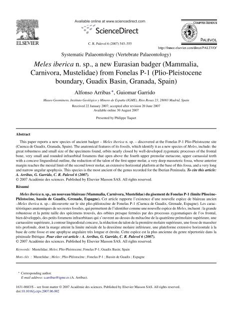
Abstract<br />
Available online at www.sciencedirect.com<br />
C. R. Palevol 6 (2007) 545–555<br />
Systematic Palaeontology (Vertebrate Palaeontology)<br />
<strong>Meles</strong> <strong>iberica</strong> n. sp., a new Eurasian badger (Mammalia,<br />
Carnivora, Mustelidae) from Fonelas P-1 (Plio-Pleistocene<br />
boundary, Guadix Basin, Granada, Spain)<br />
Alfonso Arribas ∗ , Guiomar Garrido<br />
Museo Geominero, <strong>Instituto</strong> <strong>Geológico</strong> y <strong>Minero</strong> <strong>de</strong> <strong>España</strong> (IGME), Ríos Rosas 23, 28003 Madrid, Spain<br />
Received 22 January 2007; accepted after revision 20 June 2007<br />
Available online 30 August 2007<br />
Presented by Philippe Taquet<br />
This paper reports a new species of ancient badger – <strong>Meles</strong> <strong>iberica</strong> n. sp. – discovered at the Fonelas P-1 Plio-Pleistocene site<br />
(Cuenca <strong>de</strong> Guadix, Granada, Spain). The anatomical features of its fossils, which i<strong>de</strong>ntify it as a new species of <strong>Meles</strong>, inclu<strong>de</strong>: the<br />
great robustness and small size of the specimens found, orbits nearly closed by well-<strong>de</strong>veloped zygomatic processes of the frontal<br />
bone, very small and roun<strong>de</strong>d infraorbital foramens that open above the fourth upper premolar metacone, upper carnassial teeth<br />
with a concave linguodistal outline, the reduction of the talon of the first upper molar, a very <strong>de</strong>ep masseteric fossa, whose anterior<br />
margin reaches the mesial limit of the second lower molar, an extensive horizontal platform at the base of this fossa, and a very long<br />
and narrow angular apophysis. This species is the most ancient of the genus recor<strong>de</strong>d for the Iberian Peninsula. To cite this article:<br />
A. Arribas, G. Garrido, C. R. Palevol 6 (2007).<br />
© 2007 Académie <strong>de</strong>s sciences. Published by Elsevier Masson SAS. All rights reserved.<br />
Résumé<br />
<strong>Meles</strong> <strong>iberica</strong> n. sp., un nouveau blaireau (Mammalia, Carnivora, Mustelidae) du gisement <strong>de</strong> Fonelas P-1 (limite Pliocène-<br />
Pléistocène, bassin <strong>de</strong> Guadix, Grena<strong>de</strong>, Espagne). Cet article rapporte l’existence d’une nouvelle espèce <strong>de</strong> blaireau ancien<br />
–<strong>Meles</strong> <strong>iberica</strong> n. sp.– découverte sur le site plio-pléistocène <strong>de</strong> Fonelas P-1 (Cuenca <strong>de</strong> Guadix, Grena<strong>de</strong>, Espagne). Les caractéristiques<br />
anatomiques <strong>de</strong> ses restes fossiles, qui permettent <strong>de</strong> l’i<strong>de</strong>ntifier comme une nouvelle espèce <strong>de</strong> <strong>Meles</strong>, incluent : la gran<strong>de</strong><br />
robustesse et la petite taille <strong>de</strong>s spécimens trouvés, <strong>de</strong>s orbites presque fermées par <strong>de</strong>s processus zygomatiques <strong>de</strong> l’os frontal,<br />
bien développés, <strong>de</strong>s petits foramens infraorbitaux qui s’ouvrent au-<strong>de</strong>ssus du métacône <strong>de</strong> la quatrième prémolaire supérieure, une<br />
carnassière supérieure, à contour linguodistal concave, la réduction du talon <strong>de</strong> la première molaire supérieure, une fosse <strong>de</strong> masséter<br />
très profon<strong>de</strong>, dont la marge atteint la limite mésiale <strong>de</strong> la <strong>de</strong>uxième molaire inférieure, une plateforme extensive horizontale àla<br />
base <strong>de</strong> cette fosse et une apophyse angulaire très longue et étroite. Cette espèce est la plus ancienne du genre répertoriée dans la<br />
péninsule Ibérique. Pour citer cet article : A. Arribas, G. Garrido, C. R. Palevol 6 (2007).<br />
© 2007 Académie <strong>de</strong>s sciences. Published by Elsevier Masson SAS. All rights reserved.<br />
Keywords: Mustelidae; <strong>Meles</strong>; Plio-Pleistocene; Fonelas P-1; Guadix Basin; Spain<br />
Mots clés : Mustelidae ; <strong>Meles</strong> ; Plio-Pléistocène ; Fonelas P-1 ; Bassin <strong>de</strong> Guadix ; Espagne<br />
∗ Corresponding author.<br />
E-mail address: a.arribas@igme.es (A. Arribas).<br />
1631-0683/$ – see front matter © 2007 Académie <strong>de</strong>s sciences. Published by Elsevier Masson SAS. All rights reserved.<br />
doi:10.1016/j.crpv.2007.06.002
546 A. Arribas, G. Garrido / C. R. Palevol 6 (2007) 545–555<br />
1. Introduction<br />
The Fonelas P-1 site, which was discovered in<br />
2000 and has been explored since 2001 as part of the<br />
Proyecto Fonelas [1], has become the most important<br />
site in the Iberian Peninsula with respect to research<br />
into the Plio-Pleistocene transition in a continental setting<br />
(http://www.igme.es/internet/museo/investigacion/<br />
paleontologia/fonelas/in<strong>de</strong>x.htm).<br />
The sedimentary environment and the genetic mo<strong>de</strong>l<br />
of the site (Son<strong>de</strong>o B) have been established [26],<br />
and the biological processes associated with the genesis<br />
of its fossil association (a <strong>de</strong>n/eating place of<br />
hyenids belonging to the species Pachycrocuta brevirostris)<br />
and the structure of the sedimentary matrix<br />
surrounding the fossils have been characterised. The<br />
large mammals represented [2,7] inclu<strong>de</strong> native European<br />
species (Mammuthus meridionalis, Stephanorhinus<br />
etruscus, Eucladoceros sp., Metacervoceros rhenanus<br />
cf. phillisi, Gazellospira torticornis ssp., Homotherium<br />
lati<strong>de</strong>ns, Acinonyx pardinensis, Megantereon cultri<strong>de</strong>ns<br />
ssp., Croizetoceros ramosus ssp., Lynx pardinus<br />
spelaeus, Vulpes alopecoi<strong>de</strong>s, Canis n. sp. and <strong>Meles</strong><br />
<strong>iberica</strong> n. sp.), as well as immigrant species from Asia<br />
and Africa (Leptobos etruscus, Equus cf. major, Canis<br />
etruscus, Mitilanotherium n. sp., Praeovibos sp., Hyaena<br />
brunnea, Potamochoerus n. sp., Pachycrocuta brevirostris,<br />
Canis falconeri and Capra n. sp.). No other such<br />
palaeobiological association is known in Eurasia.<br />
Analysis of the last and first appearance data (LADs<br />
and FADs) for the site’s most important taxa assign the<br />
fossil association a position between the Olivola and<br />
Tasso Faunal Units (FU) [20], or, if the indications of<br />
other authors regarding French fossil associations [15]<br />
are taken into account, in zone MNQ-18. Irrespective<br />
of the biochron used, the Fonelas P-1 site dates from<br />
the Plio-Pleistocene transition; i.e., it is some 1.7–1.9<br />
million years old. However, the fossil association of<br />
the Fonelas P-1 site does not exactly match the associations<br />
of large mammals characteristic of any of the<br />
above-mentioned FUs or MNQs [2].<br />
Eleven species of carnivores are found at the site,<br />
among which there is a single mustelid – a Eurasian<br />
badger – i<strong>de</strong>ntified from three fossils representing two<br />
adults. The genus <strong>Meles</strong> arose in Asia [22] and reached<br />
Europe at the beginning of the Pliocene. The species<br />
<strong>Meles</strong> gennevauxi Viret, 1939, is the most ancient member<br />
of the genus known in Europe; a fossil from the<br />
Lower Pliocene was found in Montpellier (France).<br />
Some authors, however, inclu<strong>de</strong> it in the genus Arctomeles<br />
[8,21]. In contrast to that indicated by Pilgrim [17],<br />
some authors [13,16] propose the genus <strong>Meles</strong> to have<br />
<strong>de</strong>veloped from the genus Melodon during the Upper<br />
Miocene.<br />
To date, the Eurasian badgers of the Plio-Pleistocene<br />
transition were known only by the species <strong>Meles</strong> thorali<br />
Viret, 1950, which has only been found at the<br />
Upper Pliocene sites of Saint-Vallier (France) and Vatera<br />
(Greece), and <strong>Meles</strong> dimitrius Koufos, 1992, discovered<br />
at the Upper Pliocene site of Gerakarou and the Lower<br />
Pleistocene site of Apollonia-1, both in the Mygdonian<br />
basin (Greece). In the Iberian Peninsula, the only known<br />
specimen of <strong>Meles</strong> for this period comes from the Lower<br />
Pleistocene site of Venta Micena. This citation of Cf.<br />
<strong>Meles</strong> sp. [18] was possible due to the finding of a canine<br />
tooth and a fragment of humerus; unfortunately, both<br />
Fig. 1. The skull of <strong>Meles</strong> <strong>iberica</strong> n. sp., holotype FP1-2001-0564.<br />
(A): Oblique, right lateral view; (B): caudal view; (C): oblique left<br />
ventral view; (D): sagittal view. Scale bar = 1 cm.<br />
Fig. 1. Crâne <strong>de</strong> <strong>Meles</strong> <strong>iberica</strong> n. sp., holotype FP1-2001-0564. (A):<br />
Vue oblique latérale droite. (B) : vue caudale ; (C) : vue oblique ventrale<br />
gauche ; (D) : vue sagittale. Barre d’échelle=1cm.
have been lost. Recently, <strong>Meles</strong> sp. has been cited at the<br />
Fuente-Nueva-3 and Barranco León-5 [14] sites, but no<br />
<strong>de</strong>scription nor figure of the material evi<strong>de</strong>nce has been<br />
published.<br />
2. Systematic palaeontology<br />
Or<strong>de</strong>r Carnivora Bowdich, 1821<br />
Family Mustelidae Fischer, 1817<br />
Subfamily Melinae Bonaparte, 1838<br />
Genus <strong>Meles</strong> Brisson, 1762<br />
<strong>Meles</strong> <strong>iberica</strong> n. sp.<br />
Etymology. From Iberica [coming from Iberia], a<br />
Greek word used as a toponym by Herodotus (5th century<br />
B.C.) to <strong>de</strong>signate the Iberian Peninsula, currently<br />
occupied by Spain and Portugal, and home to the Fonelas<br />
P-1 site.<br />
Holotype. FP1-2001-0564, skull with P2-M1 in both<br />
<strong>de</strong>ntal series (Figs. 1–3). Held at the Museo Geominero<br />
(<strong>Instituto</strong> <strong>Geológico</strong> y <strong>Minero</strong> <strong>de</strong> <strong>España</strong>, Ministerio <strong>de</strong><br />
Educación y Ciencia), Madrid, Spain.<br />
Paratypes. FP1-2001-0704, left hemimandible with<br />
i1, c, p2-m1 (Fig. 4); FP1-2002-1446, fragment of left<br />
mandible with right i1 and left i1-i3, c, p1-m1 (Fig. 5).<br />
Held at the Museo Geominero (<strong>Instituto</strong> <strong>Geológico</strong> y<br />
<strong>Minero</strong> <strong>de</strong> <strong>España</strong>, Ministerio <strong>de</strong> Educación y Ciencia),<br />
Madrid, Spain.<br />
A. Arribas, G. Garrido / C. R. Palevol 6 (2007) 545–555 547<br />
Fig. 3. Upper left <strong>de</strong>ntition of <strong>Meles</strong> <strong>iberica</strong> n. sp., holotype FP1-2001-<br />
0564. Scale bar = 1 cm.<br />
Fig. 3. Dentition supérieure gauche <strong>de</strong> <strong>Meles</strong> <strong>iberica</strong> n. sp., holotype<br />
FP1-2001-0564. Barre d’échelle=1cm.<br />
Type locality. Fonelas P-1, Guadix Basin, Granada,<br />
Spain. See [1,26] for <strong>de</strong>tails.<br />
Stratigraphic level. Facies association E (Son<strong>de</strong>o B),<br />
Unit VI (Sistema Axial) at the Guadix Formation, in<br />
accordance with [26].<br />
Geological age. Plio-Pleistocene boundary,<br />
c1.7–1.9 Ma, based on comparisons of the faunal<br />
association with ol<strong>de</strong>r faunas from the Upper Pliocene<br />
of Saint-Vallier (MNQ-17) or Senèze (MNQ-18),<br />
contemporaneous faunas from the Plio-Pleistocene<br />
boundary (Olivola-Tasso Italian FUs), and younger<br />
faunas from the Lower Pleistocene (MNQ-19)<br />
[1,2,7].<br />
Fig. 2. Comparison (frontal view) of the skull of <strong>Meles</strong> <strong>iberica</strong> n. sp. (B) with one of mo<strong>de</strong>rn-day M. meles (A) from Granada (Spain) and one<br />
of a fossil M. meles (C) (Holocene) from the Cueva <strong>de</strong> Los Torrejones (Guadalajara, Spain). Black arrows indicate the position of the infraorbital<br />
foramens (note the large size of these structures in the representatives of M. meles and the small dimensions in <strong>Meles</strong> <strong>iberica</strong> n. sp.). White arrows<br />
indicate the position of the zygomatic process of the frontal bone; note the great <strong>de</strong>velopment and projection of this process in the badger from<br />
Fonelas P-1. The small arrow indicates the position of the sagittal crest (note the height reached by this crest and, especially, its great width in <strong>Meles</strong><br />
<strong>iberica</strong> n. sp.). Specimens A and B represent senile individuals with a great <strong>de</strong>gree of tooth wear, while specimen C represents a senile individual<br />
with mo<strong>de</strong>rate <strong>de</strong>ntal wear. Scale bar = 1 cm.<br />
Fig. 2. Comparaison (vue frontale) du crâne <strong>de</strong> <strong>Meles</strong> <strong>iberica</strong> n. sp. (B) avec <strong>de</strong>s crânes <strong>de</strong> M. meles actuel (A) <strong>de</strong> Grena<strong>de</strong> (Espagne) et <strong>de</strong> M. meles<br />
fossile (C) (Holocène) <strong>de</strong> la Cueva <strong>de</strong> los Torrejones (Guadalajara, Espagne). Les flèches noires indiquent la position <strong>de</strong>s foramens infraorbitaux (à<br />
noter le gran<strong>de</strong> taille <strong>de</strong> ces structures chez M. meles et leurs faibles dimensions chez <strong>Meles</strong> <strong>iberica</strong> n. sp.). Les flèches blanches indiquent la position<br />
du processus zygomatique <strong>de</strong> l’os frontal ; à noter le grand développement et la projection <strong>de</strong> ce processus chez le blaireau <strong>de</strong> Fonelas P-1 La petite<br />
flèche indique la position <strong>de</strong> la crête sagittale (à noter la hauteur atteinte par cette crête et tout spécialement sa gran<strong>de</strong> largeur chez <strong>Meles</strong> <strong>iberica</strong><br />
n. sp.). Les spécimens (A) et(B) représentent <strong>de</strong>s individus séniles avec un grand <strong>de</strong>gré d’usure <strong>de</strong>ntaire, alors que le spécimen (C) représente un<br />
individu sénile à usure <strong>de</strong>ntaire modérée. Barre d’échelle=1cm.
548 A. Arribas, G. Garrido / C. R. Palevol 6 (2007) 545–555<br />
Fig. 4. Hemimandible showing lower left <strong>de</strong>ntition of <strong>Meles</strong> <strong>iberica</strong><br />
n. sp., paratype FP1-2001-0704. (A): Labial view; (B): lingual view;<br />
(C): occlusal view. Scale bar = 1 cm.<br />
Fig. 4. Hémimandibule montrant la <strong>de</strong>ntition inférieure gauche <strong>de</strong><br />
<strong>Meles</strong> <strong>iberica</strong> n sp., paratype FP1-2001-0704. (A) : Vue labiale ; (B):<br />
vue linguale ; (C) : vue occlusale. Barre d’échelle=1cm.<br />
Diagnosis. A small very robust badger; the skull<br />
possesses a broad sagittal crest with a strongly convex<br />
curvature; frontonasal profile mildly concave; orbits<br />
nearly closed by well-<strong>de</strong>veloped zygomatic processes of<br />
the frontal bone; infraorbital foramens very small and<br />
circular and open above the fourth upper premolar metacone;<br />
zygomatic arches with a mild curvature and close<br />
to the skull; palatine fissures in a very forward position.<br />
Presence of P1/p1. p2 with two roots. Noticeable in the<br />
upper <strong>de</strong>ntition are a linguodistally concave P4, a short<br />
M1 with a paracone clearly larger than the metacone,<br />
a lingual notch between the protocone and hypocone, a<br />
labial notch between the metacone and metaconule, and<br />
a talon, little <strong>de</strong>veloped in its distal region. The jaw of<br />
this new species shows a <strong>de</strong>ep, very well <strong>de</strong>veloped masseteric<br />
fossa, the anterior margin of which reaches the<br />
mesial limit of the second lower molar and at its base has<br />
a bony crest that forms an extensive horizontal platform.<br />
The angular apophysis is very large and narrow. The<br />
coronoid process is very graceful with very fine edges.<br />
No supernumerary cusps are seen for the first lower<br />
molar.<br />
Fig. 5. Fragment of a hemimandible and lower left <strong>de</strong>ntition of <strong>Meles</strong><br />
<strong>iberica</strong> n. sp., paratype FP1-2002-1446. (A): Labial view; (B): lingual<br />
view; (C): occlusal view. Scale bar = 1 cm.<br />
Fig. 5. Fragment d’une hémimandibule et <strong>de</strong> la <strong>de</strong>ntition <strong>de</strong> <strong>Meles</strong><br />
<strong>iberica</strong> n. sp. Paratype FP1-2002-1446. (A) : Vue labiale ; (B) : vue<br />
linguale ; (C) : vue occlusale. Barre d’échelle=1cm.<br />
3. Description<br />
3.1. Skull (Figs. 1 and 2)<br />
(Measurements: see Table 1). The skull (specimen<br />
FP1-2001-0564) is probably that of an old individual;<br />
this would seem evi<strong>de</strong>nt from the extreme <strong>de</strong>velopment<br />
of the sagittal crest and the wear shown by the teeth,<br />
which on their occlusal si<strong>de</strong> have lost much of their<br />
enamel. This prevented the position of the majority of<br />
the cusps and their <strong>de</strong>velopment from being <strong>de</strong>termined.<br />
Although the skull is that of an adult, it is very small (total<br />
length = 121.02 mm; condylobasal length = 114.77 mm).<br />
This fossil un<strong>de</strong>rwent a great <strong>de</strong>al of lateral diagenetic<br />
compression. This inclined the sagittal axis of the<br />
skull towards the left (as seen in caudal view), leaving<br />
this si<strong>de</strong> completely <strong>de</strong>formed. In contrast, the right si<strong>de</strong><br />
conserves its original proportions; the following anatomical<br />
notes therefore refer to this si<strong>de</strong>. From a dorsal view,<br />
the most obvious anatomical feature is the great height<br />
(maximum: 7.7 mm) and thickness (maximum: 4.4 mm)<br />
of the sagittal crest, the lateral profile of which is strongly<br />
convex. In its anterior region, this crest gives rise to two
A. Arribas, G. Garrido / C. R. Palevol 6 (2007) 545–555 549<br />
Table 1<br />
Comparison of the condylobasal length of the skull of <strong>Meles</strong> <strong>iberica</strong> nov. sp. with that of mo<strong>de</strong>rn-day M. meles from Europe (in mm)<br />
Tableau 1<br />
Comparaison <strong>de</strong>s mesures <strong>de</strong> longueur condylobasale du crâne (en mm) chez les espèces <strong>Meles</strong> <strong>iberica</strong> nov. sp. et M. meles actuel d’Europe<br />
Origin n Min Max ¯x S.D.<br />
<strong>Meles</strong> <strong>iberica</strong> n. sp. Fonelas P-1 1 114.77<br />
<strong>Meles</strong> thorali Saint-Vallier a 1 120<br />
<strong>Meles</strong> dimitrius Apollonia 1 b 1 125<br />
<strong>Meles</strong> meles Sierra <strong>de</strong> Aralar (Spain) c 19 120.6 135.7 127.27 4.1<br />
Doñana (Spain) d 63 125.90<br />
Ciudad Real (Spain) d 28 126.5<br />
Lugo–Santan<strong>de</strong>r (Spain) d 30 125.6<br />
Great Britain (♂) e 14 121.69 136.74 127.7 4.41<br />
Great Britain (♀) e 9 116.97 133.99 125.73 6.25<br />
Ireland (♂) e 21 122.99 132.92 126.92 2.61<br />
Ireland (♀) e 31 114.63 132.61 125.39 4.18<br />
Poland d 32 130.87<br />
Switzerland d 54 132.96<br />
Italy d 11 125.4<br />
a From [24] ; d’après [24].<br />
b From [11] ; d’après [11].<br />
c From [30] ; d’après [30].<br />
d From [25] ; d’après [25].<br />
e From [6] ; d’après [6].<br />
prominent frontal crests. On its back edge, the sagittal<br />
crest is continuous, with a well-<strong>de</strong>veloped nuchal crest.<br />
The profile of the cranial case cannot be traced accurately,<br />
since compression caused the collapse of part of<br />
this region.<br />
The zygomatic processes of the frontal bone are very<br />
<strong>de</strong>veloped and project strongly towards the lateral region,<br />
leaving at least three quarters of the orbit closed. From<br />
a dorsal view, the zygomatic arch has a mildly convex<br />
profile and lies relatively close to the skull. Behind<br />
this arch lies the opening of the external ear canal.<br />
The frontonasal profile forms a subtly concave plane.<br />
The infraorbital foramen, which is circular in shape,<br />
is very small (transversal width: 2.8 mm; dorsoventral<br />
height: 3.3 mm) and opens above the metacone of<br />
P4.<br />
The tympanic bullae appear too collapsed to infer<br />
any <strong>de</strong>tails of their morphology, although the right<br />
tympanic bulla is triangular-shaped. The main axis is<br />
oblique to the sagittal axis of the skull. The postglenoid<br />
(retroarticular or tympano-occipital) foramen<br />
opens anterior to the external ear canal. The rest of<br />
the ventral foramens cannot be seen due to the great<br />
compaction of the skull in this region. The foramen<br />
magnum appears too <strong>de</strong>formed for its perimeter to be<br />
<strong>de</strong>scribed.<br />
The palatine bone, which also appears very compressed,<br />
shows two fissures close to the sockets of the<br />
first incisors.<br />
3.2. Upper <strong>de</strong>ntition (Figs. 3 and 6)<br />
(Measurements: see Table 2). The upper <strong>de</strong>ntition<br />
shows a great <strong>de</strong>al of wear, ren<strong>de</strong>ring it impossible to<br />
recognise the morphotypes of P4 and M1 established<br />
for the genus [3]. However, M1 shows a well-<strong>de</strong>veloped<br />
labial incision between the metacone and the metaconule<br />
(morphotypes B1 or B2 according to [3]). This wear is<br />
particularly true of P4 and M1, which have completely<br />
lost the enamel of their occlusal surfaces [L (length) C-<br />
M1: 39.0 mm; L P2-M1: 30.6 mm; L P2–P4: 18.9 mm].<br />
Neither the canines nor the incisors have been preserved.<br />
The first upper premolar is also missing, although a very<br />
small socket corresponding to it is visible. The remaining<br />
premolars show two roots, except for P4, which has<br />
three.<br />
The second upper premolar is narrow, with a pyriform<br />
outline, and is mesially thickened. It has a high protocone<br />
in its mesial portion. The third upper premolar has<br />
an oval outline and a high protocone that is more centred<br />
than that of P2, and a well-<strong>de</strong>veloped linguodistal<br />
cingulum that forms a small platform. The distribution<br />
of the cusps in the upper carnassial (which has a triangular<br />
outline) cannot be ma<strong>de</strong> out, although three smooth<br />
notches can be seen at the tooth perimeter that serve to<br />
separate them. One of these notches lies in a labial position<br />
approximately at the centre of the tooth, separating<br />
the paracone and metacone; another lies in a mesial position<br />
between the paracone and protocone, and one lies
550 A. Arribas, G. Garrido / C. R. Palevol 6 (2007) 545–555<br />
Table 2<br />
Comparison of dimensions (in mm) of the upper teeth of <strong>Meles</strong><br />
Tableau 2<br />
Comparaison <strong>de</strong>s mesures <strong>de</strong>ntaires (en mm) chez les espèces <strong>de</strong> <strong>Meles</strong><br />
Upper teeth P2 P3 P4 M1<br />
L w L w L w L Lext w<br />
<strong>Meles</strong> <strong>iberica</strong> n. sp. FP1-2001-0564 4.4 2.9 5.1 3.6 8.9 7.9 12.7 8.7 11.2<br />
<strong>Meles</strong> thorali a QSV 1 5.3 3.7 5.5 4.5 10.6 8.5 14.5 9.6 12.3<br />
QSV 2 5.1 3.8 6.1 3.9 9.2 7.9 13.5 9.1 12.9<br />
QSV 3 13.4 8.2 11.8<br />
QSV 17 6 3.5 6 4.9 10.5 8.5 15 9.2 12<br />
QSV 18 14.2 8.9 12<br />
QSV 1052 13.3 8.8 12<br />
QSV 1055 15 9 12.3<br />
QSV 1056 13.1 8.5 12<br />
<strong>Meles</strong> thorali spelaeus b LV. 92.930 5 3.2 6.2 4.1 10.5 7.5 12.8 9.3 12<br />
<strong>Meles</strong> dimitrius c GER-159 8 7.8 13.3 8.5 12.1<br />
GER-160 5.9 3.9 8.2 8.2 13<br />
GER-163 7 5<br />
<strong>Meles</strong> dimitrius d APL-544 6.2 4.1 8.8 8.1 13.6 8.8 12<br />
APL-545 5.7 4.2 8.9 7.8 13.5 8.5 12<br />
<strong>Meles</strong> hollitzeri e IQW 24619 5.1 2.6 8.3 6.8 13.3 9.7<br />
(n = 3) 8.6 9.3 16.4 10.6 12.7<br />
<strong>Meles</strong> meles (¯x) f<br />
<strong>Meles</strong> meles (¯x) g (n = 23) 8.8 7.5 13.9 11.8<br />
L = total length; w = maximum width; Lext = external length.<br />
L = longueur totale ; w = largeur totale ; Lext = longueur externe.<br />
a Saint-Vallier, France [24].<br />
b Lunel Viel, France [4].<br />
c Gerakarou, Greece [10] ; Gerakarou, Grèce [10].<br />
d Apollonia, Greece [11] ; Apollonia, Grèce [11].<br />
e Untermassfeld, Germany [29] ; Untermassfeld, Allemagne [29].<br />
f Cueva <strong>de</strong> Los Torrejones (Holocene), Spain [5] ; Cueva <strong>de</strong> Los Torrejones (Holocène), Espagne [5].<br />
g Current, Sierra <strong>de</strong> Aralar, Spain [30] ; actuel, Sierra <strong>de</strong> Aralar, Espagne [30].<br />
in a more linguodistal position between the protocone<br />
and metacone. The protocone is very well <strong>de</strong>veloped<br />
lingually and slightly individualized with respect to the<br />
other cusps due to the above-mentioned notches. The<br />
profile of its linguodistal edge is concave. A labial cingulum<br />
can be seen, which at its mesiolabial edge forms<br />
a minuscule cusp continuous with the mesial cingulum.<br />
The first molar has a nearly square outline with a<br />
short, distal projection. The distribution of its cusps can<br />
be appreciated because of the notches that separate them.<br />
That which separates the paracone from the protocone is<br />
well marked at the base of the tooth, while that which separates<br />
the protocone from hypocone is less pronounced.<br />
Although the paracone and metacone are clearly separated,<br />
as are the metacone and the metaconule, the<br />
responsible notches disappear at the base of the tooth.<br />
It is important to note that the paracone is larger than the<br />
metacone, appreciably so from a labial view. The only<br />
cingulum that can be seen is in a labial position; the rest<br />
of the tooth is so worn that, if there ever were any other<br />
features, they have disappeared. The distal edge of the<br />
tooth is straight.<br />
3.3. Lower jaw (Figs. 4 and 5)<br />
The preserved hemimandibles belong to the left si<strong>de</strong><br />
of two individuals at different stages of <strong>de</strong>velopment.<br />
The teeth of specimen FP1-2001-0704 are much worn<br />
(L c-m1: 41.3 mm; L p2-m1: 31.2 mm; L p2–p4:<br />
16.7 mm), as seen in the fossil skull FP1-2001-0564;<br />
they are also of matching size and therefore probably<br />
belong to the same animal (in<strong>de</strong>ed, both fossils were<br />
recovered at the contact between excavation grid squares<br />
E2 and E3). Fossil jaw FP1-2002-1446 shows teeth that<br />
are not very worn (L c-m1: 37.4 mm; L p2-m1: 29.0 mm;<br />
L p2–p4: 15.9 mm). A biostratinomic breakage after m1<br />
prevents observation of the ascending branch’s anatomical<br />
features. However, hemimandible FP1-2001-0704<br />
is completely preserved. In general, the badgers to<br />
which these jaws belonged were of robust composition
Fig. 6. Graphs comparing the total length (L) and maximum width (w)<br />
of the first upper molar (A) and first lower molar (B) of the Fonelas P-1<br />
specimens, the fossil species M. thorali, M. dimitrius and M. hollitzeri,<br />
and two Iberian specimens of M. meles (one mo<strong>de</strong>rn-day, one fossil).<br />
The hemimandible FP1-2001-0704 belongs to the same individual as<br />
skull FP1-2001-0564. The representation of M. thorali is based on<br />
data from Saint-Vallier [24] and Vatera [27], M. dimitrius on data from<br />
Gerakarou [10] and Apollonia [11], and M. hollitzeri on data from<br />
Untermassfeld [29]. The data for M. meles * come from mo<strong>de</strong>rn-day<br />
populations in the Sierra <strong>de</strong> Aralar [30], and those for M. meles ** from<br />
Holocene fossils found in the Cueva <strong>de</strong> Los Torrejones [5].<br />
Fig. 6. Graphiques comparant la longueur totale (L) et la largeur maximum<br />
(w) <strong>de</strong> la première molaire supérieure (A) et <strong>de</strong> la première<br />
molaire inférieure (B) <strong>de</strong>s spécimens <strong>de</strong> Fonelas P-1, <strong>de</strong>s espèces fossiles<br />
M. thorali, M. dimitrius et M. hollitzeri, et <strong>de</strong> <strong>de</strong>ux spécimens<br />
espagnols <strong>de</strong> M. meles (un actuel et un fossile). L’hémi-mandibule FP1-<br />
2001-0704 appartient au même individu que le crâne FP1-2001-0564.<br />
La représentation <strong>de</strong> M. thorali est basée sur les données <strong>de</strong> Saint-<br />
Vallier [24] et <strong>de</strong> Vatera [27], celle <strong>de</strong> M. dimitrius sur les données<br />
<strong>de</strong> Gerakarou [10] et d’Apollonia [11], et celle <strong>de</strong> M. hollitzeri sur<br />
les données d’Untermassfeld [29]. Les données relatives à M. meles *<br />
proviennent <strong>de</strong> populations actuelles <strong>de</strong> la Sierra <strong>de</strong> Aralar [30], et<br />
celles relatives à M. meles ** <strong>de</strong> fossiles holocènes trouvés dans la<br />
Cueva <strong>de</strong> Los Torrejones [5].<br />
and were quite small [mandible height between p4-m1:<br />
12.5 mm (FP1-2001-0704); 12.3 mm (FP1-2001-0564)].<br />
Along the ventral edge of the jaw, and in the<br />
area where the ascending branch begins, the horizon-<br />
A. Arribas, G. Garrido / C. R. Palevol 6 (2007) 545–555 551<br />
tal branch shows a curvature with an inflexion point at<br />
the distal limit of m2. The lingual face of the horizontal<br />
branch shows strong longitudinal thickenings for the<br />
insertion of the mylohyoid and digastric muscles, which<br />
are separated by a smooth furrow. Behind the socket of<br />
m2, there is a vertical lingual crest forming a small platform<br />
that increases the basal surface of the ascending<br />
branch (i.e., it forms a <strong>de</strong>veloped retromolar platform).<br />
The mandibular foramen lies at the central point of the<br />
main axis of the ascending branch. Labially, the horizontal<br />
branch shows a small mental foramen at the distal<br />
boundary of the second lower premolar. Specimen FP1-<br />
2002-1446 also shows a second mental foramen beneath<br />
the contact between the third and fourth lower premolars.<br />
The masseteric fossa is <strong>de</strong>ep and its mesial margin lies<br />
below the mesial limit of the second lower molar. This<br />
fossa is limited in its upper region by a curved crest that<br />
follows the line of the coronoid apophysis. At its lower<br />
edge, the masseteric fossa appears to be boun<strong>de</strong>d by a<br />
crest, very <strong>de</strong>veloped laterally, which forms a wi<strong>de</strong> basal<br />
platform (<strong>de</strong>ep: 5.4 mm). In its posterior region, this<br />
platform has an extensive concavity below the mandibular<br />
condyle where the pterygoid muscle was inserted.<br />
The latter must have been very <strong>de</strong>veloped, since the<br />
angular apophysis is very long (5.4 mm) and narrow<br />
(2.9 mm), and curves slightly upwards. The coronoid<br />
apophysis is very narrow and graceful, with very fine<br />
edges.<br />
3.4. Lower <strong>de</strong>ntition (Figs. 4–6)<br />
(Measurements: see Table 3). The main observations<br />
on the lower <strong>de</strong>ntition were ma<strong>de</strong> using the fossil whose<br />
cusps were least worn (FP1-2002-1446). The canine is<br />
robust, but its crown is very low (height: 8.5 mm). It<br />
is strongly curved and has a sharp vertical, linguodistal<br />
crest, and a less sharp mesiolingual crest. The base<br />
of this tooth is notably thickened mesiodistally. The<br />
first lower premolar is present in specimen FP1-2002-<br />
1446, in which it appears as a small, simple, vestigial<br />
tooth slightly displaced towards the lingual face of the<br />
jaw. Specimen FP1-2001-0704 shows an alveolus for p1.<br />
The second lower premolars show two roots. The second,<br />
third and fourth lower premolars each have a very<br />
high, acute protocone and a distal cingulum that forms a<br />
small platform. The third lower premolar has a mediumsized<br />
mesial cingulum; this is more <strong>de</strong>veloped in p4,<br />
but absent in p2. The second and third lower premolars<br />
are therefore very asymmetric teeth with mesial margins<br />
that fall more abruptly than that of p4, in which<br />
the mesial edge is softened by the presence of the cingulum.<br />
While p3 and p4 are implanted parallel to the
552 A. Arribas, G. Garrido / C. R. Palevol 6 (2007) 545–555<br />
Table 3<br />
Comparison of dimensions (in mm) of lower teeth of <strong>Meles</strong><br />
Tableau 3<br />
Comparaison <strong>de</strong> mesures <strong>de</strong>ntaires inférieures (en mm) chez les espèces <strong>de</strong> <strong>Meles</strong><br />
Lower teeth p2 p3 p4 m1<br />
L w L w L w L Ltrig Ltal w<br />
<strong>Meles</strong> <strong>iberica</strong> n. sp. FP1-2001-0704 4.7 3.5 5.6 3.6 6.1 3.8 15.0 8.8 6.2 6.7<br />
FP1-2002-1446 4.2 3.1 5.2 3.2 6.2 3.4 13.3 7.9 5.6 5.7<br />
<strong>Meles</strong> thorali a QSV 8 4.8 4.1 6.3 3.5 6.5 3.5 15.6 7.5<br />
QSV 13 16.4 9.4 7.2<br />
QSV 16 16.2 9.5 7.1<br />
QSV 19 16.6 9.5 7.7<br />
QSV 1051 17 10 7.8<br />
<strong>Meles</strong> thorali b PO 630 F 17.3 9.4 7<br />
<strong>Meles</strong> dimitrius c GER-161 4.7 3.1 6.8 3.8 15.1 8.5 7.1<br />
GER-162 14.8 8.3 7.2<br />
<strong>Meles</strong> dimitrius d APL-15 4 5.5 3.3 6.3 3.4 15.3 8.5 6.3<br />
APL-546 5.4 3.2 5.8 3.5 7 3.9 16.2 8.8 7.1<br />
<strong>Meles</strong> hollitzeri e IQW 17971 16.4 9.6 7.4 7.3<br />
(n = 4) 4.6 4.0 5.5 3.6 6.5 4.1 17.5 9.3 8.5 8.3<br />
<strong>Meles</strong> meles (¯x) f<br />
<strong>Meles</strong> meles (¯x) g (n = 26) 16.7 7.7<br />
L = total length; w = maximum width; Ltrig = length of the trigonid; Ltal = length of the talonid.<br />
L = longueur totale ; w = largeur totale ; Ltrig = longueur du trigoni<strong>de</strong> ; Ltal = longueur du taloni<strong>de</strong>.<br />
a Saint-Vallier, France [24].<br />
b Vatera, Greece [27]; Vatera, Grèce [27].<br />
c Gerakarou, Greece [10] ; Gerakarou, Grèce [10].<br />
d Apollonia, Greece [11] ; Apollonia, Grèce [11].<br />
e Untermassfeld, Germany [29] ; Untermassfeld, Allemagne [29].<br />
f Cueva <strong>de</strong> Los Torrejones (Holocene), Spain [5] ; Cueva <strong>de</strong> Los Torrejones (Holocène), Espagne [5].<br />
g Current, Sierra <strong>de</strong> Aralar, Spain [30] ; actuel, Sierra <strong>de</strong> Aralar, Espagne [30].<br />
longitudinal axis of the jaw, p2 is slightly oblique to<br />
this, with its distal region displaced towards the lingual<br />
face.<br />
The first lower molar is long and has an amoeboid<br />
outline; it is narrower in its mesial section. It has a trigonid<br />
with a labial protoconid that forms the highest cusp<br />
of the tooth, a mesial paraconid, and a well-<strong>de</strong>veloped<br />
lingual metaconid. No cusp is seen between the protoconid<br />
and the hypoconid. Between the paraconid and<br />
the metaconid, there is a wi<strong>de</strong> space. The talonid of both<br />
specimens is worn, although a small, labial notch can<br />
be seen to separate the hypoconid and the hypoconulid.<br />
Lingually, the entoconid appears worn, as does the entoconulid<br />
and the characteristic small linguodistal cusps.<br />
Surroun<strong>de</strong>d by the hypoconid, hypoconulid, entoconid,<br />
entoconulid, and the set of linguodistal cusps, there is<br />
an almost inappreciable <strong>de</strong>pression due to the advanced<br />
wear suffered by the tooth. The second lower molar has<br />
not been preserved in either specimen, although in FP1-<br />
2001-0704 there is a long, mesiodistal socket that can<br />
hold a fused double root; the root in the mesial position<br />
is larger than that in the distal position (m2 alveolus L:<br />
5.0 mm; w :2.5 mm).<br />
4. Comparisons and discussion<br />
The badger fossils from the Fonelas P-1 site were<br />
compared with fossil and contemporary specimens of<br />
<strong>Meles</strong> meles Linnaeus, 1758 (which appears not to show<br />
sexual dimorphism in its jugal <strong>de</strong>ntition [3]), and with<br />
fossils of M. thorali [23,24], M. dimitrius [10,11], <strong>Meles</strong><br />
hollitzeri Rabe<strong>de</strong>r, 1976 [19,29], <strong>Meles</strong> thorali spelaeus<br />
[4], and <strong>Meles</strong> atavus Kormos, 1914 [9,28].<br />
The cranial profile of the studied skull, with its prominent<br />
frontal crests that join and continue as a powerful<br />
sagittal crest, its large and elongated cranial case, small<br />
orbits, subtriangular tympanic bullae, and a <strong>de</strong>ntition<br />
clearly adapted to an omnivorous diet (<strong>de</strong>ntal formula<br />
3-1-4-1/3-1-4-2) show its owner to have been a badger.<br />
In addition, the <strong>de</strong>ntition has carnassial teeth that have<br />
lost their cutting function, the premolars are very small,<br />
the second and third lower and upper premolars have a<br />
mesial protocone/protoconid, P4 is triangular in section,<br />
and M1 rhomboid. The latter is larger than P4 and its<br />
labial face is shorter than its lingual face. The first lower<br />
molar is long and has a large talonid. These data further<br />
confirm the animal to belong to the genus <strong>Meles</strong>.
However, this animal shows certain peculiarities. The<br />
skull (FP1-2001-0564) has a substantially higher and<br />
thicker sagittal crest (Figs. 1B, 1D and 2B), which is<br />
more markedly convex than that seen in mo<strong>de</strong>rn or fossil<br />
senile specimens of M. meles (sagittal crest, maximum<br />
height: 4.4, 7.3, 8.1 mm; maximum thickness: 2.2, 2.4,<br />
2.7 mm; n = 3), or than that seen in fossil specimens of<br />
M. thorali [23] or M. dimitrius [11]. In addition, the<br />
great <strong>de</strong>velopment shown by the zygomatic process of<br />
the frontal bone was not seen in any of the other current<br />
or fossil taxa analysed; certainly, M. meles, M. thorali<br />
and M. dimitrius have a more open orbit. Neither is the<br />
flattened, subtly concave frontonasal profile seen in any<br />
known fossil badgers, in which this region is slightly convex,<br />
as it is in the mo<strong>de</strong>rn badger M. meles. In addition,<br />
the infraorbital foramens of the studied fossil are much<br />
smaller than those seen in senile M. meles (transversal<br />
width: 8.5, 7.3, 8.8 mm, n = 3; dorsoventral height: 9.2,<br />
7.1, 8.9 mm, n =3) or M. thorali [23], and the palatine<br />
fissures of FP1-2001-0564 are closer to the first incisors<br />
than in M. meles or M. thorali [23].InM. dimitrius, this<br />
region could not be i<strong>de</strong>ntified.<br />
The studied skull also shows zygomatic arches with<br />
a smaller lateral projection than those seen in M. meles<br />
or M. dimitrius [11], although they resemble those of<br />
M. thorali [23,24]. A further similarity with the latter<br />
species is the double rooted P2; this is also seen in M.<br />
hollitzeri [29]. In the mo<strong>de</strong>rn badger, and in M. thorali<br />
spelaeus [4], these roots are fused.<br />
Given the <strong>de</strong>formation of the skull of M. dimitrius<br />
[11], the relative dimensions of the infraorbital foramens<br />
could not be <strong>de</strong>termined, nor the position of the palatine<br />
fissures, nor the number of roots on P2.<br />
It is difficult to <strong>de</strong>termine the distribution of the cusps<br />
of the upper <strong>de</strong>ntition due to their <strong>de</strong>gree of wear. However,<br />
some differences can be seen between this ancient<br />
badger’s <strong>de</strong>ntition and that of other fossil badgers and<br />
of the mo<strong>de</strong>rn Eurasian species. In the studied fossil, the<br />
linguodistal face of P4 is clearly concave, while those of<br />
M. meles and M. thorali are convex. In M. dimitrius,itis<br />
intermediate between the latter forms [11]; its profile is<br />
more or less straight, similar to that seen in M. hollitzeri<br />
[29]. In addition, the M1/P4 ratio is small in the Fonelas<br />
P-1 (<strong>Meles</strong> <strong>iberica</strong> n. sp.) compared to those seen in the<br />
other badger species analysed (Table 2).<br />
The anatomical <strong>de</strong>tails of M1 are difficult to see; only<br />
the general shape can be appreciated due to the wear<br />
it suffered during the animal’s lifetime. This prevents<br />
a proper appreciation of its cusp distribution. The main<br />
difference between this tooth in the studied fossil and that<br />
of M. thorali is the shape of the occlusal face. In the latter<br />
species, the talon is very well <strong>de</strong>veloped and projects<br />
A. Arribas, G. Garrido / C. R. Palevol 6 (2007) 545–555 553<br />
Fig. 7. Outlines of the occlusal faces of the first upper molars belonging<br />
to different members of the genus <strong>Meles</strong> and the new species from<br />
Fonelas P-1. (A): M. thorali;(B): M. hollitzeri;(C): M. meles;(D): M.<br />
dimitrius; (E): <strong>Meles</strong> <strong>iberica</strong> n. sp. Not to scale.<br />
Fig. 7. Aspect <strong>de</strong>s faces occlusales <strong>de</strong>s premières molaires supérieures<br />
appartenant à différents membres du genre <strong>Meles</strong> et à l’espèce nouvelle<br />
<strong>de</strong> Fonelas P-1. (A): M. thorali ;(B): M. hollitzeri ;(C): M. meles ;<br />
(D):M. dimitrius ;(E):<strong>Meles</strong> <strong>iberica</strong> n. sp. Pas àl’échelle.<br />
distally, while in FP1-2001-0564 it is much shorter and<br />
more like that of M. dimitrius (Fig. 7). In general terms,<br />
the labial contact between the trigon and the talon is<br />
more similar to that seen in M. thorali (which has a well<strong>de</strong>fined<br />
notch) or M. dimitrius than to that seen in M.<br />
meles or M. hollitzeri (in which the contact is smoother<br />
and there is no notch) [19]. The studied M1 also shows a<br />
lingual notch between the protocone and hypocone that<br />
is hardly visible in M. thorali, yet quite visible in M.<br />
dimitrius. Another outstanding feature of this tooth in<br />
the studied fossil is that the paracone is much larger than<br />
the metacone, as seen in M. hollitzeri and in the mo<strong>de</strong>rn<br />
M. meles. In contrast, in M. thorali and M. dimitrius,<br />
both cusps are about the same size (Fig. 7).<br />
The jaw of the proposed new species differs from<br />
those of other badgers in terms of the <strong>de</strong>pth and <strong>de</strong>gree<br />
of the mesial projection of the masseteric fossa. In the<br />
studied animal, this projection reaches the mesial limit<br />
of m2, while in M. thorali, M. dimitrius and M. meles, the<br />
fossa closes just after the distal boundary of m2 [11,23].<br />
In M. hollitzeri, it ends un<strong>de</strong>r the mid point of m2 [29].<br />
The extensive basal platform of this fossa is more <strong>de</strong>veloped<br />
in specimen FP1-2001-0704 than in M. thorali or<br />
M. dimitrius, and very much more <strong>de</strong>veloped than in<br />
fossil or current M. meles specimens. In addition, the<br />
mandibular foramen in the Fonelas P-1 fossil lies further<br />
forward than in M. thorali or M. dimitrius [11,23]. The<br />
labial face shows one or two mental foramens. M. thorali<br />
and M. dimitrius both show two aligned foramens, while<br />
in M. meles between 2 and 4 can be seen. The angular<br />
apophysis in the studied jaw fossils is very long and<br />
narrow, unlike in M. thorali, M. dimitrius or M. meles<br />
(fossil or present-day senile specimens: angular apophysis<br />
length: 2.2, 4.5, 2.8 mm; width: 4.5, 4.7, 5.1 mm;<br />
n = 3), in which it is short and wi<strong>de</strong>.<br />
The lower <strong>de</strong>ntition of the studied animal bears no<br />
great morphological differences to that of the other badger<br />
species analysed; other authors also indicate m1<br />
and m2 to be of little help in making diagnoses [3].
554 A. Arribas, G. Garrido / C. R. Palevol 6 (2007) 545–555<br />
The M. thorali fossil from Saint-Vallier shows diastemas<br />
between p2 and p3, and between p3 and p4, which are not<br />
seen in the Fonelas P-1 animal, nor in M. dimitrius [11],<br />
nor in M. meles (either fossil or current). The presence<br />
of p1, and that of two roots for p2, are primitive characteristics<br />
shared with M. thorali, M. hollitzeri, and M.<br />
meles [3]. The accessory cusp between the protoconid<br />
and hypoconid of m1 in M. atavus [9,28] is not visible in<br />
the proposed new species. However, variability has been<br />
recor<strong>de</strong>d in the absence/presence of this supernumerary<br />
cusp in specimens of <strong>Meles</strong> from Gombaszög [12].Given<br />
that this is part of the normal variation seen in M. meles,<br />
it has been proposed that M. atavus be recognised as M.<br />
meles atavus, a subspecies of the nominal taxon. The<br />
socket of m2 in the studied animal is long; its mesiodistal<br />
diameter is greater than its buccolingual diameter,<br />
as seen in M. thorali [23].InM. thorali spelaeus and M.<br />
meles, however, the reverse is true, and this tooth is much<br />
wi<strong>de</strong>r than it is long [4]. The same is seen in M. hollitzeri<br />
[29]. The presence of two fused roots, of which the more<br />
mesial is the larger, is very different to that seen in M.<br />
meles, which has three or more roots. In this respect, the<br />
studied animal is more like M. thorali.<br />
In summary, the studied badger shows a number of<br />
cranial, mandibular, and <strong>de</strong>ntal features that allow it to be<br />
differentiated from other fossil or the mo<strong>de</strong>rn Eurasian<br />
species M. meles, although it shows more anatomical<br />
resemblance to M. thorali. The new animal is the smallest<br />
fossil badger known to date (Tables 1–3, Fig. 6), even<br />
though its fossils are those of robust adults. This size difference<br />
is evi<strong>de</strong>nt in terms of both the length of the skull<br />
(Table 1) and the dimensions of the upper and lower<br />
<strong>de</strong>ntitions (Fig. 6). This contrasts with the similarity in<br />
size between the fossil species M. dimitrius and M. thorali<br />
(the latter is proposed as the ancestor of the nominal<br />
Euroasiatic species [3]) and the fossil and mo<strong>de</strong>rn representatives<br />
of M. meles. The similarity in size between M.<br />
thorali (from the French Upper Pliocene) and M. meles<br />
was first mentioned in the former’s original diagnosis<br />
[23].<br />
5. Conclusions<br />
This paper reports a new, ancient species of Eurasian<br />
badger classified as <strong>Meles</strong> <strong>iberica</strong>. This animal was<br />
particularly robust and small, much more so than any<br />
other European badgers from the Late Pliocene, from<br />
the Plio-Pleistocene boundary, or in<strong>de</strong>ed than the mo<strong>de</strong>rn<br />
Eurasian species M. meles. This new species shows<br />
primitive characteristics in common with M. thorali,<br />
from which it is <strong>de</strong>scen<strong>de</strong>d (some of these characteristics<br />
are also shared with M. meles), along with a mosaic<br />
of specific characteristics very probably related to life<br />
in a warm environment and on soft ground. It has been<br />
known since the first half of the 20th century that, within<br />
the mo<strong>de</strong>rn species M. meles, the Iberian representatives<br />
[which belong to the variety M. meles marianensis<br />
(Graells, 1897)] are smaller and more robust than their<br />
European counterparts, although they do not differ in<br />
terms of their cranial, mandibular or <strong>de</strong>ntal anatomy.<br />
The fossil badger unearthed at Fonelas P-1 represents<br />
a new species, <strong>Meles</strong> <strong>iberica</strong>, the most ancient member of<br />
the genus known in the Iberian Peninsula. Until now, the<br />
ol<strong>de</strong>st fossils of Eurasian badgers found in the Peninsula<br />
were of M. meles from karstic Mid-Pleistocene sites, 1.4<br />
million years younger than the Fonelas P-1 population.<br />
The fossil record for this genus in the Iberian Peninsula<br />
(extension 582,925 km 2 ) suggests that the members of<br />
this species were genetically isolated from other European<br />
populations; <strong>Meles</strong> <strong>iberica</strong> n. sp. is therefore an<br />
en<strong>de</strong>mic species of this Peninsula.<br />
For the time being, the distribution of this new<br />
species is restricted to the Iberian Peninsula, its fossil<br />
remains forming part of the E facies fossil association<br />
(Son<strong>de</strong>o B) of the Plio-Pleistocene boundary at the<br />
Fonelas P-1 site (Guadix Basin, Spain).<br />
Acknowledgements<br />
This work was fun<strong>de</strong>d by research projects IGME<br />
2001-016, IGME 2005-009, and by the ‘Consejería<br />
<strong>de</strong> Cultura <strong>de</strong> la Junta <strong>de</strong> Andalucía’ (project: ‘Estudio<br />
estratigráfico, taxonómico, tafonómico y paleoecológico<br />
<strong>de</strong>l yacimiento <strong>de</strong> macromamíferos <strong>de</strong> Fonelas<br />
(Granada) en el marco faunístico y ambiental <strong>de</strong>l Plio-<br />
Pleistoceno europeo’). The authors thank the members<br />
of the Proyecto Fonelas, whose efforts and enthusiasm<br />
were much appreciated during the excavation work. We<br />
sincerely thank Manuel Berbel Leyva for permission<br />
to work on his property. Emilo Virgós kindly provi<strong>de</strong>d<br />
unpublished data on mo<strong>de</strong>rn badgers. We also thank<br />
Adrian Burton for linguistic assistance. We are greatly<br />
in<strong>de</strong>bted to two anonymous reviewers for their helpful<br />
comments.<br />
References<br />
[1] A. Arribas, J.A. Riquelme, P. Palmqvist, G. Garrido,<br />
R. Hernán<strong>de</strong>z, C. Laplana, J.M. Soria, C. Viseras, J.J. Durán,<br />
P. Gumiel, F. Robles, J. López-Martínez, J. Carrión, Un nuevo<br />
yacimiento <strong>de</strong> gran<strong>de</strong>s mamíferos villafranquienses en la Cuenca<br />
<strong>de</strong> Guadix-Baza (Granada): Fonelas P-1, primer registro <strong>de</strong> una<br />
fauna próxima al límite Plio-Pleistoceno en la Península Ibérica,<br />
Bol. Geol. Miner. 112 (4) (2001) 3–34.
[2] A. Arribas, E. Baeza, D. Bermú<strong>de</strong>z, S. Blanco, J.J. Durán, G. Garrido,<br />
J.C. Gumiel, R. Hernán<strong>de</strong>z, J.M. Soria, C. Viseras, Nuevos<br />
registros paleontológicos <strong>de</strong> gran<strong>de</strong>s mamíferos en la Cuenca <strong>de</strong><br />
Guadix-Baza (Granada): aportaciones <strong>de</strong>l Proyecto Fonelas al<br />
conocimiento sobre las faunas continentales <strong>de</strong>l Plio-Pleistoceno<br />
europeo, Bol. Geol. Miner. 115 (3) (2004) 567–581.<br />
[3] G.F. Baryshnikov, A.Y. Puzachenco, A.V. Abramov, New analysis<br />
of variability of cheek teeth in Eurasian badgers (Carnivora,<br />
Mustelidae, <strong>Meles</strong>), Russ. J. Theriol. 1 (2) (2002) 133–149.<br />
[4] M.F. Bonifay, Carnivores quaternaires du Sud-Est <strong>de</strong> la France,<br />
Mem. Mus. natl Hist. nat., Paris, n.s., Ser. C 21 (2) (1971) 1–377.<br />
[5] J.S. Carrión, G. Gil, E. Rodríguez, M. Gacía-Antón, A. Arribas,<br />
Palynology of badger coprolites from Central Spain, Palaeogeogr.,<br />
Palaeoclimatol., Palaeoecol. 226 (2005) 259–271.<br />
[6] T. Dayan, D. Simberloff, Character displacement, sexual dimorphism,<br />
and morphological variation among British and Irish<br />
mustelids, Ecology 75 (4) (1994) 1063–1073.<br />
[7] G. Garrido, Paleontología sistemática <strong>de</strong> gran<strong>de</strong>s mamíferos<br />
<strong>de</strong>l yacimiento <strong>de</strong>l Villafranquiense superior <strong>de</strong> Fonelas P-1<br />
(Cuenca <strong>de</strong> Guadix, Granada), PhD thesis, Complutense University,<br />
Madrid, 2006 (726 p.).<br />
[8] C.H. Harington, Life at a 3.5 Million Year Old Beaver Pond in<br />
the Canadian Arctic Islands and the Mo<strong>de</strong>rn Scene, Meridian,<br />
Ottowa, 2001, pp. 11–13.<br />
[9] T. Kormos, Drei neue Raubtiere aus <strong>de</strong>n Präglazialschichten <strong>de</strong>s<br />
Somlyohegy bei Püspökfürdo, Mitt. Jahrb. Ung. Geol. Reichsanst.<br />
22 (1914) 223–247.<br />
[10] G.D. Koufos, The Pleistocene carnivores of the Mygdonia basin<br />
(Macedonia, Greece), Ann. Paleontol. 78 (4) (1992) 205–257.<br />
[11] G.D. Koufos, D. Kostopoulos, New carnivore material from the<br />
Plio-Pleistocene of Macedonia (Greece) with a <strong>de</strong>scription of a<br />
new canid, Muench. Geowiss. Abh. 34 (1997) 33–63.<br />
[12] M. Kretzoi, Die Raubtiere von Gombaszög nebst einer Übersicht<br />
<strong>de</strong>r Gesamtfauna, Ann. Hist. -Nat. Mus. Natl. Hung., Pars Mineral.,<br />
Geol. Palaeontol. 31 (1938) 88–157.<br />
[13] B. Kurtén, Pleistocene Mammals of Europe, Wei<strong>de</strong>nfeld and<br />
Nicholson, London, 1968, p. 317.<br />
[14] B. Martínez, M.P. Espigares, S. Ros, Estudio preliminar <strong>de</strong> las<br />
asociaciones <strong>de</strong> gran<strong>de</strong>s mamíferos <strong>de</strong> Fuente Nueva-3 y Barranco<br />
León-5 (Orce, Granada, <strong>España</strong>) (Informe <strong>de</strong> las campañas<br />
<strong>de</strong> 1999–2002), in: El Pleistoceno inferior <strong>de</strong> Barranco León y<br />
Fuente Nueva 3, Orce (Granada). Memoria científica campañas<br />
1999–2002, Consejería <strong>de</strong> Cultura <strong>de</strong> la Junta <strong>de</strong> Andalucía, 2003,<br />
pp. 115–136.<br />
[15] M.R. Palombo, A.M.F. Valli, Remarks on the biochronology of<br />
mammalian faunal complexes from the Pliocene to the Middle<br />
Pleistocene in France, Geol. Rom. 37 (2003-2004) 145–163.<br />
A. Arribas, G. Garrido / C. R. Palevol 6 (2007) 545–555 555<br />
[16] G. Petter, Origine, phylogénie et systématique <strong>de</strong>s blaireaux,<br />
Mammalia 35 (1971) 567–597.<br />
[17] G.E. Pilgrim, Catalogue of the Pontian Carnivora of Europe,<br />
British Museum (Nat. Hist.), London, 1931.<br />
[18] J. Pons-Moyà, Los carnívoros (Mammalia) <strong>de</strong> Venta Micena<br />
(Granada, <strong>España</strong>), Paleontologia i Evolució, mem. esp 1 (1987)<br />
109–128.<br />
[19] G. Rabe<strong>de</strong>r, Die Carnivoren (Mammalia) aus <strong>de</strong>m Altpleistozän<br />
von Deutsch-Altenburg 2. Mit Beiträgen zur Systematik einiger<br />
Musteli<strong>de</strong>n und Cani<strong>de</strong>n, Beitr. Palaeontol. Geol. Oesterr. 1<br />
(1976) 5–119.<br />
[20] P. Raia, P. Piras, T. Kotsakis, Detection of Plio-Quaternary large<br />
mammal communities of Italy. An integration of fossil faunas<br />
biochronology and similarity, Quat. Sci. Rev. 25 (2006) 846–854.<br />
[21] R.H. Tedford, C.H. Harington, An Arctic mammal fauna from the<br />
Early Pliocene of North America, Nature 425 (2003) 388–390.<br />
[22] J. Viret, Monographie paléontologique <strong>de</strong> la faune <strong>de</strong> vertébrés<br />
<strong>de</strong>s sables <strong>de</strong> Montpellier, III Carnivora fissipedia, Trav. Lab.<br />
Geol. Fac. Sci. Lyon 37 (1939) 1–26.<br />
[23] J. Viret, <strong>Meles</strong> thorali n. sp. du loess villafranchien <strong>de</strong> Saint-<br />
Vallier (Drôme), Eclog. Geol. Helv. 43 (3) (1950) 274–287.<br />
[24] J. Viret, Le lœss à bancs durcis <strong>de</strong> Saint-Vallier (Drôme) et sa<br />
faune <strong>de</strong> mammifères villafranchiens, Nouv. Arch. Mus. Hist.<br />
nat. Lyon 4 (1954) 1–200.<br />
[25] E. Virgós, Tejón–<strong>Meles</strong> meles, in: L.M. Carrascal, A. Salvador<br />
(Eds.), Enciclopedia Virtual <strong>de</strong> los Vertebrados Españoles,<br />
Museo Nacional <strong>de</strong> Ciencias Naturales, Madrid, 2005,<br />
http://www.vertebradosibericos.org/.<br />
[26] C. Viseras, J.M. Soria, J.J. Durán, S. Pla, G. Garrido, F. García-<br />
García, A. Arribas, A Large mammals site in a mean<strong>de</strong>ring fluvial<br />
context (Fonelas P-1, Late Pliocene, Guadix Basin, Spain). Sedimentological<br />
keys for its palaeoenvironmental reconstruction,<br />
Palaeogeogr., Palaeoclimatol., Palaeoecol. 242 (2006) 139–168.<br />
[27] J. Vos <strong>de</strong>, J. van <strong>de</strong>r Ma<strong>de</strong>, A. Athanassiou, G. Lyras, P.Y. Sondaar,<br />
M.D. Dermitzakis, Preliminary note on the Late Pliocene<br />
fauna from Vatera (Lesvos, Greece), Ann. Geol. Pays Hell. 39<br />
(A) (2002) 37–70.<br />
[28] T. Wiszniowska, Middle Pleistocene Carnivora (Mammalia) from<br />
Kozi Grzbiet in the Swietokrzyskie Mts, Poland, Acta Zool. Cracov.<br />
32 (14) (1989) 589–630.<br />
[29] M. Wolsan, Remains of <strong>Meles</strong> hollitzeri (Carnivora, Mustelidae)<br />
from the Lower Pleistocene site of Untermassfeld, in: R.D.<br />
Kahlke (Ed.), Das Pleistozän von Untermassfeld bei Meiningen<br />
(Thüringen), 2, Römisch-Germanisches Zentralmuseum, Mainz,<br />
2001, pp. 659–671.<br />
[30] J. Zabala, Biometría <strong>de</strong>l tejón (<strong>Meles</strong> meles L. 1758) en la Sierra<br />
<strong>de</strong> Aralar, Munibe 32 (3–4) (1980) 301–315.