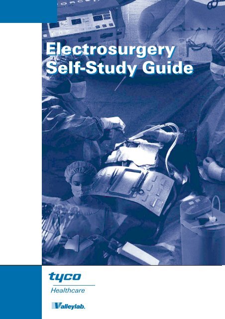
Electrosurgery Self-Study Guide
Electrosurgery Self-Study Guide
Electrosurgery Self-Study Guide

You also want an ePaper? Increase the reach of your titles
YUMPU automatically turns print PDFs into web optimized ePapers that Google loves.
<strong>Electrosurgery</strong><br />
<strong>Self</strong>-<strong>Study</strong> <strong>Guide</strong>
Brenda Cole Ulmer, RN, MN, CNOR<br />
Brenda C. Ulmer is a Senior Clinical Educator for Valleylab. In this capacity<br />
she creates and presents programs of importance to perioperative nurses<br />
and surgeons on topics focusing on patient safety in the operating room.<br />
Prior to this position, Ms. Ulmer was Director of Surgical Services at<br />
Northlake Regional Medical Center in Atlanta, Georgia. Previous work<br />
experience includes staff charge positions at Emory University Hospital in<br />
Atlanta.<br />
Ulmer received an associate degree in nursing in 1974 from DeKalb<br />
College, a bachelors in 1983 from Georgia State University, and a<br />
masters in nursing education from the University of Phoenix in 1996.<br />
She has been a certified nurse of the operating room (CNOR) since<br />
1980. Ulmer is also a member of Sigma Theta Tau’s nursing honor<br />
society.<br />
Ulmer has served on local and national committees for the<br />
Association<br />
of periOperative Registered Nurses (AORN) since 1977. She served<br />
three terms as member of the national AORN Board of Directors, and<br />
was elected President-Elect for AORN on April 1, 1999 in San<br />
Francisco. Since 1995, Ulmer has been working with the AORN,<br />
OSHA, NIOSH, ANA, physicians and nurses to facilitate the publication<br />
of workplace safety guidelines<br />
from OSHA on smoke evacuation. OSHA signed off the draft<br />
guidelines internally in the spring of 1998. They are currently under<br />
final review for publication in the Federal Register in 1999.<br />
From 1989 to 1994, Ulmer was a member of the CBPN Board of<br />
Directors (CBPN Formerly NCB: PNI). She served as President 1990-<br />
91 and 1993-94. During her tenure the Board published its first<br />
CNOR <strong>Study</strong> <strong>Guide</strong>, and started a Registered Nurses First Assistant<br />
Certification exam (CRNFA).<br />
Ms. Ulmer has presented numerous programs on nursing issues<br />
throughout the world. In 1983 she published the first nursing text on<br />
ambulatory surgery: Ambulatory Surgery: A <strong>Guide</strong> to Perioperative<br />
Nursing Care. She is widely published in nursing journals and textbooks.
ELECTROSURGERY<br />
SELF-STUDY GUIDE<br />
September, 1999<br />
A self-study guide intended to assist<br />
surgeons, perioperative nurses and<br />
other healthcare team members to<br />
provide safe and effective patient care<br />
during electrosurgery procedures.<br />
by<br />
Brenda C. Ulmer, RN, MN, CNOR<br />
<strong>Electrosurgery</strong> <strong>Self</strong>-<strong>Study</strong> <strong>Guide</strong><br />
3
<strong>Electrosurgery</strong> <strong>Self</strong>-<strong>Study</strong> <strong>Guide</strong><br />
Objectives<br />
Upon completion of this activity the participant will be able to:<br />
4<br />
1.) Relate the properties of electricity to the clinical<br />
applications of electrosurgery.<br />
2.) Review four of the variables the surgeon controls<br />
that impact surgical effect.<br />
3.) Discuss the relationship between current<br />
concentration and tissue effect.<br />
4.) Identify potential patient injuries related to electro<br />
surgery and technological advances designed to<br />
eliminate these problems.<br />
Accreditation<br />
Statements<br />
This activity has been planned and implemented in<br />
accordance with the Essentials and Standard of the<br />
Accreditation Council for Continuing Medical Education by<br />
Valleylab. Valleylab is accredited by the ACCME to provide<br />
continuing medical education for physicians<br />
Valleylab designates this educational activity for a<br />
maximum of 2 hours in Category 1 credit toward the AMA<br />
Physician’s Recognition Award. Each physician should<br />
claim only those hours of credit that he/she actually spent<br />
in the educational activity.<br />
This offering for two (2) contact hours was designed by the<br />
Clinical Education Department of Valleylab, which is an<br />
approved provider of nursing continuing education (CE) in:<br />
Colorado: Valleylab is approved as a provider of<br />
continuing education in nursing by the<br />
Colorado Nurses’ Association, which is<br />
accredited as an approver of continuing<br />
education in nursing by the American<br />
Nurses Credentialing Center’s<br />
Commission on Accreditation.<br />
Additional Valleylab provider numbers:<br />
California: #CEP 12764<br />
Iowa: #289<br />
Florida: #27I1796<br />
Kansas: #LT0134-0327<br />
Disclaimer Statement<br />
CNA “approved” refers to recognition of educational<br />
offerings only and does not imply approval or<br />
endorsement of any product of Valleylab.<br />
A certificate of completion pertains only to the participant’s<br />
completion of the self-study guide and does not, in any way,<br />
attest to the clinical competence of any participant.<br />
Verification of<br />
Completion<br />
A certificate will be provided to each person who<br />
completes the offering. The certificate pertains only to<br />
completion of the offering and is intended for recordkeeping<br />
purposes.<br />
Instructions<br />
1. Complete the test questions and check the correct<br />
responses with the key. A score of 70% or better is<br />
recommended. Please review the self-study guide<br />
portions related to missed questions, if necessary.<br />
2. Complete the registration form.<br />
3. Complete the evaluation form.<br />
4. Send the completed registration/evaluation form<br />
with the test question responses and $15.00 fee to:<br />
Clinical Education Department A50<br />
Valleylab<br />
5920 Longbow Drive<br />
Boulder, CO 80301-3299<br />
For information call: 800-255-8522 EXT. 6240<br />
5. Upon receipt of the required documents a certificate<br />
of completion for two (2) credit/contact hours will be<br />
sent. Please allow 3–4 weeks for the certificate to be<br />
mailed after submission of required paperwork.<br />
Valleylab will maintain a record of your continuing<br />
education credit and provide verification if necessary<br />
for five (5) years.<br />
Expiration Date<br />
This offering was originally produced for distribution in<br />
September 1997. It was revised in September 1999 and<br />
cannot be used after September 2002 without being<br />
updated. Therefore, credit will not be issued after<br />
September 30, 2002.<br />
Note: The terms listed in the glossary are printed in bold in<br />
the body of the text of the self-study guide.<br />
Key Concepts<br />
Electricity can be hazardous. Understanding how<br />
electricity behaves and relates to electrosurgical function<br />
and applications can contribute to its safe use. Many<br />
different types of electrosurgical technologies are in use in<br />
the surgical setting. The surgeon, perioperative nurse and<br />
other healthcare team members must be aware of the<br />
implications for use for each technology in order to ensure<br />
safe patient care.
Knowledge of the preoperative, intraoperative and<br />
postoperative medical and nursing considerations and<br />
interventions can impact positive patient outcomes.<br />
Overview<br />
<strong>Electrosurgery</strong> came into wide use because of the urgent<br />
need to control bleeding in operative procedures. There have<br />
always been safety concerns when electricity is used in a<br />
therapeutic manner. This module will cover the fundamentals<br />
of electricity, principles of electrosurgery, clinical applications,<br />
technologies, recommended practices and nursing care<br />
during electrosurgery.<br />
Introduction<br />
The use of heat to stop bleeding goes back hundreds of years.<br />
Over the years researchers constructed a variety of devices<br />
which used electricity as a means to heat tissue and control<br />
bleeding. <strong>Electrosurgery</strong> did not come into general use until<br />
the late 1920’s. A neurosurgeon, Harvey Cushing, worked<br />
with a physicist, William T. Bovie, in efforts to develop a<br />
means to stop hemorrhage with electricity. Cushing had tried<br />
implanting muscle tissue, using bone wax, fibrin clots, and<br />
silver clips, but still had patients who could not be operated<br />
on safely because of the threat of uncontrolled bleeding. In<br />
1926, using Bovie’s device, Cushing applied high frequency<br />
current during a neurosurgical procedure. The results were<br />
excellent. Cushing then started bringing back his previously<br />
inoperable patients because he at last had a means to prevent<br />
death from hemorrhage in the operating room (Pearce,<br />
1986). Cushing and Bovie widely published their work. As a<br />
result electrosurgical units came into general use in operating<br />
rooms around the world. The early units were big, ground<br />
referenced units called Bovie’s. The technology remained<br />
virtually unchanged until 1968 when a small, solid state unit<br />
was developed by Valleylab. The solid state units heralded<br />
the modern era of isolated outputs, complex waveforms, and<br />
hand-activated controls (Pearce).<br />
Fundamentals<br />
of Electricity<br />
Electricity is a phenomenon arising from the existence of<br />
positively and negatively charged particles, which make<br />
up all matter. All matter is made up of atoms. Atoms<br />
consist of electrons (negatively charged), protons<br />
(positively charged), and neutrons (neutral) particles.<br />
Atoms that contain equal numbers of electrons and<br />
protons are charge neutral. When forces are introduced<br />
that cause electrons to leave their base atoms and move<br />
to other atoms, the charges of the atom are changed:<br />
those with fewer electrons than protons become<br />
<strong>Electrosurgery</strong> <strong>Self</strong>-<strong>Study</strong> <strong>Guide</strong><br />
positively charged, and atoms with more electrons than<br />
protons become negatively charged. During movement,<br />
like charges repel each other and unlike charges attract.<br />
This electron movement is termed electricity. There are<br />
two natural properties of electricity that can impact<br />
patient care in the operating room. Electricity, which<br />
moves at nearly the speed of light, will, (1) always follow<br />
the path of least resistance; and, (2) always seek to return<br />
to an electron reservoir like the ground (Chernow, 1993).<br />
Electrical current is produced as electrons flow through a<br />
conductor. Electron flow is measured in amperes, or<br />
amps. The path electricity follows as it makes its way<br />
through a conductor and back to ground is the electrical<br />
circuit.<br />
Electricity Variables<br />
There are variables associated with electricity that must<br />
be considered when using it in a therapeutic manner.<br />
Current<br />
The two types of current that are used in the operating<br />
room are direct current (DC), and alternating current (AC).<br />
Direct current uses a simple circuit and electron flow is<br />
only in one direction. Batteries employ this simple circuit.<br />
Energy flows from one terminal on the battery and must<br />
return to the other terminal to complete the circuit.<br />
Alternating currents (AC) switch, or alternate, direction of<br />
electron flow. The frequency of these alterations is<br />
measured in cycles per second or Hertz (Hz), with one<br />
Hertz being equal to one cycle per second. Household<br />
current, in the United States, alternates at 60 cycles per<br />
second, as does much of the electrical equipment used in<br />
operating rooms. Alternating current at 60 Hertz causes<br />
tissue reaction and damage. Neuromuscular stimulation<br />
ceases at about 100,000 Hertz, as the alternating currents<br />
move into the radio frequency (RF)range (Grundemann,<br />
1995).<br />
Resistance<br />
Resistance is the opposition to the flow of current.<br />
Resistance or impedance is measured in ohms. During<br />
the use of electrosurgery one source of resistance or<br />
impedance is the patient.<br />
5
<strong>Electrosurgery</strong> <strong>Self</strong>-<strong>Study</strong> <strong>Guide</strong><br />
Voltage<br />
Voltage is the force that will cause one amp to flow<br />
through one ohm of resistance. It is measured in volts.<br />
The voltage in an electrosurgical generator provides the<br />
electromotive force that pushes electrons through the<br />
circuit.<br />
Power<br />
Power is the energy produced. The energy is measured in<br />
watts. In electrosurgical generators the power setting<br />
used by the surgeon is either displayed on an LED screen<br />
in watts; or, a percentage of the total wattage as indicated<br />
on a numerical dial setting (Lister, 1984).<br />
Principles of<br />
<strong>Electrosurgery</strong><br />
Electrocautery<br />
The electrocautery device is the simplest electrical<br />
system used in the operating room today. It uses battery<br />
power to generate a simple direct current (DC). The<br />
current never leaves the instrument to travel through the<br />
patient’s tissue. An example is the small hand-held eye<br />
cautery (Figure 1). The battery heats up a wire loop at the<br />
end of the device. The cautery is useful in ophthalmic<br />
surgery and other very minor procedures in which very<br />
little bleeding is encountered. Its use is limited because it<br />
cannot cut tissue or coagulate large bleeders. It is further<br />
limited because the target tissue tends to stick to the<br />
electrode.<br />
The term “electrocautery,” or “cautery,” is often, and<br />
incorrectly, used to describe all types of electrosurgical<br />
devices. Its use is only appropriate to describe the simple<br />
direct current cautery device.<br />
Figure 1 – Electrocautery<br />
6<br />
<strong>Electrosurgery</strong> and Radio Frequency<br />
Current<br />
A frequently asked question is why electrosurgery<br />
generators do not shock patients. The answer is because<br />
of the higher frequencies at which generators operate.<br />
Radio frequency current alternates so rapidly that cells do<br />
not react to this current. AM radio stations operate in the<br />
550 to 1500 KiloHertz (kHz) range. <strong>Electrosurgery</strong><br />
generators operate in the 200 kHz to 3.3 megahertz<br />
(MHz) range (Figure 2). Both of these are well above the<br />
range where neuromuscular stimulation or electrocution<br />
could occur (Harris, 1978).<br />
60 Hz 100 kHz 550 - 1550 kHz 54 - 880 M<br />
Household<br />
Appliances<br />
Muscle and Nerve<br />
Stimulation Ceases<br />
<strong>Electrosurgery</strong><br />
Figure 2 – Frequency Spectrum<br />
200 kHz - 3.3 MHz<br />
AM Radio TV
Bipolar <strong>Electrosurgery</strong><br />
Bipolar electrosurgery is the use of electrical current<br />
where the circuit is completed by using two parallel poles<br />
located close together. One pole is positive and the other<br />
is negative. The flow of current is restricted between<br />
these two poles. These are frequently tines of forceps, but<br />
may also be scissors or graspers. Because the poles are in<br />
such close proximity to each other, low voltages are used<br />
to achieve tissue effect. Most bipolar units employ the cut<br />
waveform because it is a lower voltage waveform, and<br />
achieves hemostasis without unnecessary charring.<br />
Because the current is confined to the tissue between the<br />
poles of the instrument and does not flow through the<br />
patient, a patient return electrode is not needed (Figure<br />
3).<br />
Figure 3 – Bipolar Circuit<br />
Bipolar is a very safe type of electrosurgery. There are<br />
some disadvantages to the use of bipolar, especially in the<br />
microbipolar mode. Bipolar cannot spark to tissue, and<br />
the low voltage makes it less effective on large bleeders<br />
(Mitchell, 1978). However, there are newer bipolar<br />
systems that incorporate a “macro” or bipolar “cut” mode<br />
that has higher voltage and is designed for use with the<br />
new generation of bipolar cutting instruments.<br />
Bipolar, especially microbipolar, has been widely used in<br />
neurosurgery and gynecologic surgery. It may be safer to<br />
use when there is a question about the efficacy of using<br />
more powerful monopolar electrosurgical units (e.g., with<br />
pacemakers, implanted automatic cardiac defibrillators).<br />
Monopolar <strong>Electrosurgery</strong><br />
The most frequently used method of delivering<br />
electrosurgery is monopolar, because it delivers a greater<br />
range of tissue effects. In monopolar electrosurgery, the<br />
generator produces the current, which travels through an<br />
active electrode and into target tissue. The current then<br />
passes through the patient’s body to a patient return<br />
electrode where it is collected and carried safely back to<br />
<strong>Electrosurgery</strong> <strong>Self</strong>-<strong>Study</strong> <strong>Guide</strong><br />
the generator (Figure 4). This is the intended pathway for<br />
the electrical current. The type of monopolar generator<br />
used, along with appropriate surgeon and nursing<br />
interventions can help to ensure that this is the path the<br />
current takes through the patient.<br />
Figure 4 – Monopolar Circuit<br />
Current Concentration/Density<br />
The purpose of both methods of delivery is to produce<br />
heat for a desired surgical effect. When current is<br />
concentrated, heat is produced. The amount of heat<br />
produced determines the extent of the tissue effect.<br />
Current concentration or density depends on the size of<br />
the area through which the current flows. A small area<br />
that concentrates the current offers more resistance,<br />
which necessitates more force to push the current<br />
through the limited space and therefore generates more<br />
heat. A large area offers less resistance to the flow of<br />
current reducing the amount of heat produced (Figure 5).<br />
Low Current<br />
Concentration<br />
Figure 5 – Current Concentration/Density<br />
High Current<br />
Concentration<br />
7
<strong>Electrosurgery</strong> <strong>Self</strong>-<strong>Study</strong> <strong>Guide</strong><br />
Electrosurgical Mode<br />
Differentiation<br />
Electrosurgical generators can produce current in three<br />
different modes, each with distinct tissue effects. The<br />
modes are cut, fulguration and desiccation.<br />
Electrosurgical Cut<br />
The cutting current is a continuous waveform (Figure 6).<br />
Figure 6 – Cut Waveform<br />
Since the delivery of current is continuous, much lower<br />
voltages are required to achieve the desired effect of<br />
tissue vaporization. To achieve a cutting effect, the tip of<br />
the active electrode should be held just over the target<br />
tissue. The current vaporizes cells in such a way that a<br />
clean tissue cut is achieved (Figure 7). The cut mode of<br />
the generator is also a good choice for achieving<br />
coagulation of tissue through desiccation.<br />
Figure 7 – Vaporization or Cutting<br />
Blended Mode<br />
The blended mode on a generator is a function of the cut<br />
waveform. Blended currents produce voltages higher<br />
than that of the pure cut mode. The generator output is<br />
modified to a dampened waveform that produces some<br />
hemostasis during cutting (Tucker, 1998). There are<br />
several blended waveforms that provide different ratios of<br />
coagulation with the cutting current by modifying the<br />
8<br />
PURE CUT<br />
100% on<br />
duty (on/off) cycle (Figure 8). Examples of blend<br />
waveforms from different generators are:<br />
Blend 1 = 50% on/50% off<br />
Blend 2 = 40% on/60% off<br />
Blend 3 = 25% on/75% off<br />
Variations of cut to coagulation blends may occur among<br />
manufacturers. Generally, however, a higher blend<br />
number means more coagulation.<br />
Figure 8 – Blended Waveforms<br />
Fulguration<br />
BLEND 1 BLEND 2 BLEND 3<br />
50% on<br />
50% off<br />
40% on<br />
60% off<br />
Tissue fulguration is achieved with the coagulation mode<br />
on the generator. The coagulation mode produces an<br />
interrupted or dampened waveform with a duty cycle that<br />
is on about 6% of the time (Figure 9).<br />
COAG<br />
6% on<br />
94% off<br />
Figure 9 – Coagulation Waveform<br />
25% on<br />
75% off<br />
The coagulation current produces spikes of voltage as<br />
high as 5000 volts at 50 watts. The tissue is heated when<br />
the waveform spikes, and cools down in between spikes,<br />
thus producing coagulation of the cell during the 94% off<br />
cycle of he waveform.
The correct method for achieving fulguration when using<br />
coagulation is to hold the tip of the active electrode<br />
slightly above the target tissue (Figure 10).<br />
Figure 10 – Fulguration<br />
Desiccation<br />
Electrosurgical desiccation can be produced using either<br />
the cut or coagulation mode on the generator. The<br />
difference between fulguration and desiccation is that<br />
the tip of the active electrode must contact the target<br />
tissue in order to achieve desiccation (Figure 11). The<br />
desired mode to achieve tissue desiccation through direct<br />
contact is the cut waveform.<br />
CUT<br />
Low voltage<br />
waveform<br />
100% duty<br />
cycle<br />
Figure 11 – Desiccation<br />
COAG<br />
High voltage<br />
waveform<br />
6% duty cycle<br />
Other Variables That Impact Tissue Effect<br />
The electrosurgical mode that the surgeon uses has a<br />
definite impact on patient tissue. There are other variables<br />
that can also alter the outcome of the electrosurgical<br />
tissue effect:<br />
TIME – The length of time that the surgeon uses the active<br />
electrode determines the amount of tissue effect. Too long<br />
an activation time will produce wider and deeper tissue<br />
damage. Too short an activation time will not produce the<br />
desired tissue effect.<br />
<strong>Electrosurgery</strong> <strong>Self</strong>-<strong>Study</strong> <strong>Guide</strong><br />
POWER – The power setting the surgeon uses will alter<br />
tissue effect. The surgeon should always use the lowest<br />
setting to achieve the desired tissue effect. The setting will<br />
differ from patient to patient. Muscular patients of weight<br />
and height in proper portions will require lower power<br />
settings than obese or emaciated patients. The location of<br />
the return electrode in relation to the surgical site will<br />
impact the power required to overcome the amount of<br />
tissue mass (resistance) between the two (2) sites.<br />
ELECTRODE – The size of the active electrode influences<br />
the tissue effect of the generator. A large electrode needs<br />
a higher power setting than a smaller one because the<br />
current is dispersed over a wider surface area (Figure 12).<br />
A clean electrode will need less power to do the same<br />
work as a dirty one because eschar buildup has higher<br />
resistance and will hamper the passage of the current.<br />
Electrodes can be coated with Teflon (PTFE) or an<br />
elastomeric silicone coating to reduce eschar buildup.<br />
These coated blades wipe clean with a sponge and<br />
eliminate that need for a “scratch pad” which causes<br />
grooves on stainless steel electrodes that may contribute<br />
to eschar build-up. These different types of blades should<br />
be evaluated because some will enable the surgeon to use<br />
lower power settings, reducing the potential for thermal<br />
spread. In addition, some coated electrodes are bendable,<br />
have a non-flake coating and retain their cleaning<br />
properties longer than other coated electrodes.<br />
Low current<br />
concentration<br />
(density)<br />
increases<br />
Current Concentration & Heat<br />
decreases increases<br />
Electrode size<br />
Power Setting Requirement<br />
decreases<br />
High current<br />
concentration<br />
(density)<br />
Figure 12 – Current Concentration Effect on Power Settings<br />
TISSUE – Patient tissue can determine the effectiveness<br />
of the generator. The physical characteristics of the<br />
patient’s body will determine the amount of impedance<br />
encountered by the electrosurgical current as it attempts<br />
to complete the circuit through the return electrode to the<br />
generator. A lean muscular patient will conduct the<br />
electrosurgical current much better than an obese or an<br />
emaciated patient.<br />
9
<strong>Electrosurgery</strong> <strong>Self</strong>-<strong>Study</strong> <strong>Guide</strong><br />
Electrosurgical<br />
Technologies<br />
As the sophistication of surgical procedures has evolved<br />
over time, so too have electrosurgical technologies.<br />
Meeting the challenge of improved patient care is but one<br />
of the goals of the medical manufacturing partner within<br />
the health care arena. The surgeon and perioperative<br />
nurse must be familiar with both long-standing<br />
technologies and with emerging ones so that the safest<br />
and most effective care is available to patients.<br />
Ground-Referenced Generators<br />
The first generation of electrosurgery units that were<br />
developed in the early 1900’s were ground-referenced.<br />
When using these units, it is earth ground that completes<br />
the electrosurgical circuit. These units were spark-gap<br />
systems with high output and high performance<br />
(Hutchisson, 1998), making them popular with surgeons.<br />
The major hazard when using a grounded system is that<br />
current division can occur. If the electrical current finds<br />
an easier (lower resistance) and quicker way to return to<br />
ground, and if the current were sufficiently concentrated,<br />
the patient could be burned at any point where the current<br />
exits the patient’s body (Figure 13).<br />
Figure 13 – Current Division<br />
This could be where the patient’s hand touches the side of<br />
the OR bed, a knee touches a stirrup, or any number of<br />
alternate exit sites (Figure 14).<br />
10<br />
Figure 14 – Alternate Site Burn<br />
In early models of ground-referenced generators, the<br />
surgeon could use electrosurgery regardless of whether a<br />
patient return electrode was attached to the patient. This<br />
resulted in patient burns. Later models offered a cord fault<br />
alarm, which alerted the staff if the patient return<br />
electrode was not plugged into the generator. The<br />
disadvantage was that electrosurgery could still be used<br />
even if the patient return electrode was not on the<br />
patient. Burns could also occur at the return electrode site.<br />
In 1995 the Emergency Care Research Institute (ECRI)<br />
stated that “spark-gap units are outdated and have been<br />
largely superseded by modern technology”. The Institute<br />
recommended that hospitals with surgeons who still insist<br />
on using ground-referenced units should obtain signed<br />
statements from these surgeons acknowledging the risk<br />
of using spark-gap electrosurgical units (Kirshenbaum,<br />
1996).<br />
Solid-State Generators<br />
Solid-state generators were introduced in 1968. These<br />
units were much smaller and used “isolated” circuitry. In<br />
isolated units, the electrical current produced by the<br />
generator is referenced to the generator and will ignore all<br />
grounded objects that may touch the patient except the<br />
return electrode. With isolated generators current<br />
division cannot occur and there is no possibility of<br />
alternate site burns (Figure 15).
Figure 15 – Isolated Circuit<br />
An isolated generator will not work unless the patient<br />
return electrode is attached to the patient. However,<br />
without additional safety features, the generator cannot<br />
determine the status of the contact at the pad/patient<br />
interface. Should the patient return electrode be<br />
compromised in the quantity or quality of the pad/patient<br />
interface in some way during surgery, a return electrode<br />
burn could occur (Figure 16). The perioperative nurse<br />
must be certain that the patient return electrode is in<br />
good contact with the patient throughout the surgical<br />
procedure.<br />
Figure 16 – Pad Site Burn<br />
Contact Quality Monitoring<br />
Patient return electrodes employing a contact quality<br />
monitoring system were introduced in 1981. Contact<br />
quality monitoring uses a split pad system whereby an<br />
interrogation current constantly monitors the quality of the<br />
contact between the patient and the patient return<br />
electrode (Figure 17).<br />
<strong>Electrosurgery</strong> <strong>Self</strong>-<strong>Study</strong> <strong>Guide</strong><br />
Figure 17 – Return Electrode Monitoring System<br />
If a condition develops at the patient return electrode<br />
site that could result in a patient burn, the system<br />
inactivates the generator while giving audible and visual<br />
alarm signals. This represents a major safety device for<br />
patients and perioperative personnel since return<br />
electrode burns account for a majority of patient burns<br />
during electrosurgery. According to ECRI many<br />
electrosurgery burns could be eliminated by a patient<br />
return electrode quality contact monitoring system (ECRI,<br />
1999).<br />
Monopolar Electrosurgical Hazards<br />
During Minimally Invasive Surgery<br />
In the past few years the number of laparoscopic<br />
procedures has risen dramatically and is expected to<br />
continue (Figure 18) (MDI, 1996).<br />
1800<br />
1400<br />
1200<br />
1000<br />
800<br />
600<br />
Procedure Volume in 000’s 1600<br />
400<br />
200<br />
0<br />
Estimated Increase in Laparoscopy for the Six Leading<br />
General Surgical Procedures by Volume<br />
1994 2000<br />
Open<br />
Laparoscopic<br />
Figure 18 – Estimated Increase in Laparoscopy for the Six<br />
Leading General Surgical Procedures by Volume<br />
11
<strong>Electrosurgery</strong> <strong>Self</strong>-<strong>Study</strong> <strong>Guide</strong><br />
This increase has brought concerns about monopolar<br />
electrosurgery when used during minimally invasive<br />
procedures. There have been reports of severe illness and<br />
death following laparoscopy where use of electrosurgery<br />
has been implicated as a cause (ECRI, 1995). The hazards<br />
associated with endoscopic electrosurgery use are:<br />
12<br />
• direct coupling<br />
• insulation failure<br />
• capacitive coupling<br />
Each of these can cause severe patient injury if the current<br />
is highly concentrated at the point of contact.<br />
In determining when and how these hazards occur, it is<br />
useful to divide the active electrode and cannula system<br />
into four zones (Figure 19).<br />
One intended<br />
Three unintended<br />
Zone 1 Zone 2 Zone 3 Zone 4<br />
Figure 19 – Four Zones of Injury<br />
Zone 1 is the area at the tip of the active electrode in<br />
view of the surgeon. Zone 2 encompasses the area just<br />
outside the view of the surgeon to the end of the cannula.<br />
Zone 3 is the area of the active electrode covered by the<br />
cannula system. This is also outside the view of the<br />
surgeon. Zone 4 is the portion of the electrode and<br />
cannula that is outside the patient’s body. The greatest<br />
concern is the unseen incidence of stray electrosurgery<br />
current in Zones 2 and 3 (Harrell, 1998).<br />
Direct coupling occurs when the active electrode is<br />
activated in close proximity or direct contact with other<br />
conductive instruments within the body. This can occur<br />
within Zones 1, 2, or 3. If it occurs outside the field of<br />
vision and the current is sufficiently concentrated a patient<br />
injury can occur.<br />
Insulation failure occurs when the coating that is applied<br />
to insulate the active electrode is compromised. This can<br />
happen in multiple ways that range from repeated uses to<br />
rough handling to using very high voltage current. There is<br />
also concern that some active electrodes may not meet<br />
the standards for electrosurgical devices set by the<br />
Association for the Advancement of Medical<br />
Instrumentation (AAMI), and the American National<br />
Standards Institute (ANSI). Insulation failure that occurs<br />
in Zones 2 or 3 could escape detection by the surgeon<br />
and cause injury to adjacent body structures if the current<br />
is concentrated (Figure 20).<br />
Figure 20 – Insulation Failure<br />
Capacitive coupling is the least understood of the<br />
endoscopic electrosurgical hazards. Anytime two<br />
conductors are separated by an insulator a capacitor is<br />
created. For example, a capacitor is created by inserting<br />
an active electrode, surrounded by its insulation, down a<br />
metal cannula (Figure 21).<br />
Abdominal Wall<br />
Laparoscopic View<br />
Conductor<br />
(Electrode Tip)<br />
Insulator<br />
(Electrode Insulation)<br />
Conductor<br />
(Metal Cannula)<br />
Figure 21 – Instrument/Metal Cannula Configuration<br />
Capacitively coupled electrical current can be transferred<br />
from the active electrode, through intact insulation and<br />
into the conductive metal cannula. Should the cannula<br />
then come in contact with body structures, that energy<br />
can be discharged into these structures and damage them<br />
(Harrell). With an all-metal cannula, electrical energy<br />
stored in the cannula will tend to disperse into the patient<br />
through the relatively large contact area between the<br />
cannula and the body wall. Because the contact area is<br />
relatively large, the electrical energy is less concentrated<br />
and less dangerous. For this reason it is unwise to use
plastic anchors to secure the cannula because it isolates<br />
the current from the body wall. Some institutions use<br />
plastic trocar cannula systems because they wrongly<br />
believe them to be safer. The plastic systems can also be<br />
dangerous because the patient’s own conductive tissue<br />
within the body can form the second conductor, creating a<br />
capacitor. The patient’s omentum or bowel draped over<br />
the plastic cannula could discharge stored energy to<br />
adjacent body structures.<br />
Recommendations to avoid electrosugical complications<br />
during minimally invasive surgery are:<br />
• Inspect insulation carefully<br />
• Use lowest possible power setting<br />
• Use low voltage (cut) waveform<br />
• Use brief intermittent activation vs. prolonged<br />
activation<br />
• Do not activate in open circuit<br />
• Do not activate electrode in close proximity or direct<br />
contact with metal/conductive object<br />
• Use bipolar electrosurgery when appropriate<br />
• In the operative channel for activated electrodes<br />
- Select an all metal cannula system as the safest<br />
choice<br />
- Do not use a hybrid system (metal and plastic<br />
components)<br />
• Utilize available technology<br />
- Tissue response generator to reduce capacitive<br />
coupling in the low voltage waveform<br />
- Active electrode monitoring system to eliminate<br />
concerns with insulation failure and capacitive<br />
coupling<br />
Active Electrode Monitoring<br />
The risks posed to the patient by insulation failure and<br />
capacitive coupling can be alleviated with active<br />
electrode monitoring. The active electrode monitoring<br />
system is used together with the electrosurgical unit<br />
(Figure 22). When in place, this system continuously<br />
monitors and actively shields against stray electrosurgical<br />
current. According to ECRI, active electrode monitoring is<br />
the most effective means to minimize the potential for<br />
patient injuries due to insulation failure or capacitive<br />
coupling (Kirshenbaum, 1996).<br />
Exposed Active<br />
Electrode<br />
Integrated Electrode<br />
Conductive Shield<br />
Outer Insulator<br />
Internal Active Insulator<br />
Trocar Cannula<br />
Figure 22 – Active Electrode Monitoring<br />
<strong>Electrosurgery</strong> <strong>Self</strong>-<strong>Study</strong> <strong>Guide</strong><br />
Argon Enhanced <strong>Electrosurgery</strong><br />
In the late 1980’s an argon delivery system was combined<br />
with electrosurgery to create argon enhanced<br />
electrosurgery. This modality of electrosurgery should not<br />
be confused with a laser. The argon gas shrouds the<br />
electrosurgery current in a stream of ionized gas that<br />
delivers the spark to tissue in a beam-like fashion (Figure<br />
23). Argon is an inert, nonreactive gas. It is heavier than<br />
air, and easily ionizes. Because the beam concentrates the<br />
electrosurgical current a smoother, more pliable eschar is<br />
produced. In addition, the argon disperses blood,<br />
improving visualization. Because the heavier argon<br />
displaces some of the oxygen at the surgical site, less<br />
smoke is produced. When used during surgery, argonenhanced<br />
electrosurgery can reduce blood loss and<br />
surgical time (Rothrock, 1999).<br />
Figure 23 – Argon Enhanced <strong>Electrosurgery</strong><br />
Active<br />
Return<br />
ESU<br />
Inhibit<br />
adapter<br />
AEM Monitor<br />
13
<strong>Electrosurgery</strong> <strong>Self</strong>-<strong>Study</strong> <strong>Guide</strong><br />
Tissue Response Technology<br />
The latest technology in electrosurgical generators uses a<br />
computer-controlled tissue feedback system that senses<br />
tissue impedance (resistance). This feedback system<br />
provides consistent clinical effect through all tissue types.<br />
The generator senses tissue resistance and automatically<br />
adjusts the current and output voltage to maintain a<br />
consistent surgical effect. This feedback system reduces<br />
the need to adjust power settings for different types of<br />
tissue. It also gives improved performance at lower power<br />
settings and voltages, which helps to reduce the risk of<br />
patient injury (Figure 24).<br />
Figure 24 – Tissue Response Technology<br />
Vessel Sealing Technology<br />
A specialized generator/instrument system has been<br />
developed that is designed to reliably seal vessels and<br />
tissue bundles for surgical ligation both in laparoscopic<br />
and open surgery applications. It applies a modified form<br />
of bipolar electrosurgery in combination with a regulated<br />
optimal pressure delivery by the instruments in order to<br />
fuse the vessel walls and create a permanent seal (Figure<br />
25).<br />
The output is feedback-controlled so that a reliable seal is<br />
achieved in minimal time independent of the type or amount<br />
of tissue in the jaws. The result is reliable seals on vessels up<br />
to 7 mm in diameter or tissue bundles with a single activation.<br />
The thermal spread is significantly reduced compared to<br />
traditional bipolar systems and is comparable to ultrasonic<br />
coagulation. The seal site is often translucent, allowing<br />
evaluation of hemostasis prior to cutting. The seal’s strengths<br />
are comparable to mechanical ligation techniques such as<br />
sutures and clips and are significantly stronger than other<br />
energy-based techniques such as standard bipolar or<br />
ultrasonic coagulation. The seals have been proven to<br />
withstand more than three times normal systolic blood<br />
pressure.<br />
14<br />
Tissue Resistance<br />
INPUT<br />
Computer Controlled<br />
OUTPUT<br />
Figure 25 – Vessel Sealing Technology<br />
Bipolar Generator<br />
• Low Voltage - 180V<br />
• High Amperage - 4A<br />
• Tissue Response<br />
Instruments<br />
• High pressure<br />
• Open (reposable)<br />
• Laparoscopic (disposable)<br />
System Operation<br />
• Applies optimal pressure to vessel/tissue bundle<br />
• Energy delivery cycle:<br />
- Measures initial resistance of tissue and<br />
chooses appropriate energy settings<br />
- Delivers pulsed energy with continuous<br />
feedback control<br />
• Pulses adapt as the cycle progresses<br />
- Senses that tissue response is complete and<br />
stops the cycle<br />
Figure 26 – Vessel Sealing System Operation<br />
Smoke<br />
Evacuation<br />
In 1994 the Association of Operating Room Nurses<br />
(AORN) first published recommended practices stating
that patients and perioperative personnel should be<br />
protected from inhaling the smoke generated during<br />
electrosurgery (AORN, 1999). Smoke evacuation should<br />
be part of room set-up whenever smoke or plume<br />
generating equipment is used. That could include<br />
electrosurgical units or lasers.<br />
Toxic fumes and carcinogens have been isolated from<br />
surgical smoke (Ulmer, 1997). Formaldehyde and<br />
benzene are but two of the substances that have been<br />
isolated from smoke. The Occupational Safety and Health<br />
Administration (OSHA), has issued a booklet with<br />
recommendations for protecting workers from the<br />
hazards of benzene (OSHA, 1987). Using protective<br />
equipment, such as a smoke evacuator, can decrease the<br />
risk associated from inhaling these substances. In<br />
September, 1996, the National Institute for Occupational<br />
Safety and Health (NIOSH) issued a hazard alert through<br />
the Centers for Disease Control’s (CDC) healthcare facility<br />
network. The alert recommended that laser and<br />
electrosurgical smoke be evacuated and filtered to protect<br />
healthcare workers (NIOSH, 1998).<br />
Patients, as well as surgical staff, are exposed to smoke<br />
during laparoscopic procedures. Researchers have<br />
determined that the byproducts contained in smoke are<br />
absorbed into the patient’s bloodstream, producing<br />
substances such as methemoglobin and<br />
carboxyhemoglobin that can pose a potential hazard to<br />
the patient (Ott, 1997).<br />
Before surgery the perioperative nurse should determine the<br />
volume of smoke that will be produced during the procedure,<br />
and select the appropriate smoke evacuation system. The<br />
vacuum source should be portable, easy to set up and use.<br />
Figure 29 – Smoke Evacuator System<br />
<strong>Electrosurgery</strong> <strong>Self</strong>-<strong>Study</strong> <strong>Guide</strong><br />
A filtration system with a triple filter offers the greatest<br />
protection. This system consists of a prefilter to filter out<br />
large particles; an Ultra Low Penetrating Air (ULPA) filter to<br />
capture microscopic particles; and a charcoal filter to adsorb,<br />
or bind to the toxic gases (Figure 27)<br />
Prefilter<br />
ULPA<br />
Charcoal<br />
Figure 27 – Triple Filter System<br />
The vacuum source should be able to pull 50 cubic feet per<br />
minute of air through the system. This ability provides the<br />
most effective evacuator, and will offer the nurse the<br />
flexibility to select the appropriate capture device to use with<br />
it. The surgical team may elect to use a carriage that mounts<br />
on the electrosurgical pencil to evacuate the smoke (Figure<br />
28).<br />
Figure 28 – Electrosurgical Pencil with Smoke Evacuation<br />
Attachment<br />
This is the most convenient method to evacuate smoke,<br />
but it may be less effective during procedures that<br />
produce a large volume of smoke. A system that gives the<br />
perioperative staff the option of selecting evacuation<br />
settings will be of use in a wide variety of surgical<br />
procedures (Figure 29).<br />
15
<strong>Electrosurgery</strong> <strong>Self</strong>-<strong>Study</strong> <strong>Guide</strong><br />
Electrosurgical<br />
Accessories<br />
Electrosurgical units are only part of the electrosurgical<br />
system. Another, integral, part is the accessories. These<br />
include active electrodes, holsters, and patient return<br />
electrodes (grounding pads).<br />
Active Electrodes<br />
Active electrodes deliver concentrated current to the<br />
target tissue. There is a wide assortment of active<br />
electrodes available for both bipolar and monopolar units.<br />
Active electrode pencils or forceps may be controlled by<br />
handswitches or foot pedals. Pencil tips are available in<br />
needles, blades, balls, and loops (Figure 30). Some active<br />
electrodes are a combined suction and coagulation<br />
instrument. As minimally invasive surgery has gained<br />
popularity, active laparoscopy electrodes have also been<br />
developed. They are available as disposable and reusable<br />
products.<br />
Figure 30 – Active Electrodes<br />
In 1991 an electrosurgical device was developed for use<br />
with the ultrasonic surgical aspirator (Figure 31). The<br />
ultrasonic surgical aspirator handpiece attaches to a<br />
command console. When in operation, the ultrasonic<br />
handpiece vibrates at a frequency of 23,000 times per<br />
second. That frequency is above the range of human<br />
hearing, thus the name ultrasonic. The device does not<br />
emit sound waves or light waves. Its vibrating tip works by<br />
contacting tissue. When it does, fragmentation occurs.<br />
16<br />
By itself, the ultrasonic handpiece provides no hemostasis.<br />
When an electrosurgical module is added to the<br />
handpiece, the surgeon is able to fragment, cut or<br />
coagulate tissue, simultaneously or independently.<br />
Figure 31 – Ultrasonic Surgical Aspirator Handpiece with<br />
Electrosurgical Module Attachment<br />
Holsters<br />
Holsters are a vital safety component of the<br />
electrosurgical system. When active electrodes are not in<br />
use, they should be placed in holsters where they will be<br />
easily accessible and visible to the surgical team. They<br />
should be of the<br />
type recommended by the manufacturer for use with<br />
electrosurgical active electrodes, and should meet<br />
standards for heat and fire resistance. Patient safety can<br />
be threatened by the use of pouches and other makeshift<br />
holsters not intended for use with electrosurgery active<br />
electrodes.<br />
Patient Return Electrodes<br />
Patient return electrodes, or grounding pads, collect the<br />
monopolar current that has entered the patient and allow<br />
it to be safely removed and returned to the generator.<br />
There are many types of patient return electrodes used,<br />
ranging from metal plates to dual section pads. Reusable<br />
metal plates are made of stainless steel and fit under the<br />
patient. With such plates, conductive gel must be used to<br />
improve the conductive plate/patient interface. The<br />
quality of contact depends on gravity and the amount of<br />
gel used. A disposable version of this plate is also used. It<br />
is made of cardboard and covered with foil. Neither type<br />
plate conforms to body contours and effectiveness may<br />
vary. Pre-gelled, disposable foam pads adapt well to body
contours and an adhesive edge helps to hold the pad on<br />
the patient. They come in many shapes and sizes. When<br />
using them, it is important to store cartons flat so that the<br />
gel does not accumulate on one side of the pad. During<br />
use the drier side of an improperly stored pad could<br />
contribute to an electrosurgical burn. Gelled pads can also<br />
dry out during long storage periods, thereby<br />
compromising their conductivity. When using gelled pads,<br />
care must be taken to rotate the stock and store the<br />
cartons properly.<br />
Conductive adhesive pads are similar to gel pads but<br />
instead of gel they use a layer of adhesive over the pad<br />
surface. The adhesive promotes conductivity and good<br />
contact with the patient’s skin. These pads may be a dry<br />
conductive adhesive or a high moisture conductive<br />
adhesive. Both conform well to body contours. A high<br />
moisture product will tend to increase patient skin<br />
conductivity during use. These types of pads are also<br />
available in the split dual section design. The split pad<br />
design incorporates an “interrogation circuit” which is part<br />
of a system to actively monitor the amount of impedance<br />
at the patient/pad interface because there is a direct<br />
relationship between this impedance and the contact area.<br />
The system is designed to deactivate the generator before<br />
an injury can occur, if it detects a dangerously high level of<br />
impedance at the patient/pad interface.<br />
It is important to place the return electrode properly on the<br />
patient. The patient return electrode should be as close as<br />
possible to the surgical site and should be positioned over a<br />
large muscle mass. Muscle conducts electrical current<br />
better than fatty tissue, scar tissue or bony prominences<br />
(Fairchild, 1997). Patient return electrodes should not be<br />
placed over metal prostheses because the scar tissue<br />
surrounding the implant increases resistance to the flow of<br />
electrical current. The pad site should be clean and dry, and<br />
free from excessive hair. The patient return electrode<br />
should not be placed where fluids are likely to pool during<br />
surgery. If the patient has a pacemaker, the patient return<br />
electrode should be placed as far away as possible so that it<br />
directs the current away from the pacemaker. The<br />
pacemaker manufacturer should be consulted to determine<br />
whether or not the pacemaker is susceptible to electrical<br />
interference. The pacemaker should be checked for proper<br />
function postoperatively.<br />
It is also important to read and follow the manufacturer’s<br />
recommendations for the patient return electrode being<br />
used. These recommendations are legal and binding<br />
<strong>Electrosurgery</strong> <strong>Self</strong>-<strong>Study</strong> <strong>Guide</strong><br />
instructions when using the product. Failure to follow<br />
recommended use could constitute negligence should an<br />
incident occur.<br />
Perioperative Care of the<br />
Patient<br />
Care of the patient during electrosurgery can be<br />
enhanced by following routine and systematic procedures.<br />
Points to include during perioperative care of the patient<br />
during electrosurgery include, but are not limited to:<br />
Preoperative<br />
• Know which Electrosurgical unit (ESU) will be used<br />
and how to use it. Consult the instruction manual for<br />
specific instructions or questions.<br />
• Have all equipment and accessories available and<br />
use only accessories designed and approved for use<br />
with the unit.<br />
• Check the operation of the alarm systems.<br />
• Avoid the use of flammable anesthetics.<br />
• Place EKG electrodes as far away from the surgery<br />
site as possible.<br />
• Do not use needle EKG monitoring electrodes, they<br />
may transmit leakage current (Atkinson, 1992).<br />
• Check the line cord and plug on the ESU. Extension<br />
cords should not be used.<br />
• Do not use any power or accessory cord that is<br />
broken, cracked, frayed or taped.<br />
• Check the biomedical sticker to insure the generator<br />
has undergone a current inspection.<br />
• Cover the foot pedal with a plastic bag.<br />
• Document the generator serial number on the<br />
perioperative record.<br />
• Record exact anatomical pad position and skin<br />
condition of the pad site.<br />
• Do not cut or alter a patient return electrode.<br />
Intraoperative<br />
• If alcohol based skin preparations are used, they<br />
should be allowed to dry prior to draping (Meeker,<br />
1991).<br />
• Use the lowest possible power settings that achieve<br />
the desired surgical effect. The need for abnormally<br />
high settings may indicate a problem within the<br />
system.<br />
17
<strong>Electrosurgery</strong> <strong>Self</strong>-<strong>Study</strong> <strong>Guide</strong><br />
18<br />
• Position cords so that people cannot trip over them.<br />
Do not roll equipment over electrical cords.<br />
• If the patient is moved or repositioned, check the<br />
patient return electrode to be sure that it is still in<br />
good contact with the patient. Patient return<br />
electrodes should not be repositioned. If the patient<br />
return electrode is removed for any reason, a new<br />
pad should be used.<br />
• When an active electrode is not in use, remove it<br />
from the surgical field and from contact with the<br />
patient. An insulated holster should always be used.<br />
• Do not coil active electrode cables, or grounding<br />
pad cables. This will increase leakage current and<br />
may present a potential danger to the patient.<br />
• If possible, avoid “buzzing” hemostats in a way that<br />
creates metal to metal arcing. If “buzzing” a hemostat<br />
is necessary, touch the hemostat with the active<br />
electrode and then activate the generator. This will<br />
help eliminate unwanted shocks to surgical team<br />
members.<br />
• Use endoscopes with insulated eye pieces.<br />
• Keep active electrodes clean. Eschar build-up will<br />
increase resistance, reduce performance, and require<br />
higher power settings.<br />
• Do not submerse active accessories in liquid.<br />
• Note the type of active electrode used on the<br />
perioperative record.<br />
• If an ESU alarm occurs, check the system to assure<br />
proper function. Document any alarm intervention.<br />
• Do not use the generator top as a storage space for<br />
fluids. Spills could cause malfunctions (Hutchisson,<br />
1998).<br />
Postoperative<br />
• Turn off the ESU.<br />
• Turn all dials to zero.<br />
• Disconnect all cords by grasping the plug, not the<br />
cord.<br />
• Inspect patient return electrode site to be sure it is<br />
clear of injury (AORN, 1999).<br />
• Inspect patient return electrode after removal. If an<br />
undetected problem has occurred, such as a burn,<br />
evidence of that burn may appear on the pad.<br />
• Discard all disposable items according to hospital<br />
policy.<br />
• Remove and discard the plastic bag covering the foot<br />
pedal.<br />
• Clean the ESU, foot pedal, and power cord.<br />
• Coil power cords for storage.<br />
• Clean all reusable accessories.<br />
Routine Care and Maintenance of<br />
ESU Equipment<br />
• Routinely replace all reusable cables and active<br />
electrodes at appropriate intervals, depending upon<br />
usage.<br />
• Have a qualified Biomedical Engineer inspect the unit<br />
at least every six months.<br />
• If an ESU is dropped, a Biomedical Engineer should<br />
inspect it before it is used again.<br />
• Replace adapters that do not provide tight<br />
connections.<br />
• Inspect “permanent” cords and cables for cracks in<br />
the insulation.<br />
Proper use and maintenance of electrosurgical equipment<br />
can prolong its life and reduce costly repairs.<br />
Summary<br />
Surgeons and perioperative nurses have the opportunity<br />
to combine their unique technical skills and knowledge<br />
with the latest technology to provide high quality patient<br />
care. Positive patient outcomes can be successfully<br />
achieved through good medical and nursing practice<br />
combined with careful documentation. The importance of<br />
skill and knowledge is especially critical during the use of<br />
electrosurgery. An educated surgeon or nurse is the<br />
patient’s best advocate.
Glossary<br />
Active Electrode<br />
An electrosurgical instrument or accessory that<br />
concentrates the electric (therapeutic) current at the<br />
surgical site.<br />
Active Electrode Monitoring<br />
A system that continuously conducts stray current from<br />
the laparoscopic electrode shaft back to the generator and<br />
away from patient tissue. It also monitors the level of stray<br />
current and interrupts the power should a dangerous level<br />
of leakage occur.<br />
Alternating Current<br />
A flow of electrons that reverses direction at regular<br />
intervals.<br />
Bipolar <strong>Electrosurgery</strong><br />
<strong>Electrosurgery</strong> where current flows between two bipolar<br />
electrodes that are positioned around tissue to create a<br />
surgical effect (usually desiccation). Current passes from<br />
one electrode, through the desired tissue, to another<br />
electrode, thus completing the circuit without entering<br />
any other part of the patient’s body.<br />
Bipolar Instrument<br />
Electrosurgical instrument or accessory that incorporates<br />
both an active and return electrode pole.<br />
Blend<br />
A waveform that combines features of the cut and coag<br />
waveforms; current that cuts with varying degrees of<br />
hemostasis.<br />
Capacitive Coupling<br />
The condition that occurs when electrical current is<br />
transferred from one conductor (the active electrode),<br />
through intact insulation, into adjacent conductive<br />
materials (tissue, trocars, etc.).<br />
Cautery<br />
The use of heat or caustic substances to destroy tissue or<br />
coagulate blood.<br />
Circuit<br />
The path along which electricity flows.<br />
Coagulation<br />
The clotting of blood or destruction of tissue with no<br />
cutting effect; electrosurgical fulguration and desiccation.<br />
Contact Quality Monitoring<br />
A system that actively monitors tissue impedance<br />
(resistance) at the interface between the patient’s body<br />
<strong>Electrosurgery</strong> <strong>Self</strong>-<strong>Study</strong> <strong>Guide</strong><br />
and the patient return electrode and interrupts the power<br />
if the contact quality and/or quantity is compromised.<br />
Current<br />
The number of electrons moving past a given point per<br />
second, measured in amperes.<br />
Current Density<br />
The amount of current flow per unit of surface area;<br />
current concentration directly proportional to the amount<br />
of heat generated.<br />
Current Division<br />
Electrical current leaving the intended electrosurgical<br />
circuit and following an alternate path of least resistance<br />
to ground; typically the cause of alternate site burns when<br />
using a grounded generator.<br />
Cut<br />
A low-voltage, continuous waveform optimized for<br />
electrosurgical cutting.<br />
Cutting<br />
Use of the cut waveform to achieve an electrosurgical<br />
effect that results from high current density in the tissue<br />
causing cellular fluid to burst into steam and disrupt the<br />
structure. Voltage is low and current flow is high.<br />
Desiccation<br />
The electrosurgical effect of tissue dehydration and<br />
protein denaturation caused by direct contact between<br />
the electrosurgical electrode and tissue. Lower current<br />
density/concentration than cutting.<br />
Diathermy<br />
The healing of body tissue generated by resistance to the<br />
flow of high-frequency electric current.<br />
Direct Coupling<br />
The condition that occurs when one electrical conductor<br />
(the active electrode) comes into direct contact with<br />
another secondary conductor (scopes, graspers). Electrical<br />
current will flow from the first conductor into the<br />
secondary one and energize it.<br />
Direct Current<br />
A flow of electrons in only one direction.<br />
<strong>Electrosurgery</strong><br />
The passage of high frequency electrical current through<br />
tissue to create a desired clinical effect.<br />
ESU<br />
ElectroSurgical Unit.<br />
19
<strong>Electrosurgery</strong> <strong>Self</strong>-<strong>Study</strong> <strong>Guide</strong><br />
Frequency<br />
The rate at which a cycle repeats itself; in electrosurgery,<br />
the number of cycles per second that current alternates.<br />
Fulguration<br />
Using electrical arcs (sparks) to coagulate tissue. The<br />
sparks jump from the electrode across an air gap to the<br />
tissue.<br />
Generator<br />
The machine that coverts low-frequency alternating<br />
current to high-frequency electrosurgical current.<br />
Ground, Earth Ground<br />
The universal conductor and common return point for<br />
electric circuits.<br />
Grounded Output<br />
The output on a electrosurgical generator referenced to<br />
ground.<br />
Hertz<br />
The unit of measurement for frequency, equal to one cycle<br />
per second.<br />
Insulation Failure<br />
The condition that occurs when the insulation barrier<br />
around an electrical conductor is breached. As a result,<br />
current will travel outside the intended circuit.<br />
Isolated Output<br />
The output of an electrosurgical generator that is not<br />
referenced to earth ground.<br />
Leakage Current<br />
Current that flows along an undesired path, usually to<br />
ground; in isolated electrosurgery, RF current that regains<br />
its ground reference.<br />
Monopolar <strong>Electrosurgery</strong><br />
A surgical procedure in which only the active electrode is<br />
in the surgical wound; electrosurgery that directs current<br />
through the patient’s body and requires the use of a<br />
patient return electrode.<br />
Monopolar Output<br />
A grounded or isolated output on an electrosurgical<br />
generator that directs current through the patient to a<br />
patient return electrode.<br />
Ohm<br />
The unit of measurement of electrical resistance.<br />
Pad<br />
A patient return electrode.<br />
20<br />
Patient Return Electrode<br />
A conductive plate or pad (dispersive electrode) that<br />
recovers the therapeutic current from the patient during<br />
electrosurgery, disperses it over a wide surface area, and<br />
returns it to the electrosurgical generator.<br />
Power<br />
The amount of heat energy produced per second,<br />
measured in watts.<br />
Radio Frequency<br />
Frequencies above 100 kHz that transmit radio signals;<br />
the high-frequency current used in electrosurgery.<br />
Resistance<br />
The lack of conductivity or the opposition to the flow of<br />
electric current, measured in ohms.<br />
RF<br />
Radio frequency.<br />
Tissue Response Technology<br />
An electrosurgical generator technology that continuously<br />
measures the impedance/resistance of the tissue in<br />
contact with the electrode and automatically adjusts the<br />
output accordingly to achieve a consistent tissue effect.<br />
Vessel Sealing Technology<br />
An electrosurgical technology that combines a modified<br />
form of electrosurgery with a regulated optimal pressure<br />
delivery by instruments to fuse vessel walls and create a<br />
permanent seal.<br />
Volt<br />
The unit of measurement for voltage.<br />
Voltage<br />
The force that pushes electric current through resistance;<br />
electromotive force or potential difference expressed in<br />
volts.<br />
Watt<br />
The unit of measurement for power.<br />
Waveform<br />
A graphic depiction of electrical activity that can show<br />
how voltage varies over time as current alternates.
References<br />
AORN Standards and Recommended Practices for<br />
Perioperative Nursing (1999). Denver: Association of<br />
Operating Room Nurses.<br />
Atkinson, L. J. Fortunato, N.M. (1996). Berry & Kohn’s<br />
Operating Room Technique. (8th Ed.). St. Louis: Mosby.<br />
Chernow, B.A., & Vallasi, G.A. (1993). The Columbia<br />
Encyclopedia (5th Ed). New York: Columbia University<br />
Press<br />
ECRI (1995). Are Bovie CSV and other spark-gap<br />
electrosurgical units safe to use? Health Devices 24(7). 293-<br />
294.<br />
ECRI (1999). <strong>Electrosurgery</strong>. Operating Room Risk<br />
Management (Jan, 1999). 1-15.<br />
Fairchild, S.S. (1997). Perioperative nursing: principles and<br />
practice (2nd Ed.). Boston: Little, Brown. 341-345.<br />
Grundemann, B. J., & Fernsebner, B. (Eds.). (1995).<br />
Comprehensive Perioperative Nursing (Vol.1). Boston:<br />
Jones and Bartlett.<br />
Harrell, G.J., & Kopps, D.R. (1998). Minimizing patient risk<br />
during laparoscopic electrosurgery. AORN Journal, 67(6).<br />
1194-1205.<br />
Harris, F.W. (1978). A primer on basic electricity for a<br />
better understanding of electrosurgery. Boulder: Valleylab.<br />
Henderson, M. (1999). A matter of choice: the insulscan<br />
insulation tester. Surgical Services Management, 5(8). 8-<br />
10.<br />
Hutchisson, B.; Baird, M.G.; & Wagner, S. (1998).<br />
<strong>Electrosurgery</strong> safety. AORN Journal, 68(5). 830-837.<br />
Kirshenbaum, G. & Temple, D.R. (1996). Active electrode<br />
monitoring in laparoscopy: the surgeon’s perspective.<br />
Surgical Services Management, 2(2). 46-49.<br />
Lister, E. C.(1984). Electric circuits & machines (6th Ed.).<br />
New York: McGraw-Hill.<br />
Medical Data International (MDI). US Surgical Procedure<br />
Volumes (1996) Exhibits 6-1 & 6-2.<br />
<strong>Electrosurgery</strong> <strong>Self</strong>-<strong>Study</strong> <strong>Guide</strong><br />
Meeker, J. J. , & Rothrock, J. C. (1999). Alexander’s Care<br />
of the Patient in Surgery (11th Ed.). St. Louis: Mosby.<br />
Mitchell, et al. (1978). A handbook of surgical diathermy.<br />
Bristol: John Wright & Sons.<br />
National Institute for Occupational Safety and Health<br />
(NIOSH) (Mar.,1998). Control of smoke from laser/electric<br />
surgical procedures. Cincinnati: NIOSH Department of<br />
Health and Human Services publication no. 96-128.<br />
Occupational Safety and Health Administration (1987).<br />
Hazards of Benzene (OSHA 3099). Washington, DC: US<br />
Government Printing Office.<br />
Ott, D.E. (1997). Smoke and Particulate Hazards During<br />
Laparoscopic Procedures. Surgical Services Management<br />
3(3). 11-12.<br />
Pearce J.A. (1986) <strong>Electrosurgery</strong>. New York: John Wiley<br />
& Sons.<br />
Rothrock, J.C. (1999) The RN First Assistant (3rd Ed.).<br />
Philadelphia: Lippincott. 327-333.<br />
Tucker, R.D. (1998). Understanding electrosurgery better.<br />
Contemporary Urology (October, 1998). 68-83.<br />
Ulmer, B.C. (1997). Air quality in the OR. Surgical Services<br />
Management, 3(3). 18-21.<br />
21
<strong>Electrosurgery</strong> <strong>Self</strong>-<strong>Study</strong> <strong>Guide</strong><br />
<strong>Self</strong>-<strong>Study</strong> <strong>Guide</strong> Test Questions<br />
1.) Electricity always seeks<br />
a.) the path of most resistance to return to earth ground.<br />
b.) the path of most resistance to return to its source.<br />
c.) the path of least resistance to return to earth ground.<br />
d.) the path of least resistance to return to its source.<br />
2.) The properties of electricity include<br />
a.) current, circuit, resistance and voltage.<br />
b.) current, ground, current and voltage.<br />
c.) current, resistance, hertz and voltage.<br />
d.) current, circuit, ground and amperage.<br />
3.) Neuromuscular stimulation from the flow of alternating<br />
electric current ceases at frequencies above<br />
a.) 50,000 cycles per second.<br />
b.) 100,000 cycles per second.<br />
c.) 200,000 cycles per second.<br />
d.) 350,000 cycles per second.<br />
4.) When current concentration/density is high<br />
a.) the resistance is reduced.<br />
b.) the resistance is unaffected.<br />
c.) heat is produced.<br />
d.) heat is dissipated.<br />
5.) A patient return electrode is not required when using<br />
bipolar electrosurgery because<br />
a.) the voltage is too low to cause an injury.<br />
b.) the voltage is confined to the target tissue.<br />
c.) the current is confined between the two poles of<br />
the instrument.<br />
d) the current disperses in the tissue at the tip of the<br />
instrument.<br />
6.) As the current concentration is increased<br />
a.) the power setting requirement is decreased.<br />
b.) the power setting requirement is increased.<br />
c.) the heat produced in the tissue remains the same.<br />
d.) the heat produced in the tissue is decreased.<br />
7.) The cutting waveform can be modified to provide<br />
simultaneous hemostasis (blended waveform) by<br />
a.) decreasing the voltage and increasing the duty<br />
(on/off) cycle.<br />
b.) increasing the voltage and decreasing the duty cycle.<br />
c.) decreasing the voltage and decreasing the duty cycle.<br />
d.) increasing the voltage and increasing the duty cycle.<br />
8.) Contact quality monitoring is a system that<br />
a.) constantly monitors the quality of the pad/patient<br />
interface.<br />
b.) constantly monitors the quality of the generator/pad<br />
22<br />
interface.<br />
c.) combines isolated and interrogation circuitry to<br />
monitor output.<br />
d.) combines isolated and interrogation circuitry to<br />
monitor resistance.<br />
9.) The hazards associated with endoscopic<br />
electrosurgery use include all of the following except<br />
a.) direct coupling.<br />
b.) current division.<br />
c.) insulation failure.<br />
d.) capacitive coupling.<br />
10.) Tissue response technology incorporates a<br />
computer-controlled tissue feedback system that<br />
a.) automatically changes the power settings.<br />
b.) automatically reduces the amount of power required.<br />
c.) senses the conductivity of the target tissue.<br />
d.) senses the impedance/resistance of the target<br />
tissue.<br />
11.) The smoke produced from the electrosurgical<br />
device is<br />
a.) not as harmful as laser plume and need not be<br />
evacuated.<br />
b.) not as harmful as laser plume, but should be<br />
evacuated.<br />
c.) potentially as harmful as laser plume and<br />
evacuation is recommended.<br />
d.) potentially as harmful as laser plume and<br />
evacuation is optional.<br />
12.) The site selected for the patient return electrode<br />
should be all of the following except<br />
a.) over a large muscle mass.<br />
b.) as close to the surgical site as possible.<br />
c.) protected from fluid invasion.<br />
d.) close to a pacemaker to divert the current.<br />
13.) Vessel sealing technology is incorporated into a<br />
specialized electrosurgical generator that combines<br />
a.) modified monopolar electrosurgery with<br />
regulated pressure delivery.<br />
b.) modified bipolar electrosurgery with regulated<br />
pressure delivery.<br />
c.) increased voltage electrosurgery with decreased<br />
pressure delivery.<br />
d.) decreased voltage electrosurgery with decreased<br />
pressure delivery.<br />
Test Key<br />
13.) b<br />
9.) b<br />
10.) d<br />
11.) c<br />
12.) d<br />
5.) c<br />
6.) a<br />
7.) b<br />
8.) a<br />
1.) c<br />
2.) a<br />
3.) b<br />
4.) c
Registration/Evaluation<br />
Form<br />
This form may not be duplicated or copied<br />
To receive continuing education credit, mail this form and<br />
a $15.00 check made payable to Valleylab:<br />
To: Clinical Education Department A50<br />
Valleylab<br />
5920 Longbow Drive<br />
Boulder, CO 80301-3299<br />
All sections of this form must be completed in order to<br />
receive a certificate of completion and to comply with the<br />
record-keeping requirements of an approved provider.<br />
Please type or print legibly<br />
Name MD ❑ DO ❑ RN ❑ LPN/LVN ❑ Other ❑<br />
Institution/Affiliation<br />
Business Address<br />
City State Zip Code<br />
Business Phone #<br />
Home Address<br />
City State Zip Code<br />
Social Security Number # License #<br />
Date offering was completed<br />
Evaluation<br />
We appreciate your comments and evaluation of this<br />
offering which will assist us in planning future educational<br />
programs. Yes No<br />
1. Did the activity meet the stated objectives? ❑ ❑<br />
2. Did the activity meet your personal learning ❑❑ objectives?<br />
3. Do you plan on changing any aspect of ❑❑ your practice as a result of this activity? If yes, what?<br />
________________________________________________<br />
Scoring Key: 5=Excellent 4=Good 3=Average 2=Fair 1=Poor<br />
Organization of content 5 4 3 2 1<br />
Effectiveness of this learning method 5 4 3 2 1<br />
<strong>Electrosurgery</strong> <strong>Self</strong>-<strong>Study</strong> <strong>Guide</strong><br />
Relevance of content to practice 5 4 3 2 1<br />
Relevance of test questions to content 5 4 3 2 1<br />
Overall quality 5 4 3 2 1<br />
Suggestions for additional topics to be presented in this<br />
format:<br />
Comments:<br />
Valleylab designates this educational activity for a<br />
maximum of 2 hours in Category 1 credit toward the<br />
AMA Physician’s Recognition award. Each physician<br />
should claim only those hours of credit that he/she<br />
actually spent in the educational activity.<br />
Offering accredited for 2 contact hours of continuing<br />
education for nurses.<br />
Test Question<br />
Responses<br />
Please circle the correct response.<br />
1.) a b c d 8.) a b c d<br />
2.) a b c d 9.) a b c d<br />
3.) a b c d 10.) a b c d<br />
4.) a b c d 11.) a b c d<br />
5.) a b c d 12.) a b c d<br />
6.) a b c d 13.) a b c d<br />
7.) a b c d<br />
Expiration Date<br />
This offering was originally produced for distribution in<br />
September 1997. It was revised in September 1999 and<br />
cannot be used after September 2002 without being<br />
updated. Therefore, will not be issued after September 30,<br />
2002.<br />
23
<strong>Electrosurgery</strong> <strong>Self</strong>-<strong>Study</strong> <strong>Guide</strong><br />
24
Specifications subject to change without notice. © 1999 Valleylab S-<strong>Self</strong>-<strong>Study</strong><strong>Guide</strong>/GB<br />
Tyco Healthcare UK Limited<br />
154 Fareham Road<br />
GOSPORT<br />
Hampshire<br />
PO13 0AS<br />
UK<br />
Tel: +44 (0) 1329 224114<br />
Fax: +44 (0) 1329 224390<br />
Tyco Healthcare Nederland BV<br />
Hogeweg 105<br />
5301 LL ZALTBOMMEL<br />
The NETHERLANDS<br />
Tel: +31 418 57 66 00<br />
Fax: +31 418 57 67 93<br />
TycoHealthNed@TycoHealth.com<br />
www.tycohealthcare.nl<br />
Tyco Healthcare Belgium N.V.-S.A.<br />
Generaal de Wittelaan 9/5<br />
B-2800 MECHELEN<br />
BELGIUM<br />
Tel: +32 (0) 15 29 44 50<br />
Fax: +32 (0) 15 29 44 55<br />
tycohealthcare.belgium@tycohealth.<br />
com<br />
Tyco Polska<br />
Healthcare Division<br />
Ul. Pawinskiego 5A<br />
02-106 Warszawa<br />
POLAND<br />
Tel: +48 22 668 7117<br />
Fax: +48 22 658 4061<br />
10/2002