Clinical Case Studies COULTER® GEN•S™ System Enhanced VCS ...
Clinical Case Studies COULTER® GEN•S™ System Enhanced VCS ...
Clinical Case Studies COULTER® GEN•S™ System Enhanced VCS ...

You also want an ePaper? Increase the reach of your titles
YUMPU automatically turns print PDFs into web optimized ePapers that Google loves.
General Chemistry<br />
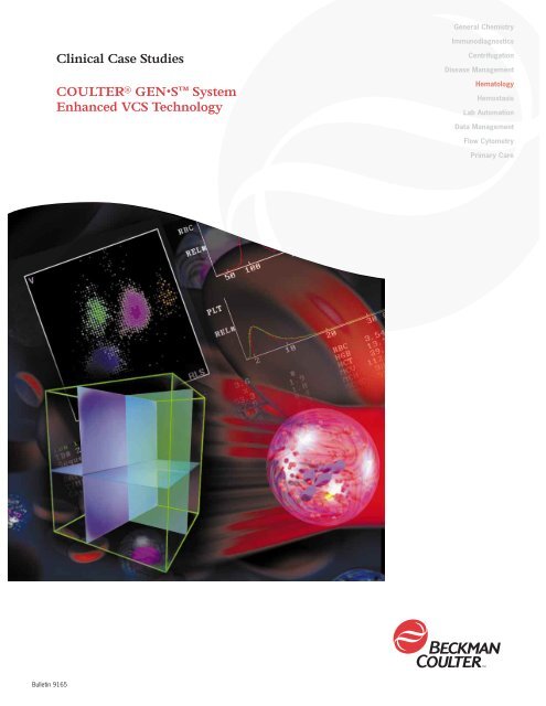
<strong>Clinical</strong> <strong>Case</strong> <strong>Studies</strong><br />
COULTER ® GEN•S <strong>System</strong><br />
<strong>Enhanced</strong> <strong>VCS</strong> Technology<br />
Immunodiagnostics<br />
Centrifugation<br />
Disease Management<br />
Hematology<br />
Hemostasis<br />
Lab Automation<br />
Data Management<br />
Flow Cytometry<br />
Primary Care<br />
Bulletin 9165
COULTER GEN•S <strong>Clinical</strong> <strong>Case</strong> Study<br />
ENHANCED <strong>VCS</strong> TECHNOLOGY ON THE COULTER GEN•S SYSTEM<br />
Coulter <strong>VCS</strong> Technology<br />
IntelliKinetics Process<br />
AccuGate Software<br />
Reticulocyte Analysis<br />
CASE STUDIES<br />
Normal Blood<br />
Left Shift<br />
Severe Infection, Post Operative<br />
Acute Myeloid Leukemia (M1) at Presentation<br />
Acute Myeloid Leukemia (M4) in Relapse<br />
Virus Infection<br />
Chronic Lymphocytic Leukemia<br />
Myelodysplastic Syndrome on Treatment<br />
Normochromic Anemia<br />
EDTA Induced Platelet Aggregation<br />
Monocytosis<br />
Eosinophilia (aged sample)
COULTER GEN•S <strong>Clinical</strong> <strong>Case</strong> Study<br />
ENHANCED <strong>VCS</strong> TECHNOLOGY ON THE COULTER GEN•S SYSTEM<br />
Beckman Coulter<br />
Technology<br />
<strong>VCS</strong> Technology is the most powerful tool available for blood cell<br />
analysis. An acronym for Volume, Conductivity and Scatter, this proprietary<br />
technology offers the greatest sensitivity, specificity and efficiency of any<br />
cell analysis system available today.<br />
The analysis begins with a properly prepared sample using a specialized<br />
reagent system. Two reagents (Erythrolyse and Stabilyse) are mixed<br />
with the blood sample in an orbital mixing chamber to remove red blood<br />
cells while leaving WBCs in an unaltered or “near native” state. First,<br />
Erythrolyse reagent is used to lyse RBCs. Stabilyse reagent is then added<br />
to stop the lytic reaction, leaving the Leukocytes ready for analysis.<br />
<strong>VCS</strong> technology uses a specially constructed flow cytometer that has been<br />
modified to provide more information on unstained cells than is possible<br />
using light scatter alone. The system contains a quartz crystal flow cell<br />
through which the Leukocytes are passed using hydrodynamic focusing to<br />
ensure that they appear in front of the detection system one at a time.<br />
(Diagram 1)<br />
<strong>VCS</strong> is the only single channel analysis that uses three independent energy<br />
sources to probe up to 8,192 cells within the flow cell. This combination of<br />
volume, conductivity and light scatter allows the direct measurement of all<br />
five normal Leukocyte classes.<br />
(Diagrams 2 a,b,c)<br />
HYDRODYNAMIC FOCUSING<br />
Sheath Stream<br />
Flow Cell<br />
Cell Stream<br />
Diagram 1<br />
2
Diagram 2a<br />
Diagram 2b<br />
Diagram 2c<br />
Volume<br />
<strong>VCS</strong> utilizes the Coulter principle of electrical impedance to measure the<br />
volume that the entire cell displaces in an isotonic diluent. This method<br />
accurately sizes all cell types regardless of their orientation in the light<br />
path. Because <strong>VCS</strong> technology uses such a highly accurate measure of<br />
cell volume, this information can be used to correct the conductivity and<br />
scatter signals to give a pair of measurements that are very powerful, and<br />
unique to Coulter. (See diagram 2a)<br />
Conductivity<br />
Alternating current in the radiofrequency (RF) range passes through a cell’s<br />
membrane, penetrating the cell. This powerful probe is used to collect<br />
information about cell size and internal structure, including chemical<br />
composition and nuclear volume. By correcting the conductivity signal so<br />
that it is no longer influenced by cell size, we obtain a measurement that is<br />
related only to the internal structure of the cell. This new measure, called<br />
opacity, allows <strong>VCS</strong> technology to separate cells of similar size, but<br />
different internal composition. It also allows the instrument to calculate<br />
the Nuclear/cytoplasmic ratio - a feature useful in distinguishing variant<br />
lymphocytes from normal lymphs. (See diagram 2b)<br />
Scatter<br />
Within the <strong>VCS</strong> system a focused elliptical light beam from a Helium-Neon<br />
Laser is used to give information about cellular granularity, nuclear<br />
lobularity and cell surface characteristics. Coulter eliminates the size<br />
component of the light scatter signals to give a new measurement called<br />
Rotated Light Scatter (RLS). In doing so we are able to determine the<br />
optimum angle of scatter for each cell type and design the scatter detector<br />
to cover this range (10-70 degrees). This allows <strong>VCS</strong> technology to<br />
accurately separate what would normally be mixed cell types (such as<br />
Neutrophils and Eosinophils) into distinct clusters without mathematical<br />
manipulation. It also enhances the separation between the non granular<br />
cell types. (See diagram 2c)<br />
<strong>VCS</strong>-Cube<br />
Results from each of the 8,000 analyzed<br />
white cells, (or 32,000 red cells in the<br />
Reticulocyte analysis) are assigned X,Y & Z<br />
co-ordinates in a 3-dimensional array based<br />
respectively on their RLS, volume and<br />
opacity (Diagram 3). Each axis of the cube<br />
Diagram 3
COULTER GEN•S <strong>Clinical</strong> <strong>Case</strong> Study<br />
ENHANCED <strong>VCS</strong> TECHNOLOGY ON THE COULTER GEN•S SYSTEM<br />
is subdivided into 256 channels, giving over 16,700,000 possible data<br />
locations within which cells with similar characteristics will form distinct<br />
clusters. The operator may visualize the cube either in 3D or as a two<br />
dimensional dataplot that shows Volume (Y axis) and Light Scatter (X axis).<br />
<strong>VCS</strong><br />
On the GEN•S <strong>System</strong>, <strong>VCS</strong> technology has been enhanced by the addition<br />
of two major modifications that allow even better differential results with an<br />
IntelliKinetics<br />
AccuGate<br />
improved level of flagging efficiency. These two new features (Intellikinetics<br />
and AccuGate) are the result of eight years’ experience with the <strong>VCS</strong><br />
Diagram 4<br />
technology. Their addition allows the GEN•S <strong>System</strong> to provide reduced<br />
levels of False Positive and False Negative differential flagging, thus saving<br />
the laboratory from performing unnecessary film reviews. <strong>Enhanced</strong> <strong>VCS</strong><br />
technology can also improve differential results with respect to aged sample<br />
stability, elimination of interferences and comparison to reference methods.<br />
The two systems are also applied to the Reticulocyte analysis on the GEN•S<br />
<strong>System</strong>, providing improved separation and reduced flagging levels in these<br />
samples as well. (See Diagram 4)<br />
IntelliKinetics Process<br />
The IntelliKinetics application is a hardware and software management tool<br />
that has been developed to assist in the control of fluctuations within the<br />
laboratory environment. Using the IntelliKinetics process, the GEN•S <strong>System</strong><br />
1<br />
Scatter<br />
2<br />
Scatter<br />
Volume<br />
Volume<br />
Volume<br />
Volume<br />
3<br />
Scatter<br />
4<br />
Scatter<br />
Diagram 5<br />
4
monitors and reacts to variables such as the ambient temperature,<br />
optimizing the instrument system to provide consistent reaction kinetics. This<br />
is achieved using intelligent management of reagent reaction temperature,<br />
exposure time, and reagent delivery volume which are controlled through<br />
automatic system adjustments. The combination of this new application with<br />
improved electronics provides a high quality data signal to the <strong>VCS</strong> probes<br />
and to the analysis algorithms, resulting in cell populations that are in a<br />
consistent location in multidimensional space.<br />
The two examples demonstrate the benefits that the IntelliKinetics process<br />
provides in terms of improved population separation. Plots 1 & 2 represent a<br />
fresh blood sample with (Plot. 2) and without (Plot. 1) the use of reaction<br />
control, Plots 3 & 4 repeat the experiment this time for a day old blood<br />
sample. Plot 3 shows the scatterplot without the use of the IntelliKinetics<br />
process, while Plot 4 has the reaction management added. (See Diagram 5)<br />
AccuGate Software<br />
List Mode data collected from the <strong>VCS</strong> probes is analyzed by the AccuGate<br />
software. This new computer algorithm uses advanced contour gating to<br />
identify and classify the WBC sub-populations in a tailored, sample specific<br />
way. AccuGate software contains adaptive statistical tools that assist in<br />
delineating overlapping populations, such as clusters of variant lymphocytes<br />
and monocytes, that are difficult to distinguish using linear gating applications.<br />
The algorithm is also capable of adapting to shifts in populations which<br />
are often manifested when morphologic abnormalities are present.<br />
The benefits of adaptive gating include new suspect flags and additional<br />
“research only” parameters, increased precision and accuracy of results,<br />
color enhancement of population density and cell category dataplots. White<br />
cell differentials on the GEN•S <strong>System</strong> can be presented in multidimensional<br />
formats; additionally an operator can hide or display any series of individual<br />
cell populations within the display.<br />
The six diagrams on the following page illustrate an example of white cell<br />
differential analysis using AccuGate software. Although shown two<br />
dimensionally, separation actually occurs in three dimensions, and we have<br />
attempted to visualize how contour gating works using the bending line in the<br />
DC (x-axis) vs. RLS (y-axis) two-dimensional views.
COULTER GEN•S <strong>Clinical</strong> <strong>Case</strong> Study<br />
ENHANCED <strong>VCS</strong> TECHNOLOGY ON THE COULTER GEN•S SYSTEM<br />
Diagram 1<br />
Diagram 2<br />
Diagram 3<br />
Diagram 4<br />
Diagram 5<br />
Diagram 6<br />
1. First, Eosinophils are separated from all other populations.<br />
2. Monocytes are identified as a separate population and<br />
classified.<br />
3. Neutrophils are separated from Lymphocytes and Basophils.<br />
4. Basophils and Lymphocytes are classified.<br />
5. Completed separation (2-dimensional).<br />
6. Actual 3-dimensional display, illustrating color separation of each white<br />
cell subtype.<br />
6
Reticulocyte Analysis<br />
The Reticulocyte analysis combines the established methodology of the New<br />
Methylene Blue procedure with the standardized analysis and greater<br />
precision of flow cytometry utilizing the <strong>VCS</strong> system. In doing so, it provides<br />
high quality results without the need for fluorescent dyes and expensive<br />
argon ion laser systems, and opens up to the user the potential for providing<br />
improved reproducible data to their clinician customers.<br />
Within the analyzer, a small segment of the blood sample is incubated in a<br />
heated chamber with a special new methylene blue solution, precipitating any<br />
residual RNA within the erythrocytes. A portion of the stained blood sample is<br />
then transferred to a second chamber together with a hypotonic clearing<br />
solution that will remove erythrocyte hemoglobin but preserve the stained<br />
RNA within the cell. Almost immediately, the sample is processed through the<br />
<strong>VCS</strong> flow cell for analysis by the same three independent probes used for<br />
differential analysis.<br />
The AccuGate algorithm system is also used for the analysis of reticulocyte<br />
samples. Again, we have illustrated the separation two dimensionally although<br />
separation actually occurs in three dimensions.<br />
Diagram 1:<br />
Platelets are classified first.<br />
Diagram 2:<br />
The mature red cell population is<br />
distinguished from the immature<br />
reticulocytes.<br />
Diagram 3:<br />
Once isolated, the level of Reticulocyte<br />
maturity can be calculated.<br />
Contour gating is of particular benefit in reticulocyte analysis because the<br />
cell population under study is a gradual continuum of increasing maturity. The<br />
non-linear separation techniques and multiresolution analysis used by the<br />
AccuGate algorithm allow the GEN•S <strong>System</strong> to provide improved precision<br />
and accuracy of results particularly in the presence of abnormal RBC types.<br />
New parameters, such as Immature Reticulocyte Fraction (IRF) and Mean<br />
Reticulocyte Volume (MRV), are now available. “Research only” parameters<br />
can also be provided, including Mean Sphered Cell Volume (MSCV) and<br />
High Light scatter Reticulocyte percent and absolute number (ALR%,<br />
HLR absolute).
COULTER GEN•S <strong>Clinical</strong> <strong>Case</strong> Study<br />
DIAGNOSIS: NORMAL BLOOD<br />
GEN•S SYSTEM FLAGS<br />
Suspect:<br />
None<br />
Definitive:<br />
None<br />
Manual Differential:<br />
Neutrophilis 65%<br />
Lymphocytes 24%<br />
Monocytes 8%<br />
Eosinophilis 2%<br />
Basophilis 1%<br />
RBC Morphology:<br />
Normal<br />
8
COULTER GEN•S <strong>Clinical</strong> <strong>Case</strong> Study<br />
DIAGNOSIS: LEFT SHIFT<br />
GEN•S SYSTEM FLAGS<br />
Suspect:<br />
Imm NE 1<br />
Definitive:<br />
None<br />
Manual Differential:<br />
Segmented Neutrophils 80%<br />
Band Neutrophils 9%<br />
Lymphocytes 6%<br />
Monocytes 3%<br />
Eosinophils 1%<br />
Metamyelocytes 1%<br />
RBC Morphology:<br />
Normal<br />
10
COULTER GEN•S <strong>Clinical</strong> <strong>Case</strong> Study<br />
DIAGNOSIS: SEVERE INFECTION, POST OPERATIVE<br />
GEN•S SYSTEM FLAGS<br />
Suspect:<br />
NE Blasts<br />
Imm NE 1<br />
Imm NE 2<br />
Definitive:<br />
Leukocytosis<br />
Neutrophilia #<br />
Eosinophilia #<br />
Erythrocytosis<br />
Anisocytosis 1+<br />
Manual Differential:<br />
Segmented Neutrophils 79%<br />
Band Neutrophils 8%<br />
Lymphocytes 4%<br />
Monocytes 2%<br />
Eosinophils 2%<br />
Metamyelocytes 1%<br />
Myelocytes 4%<br />
RBC Morphology:<br />
Anisocytosis 1+<br />
Platelet Anisocytosis<br />
12
COULTER GEN•S <strong>Clinical</strong> <strong>Case</strong> Study<br />
DIAGNOSIS: ACUTE MYELOID LEUKEMIA (M1) AT PRESENTATION<br />
GEN•S SYSTEM FLAGS<br />
Suspect:<br />
NE Blasts<br />
Imm NE 1<br />
Imm NE 2<br />
Definitive:<br />
Leukocytosis<br />
Neutrophilia #<br />
Eosinophilia #<br />
Anemia<br />
Anisocytosis 1+<br />
Manual Differential:<br />
Blasts 95%<br />
Lymphocytes 5%<br />
RBC Morphology:<br />
Anisocytosis 1+<br />
14
COULTER GEN•S <strong>Clinical</strong> <strong>Case</strong> Study<br />
DIAGNOSIS: ACUTE MYELOID LEUKEMIA (M4) IN RELAPSE<br />
GEN•S SYSTEM FLAGS<br />
Suspect:<br />
NE Blasts<br />
Imm NE 1<br />
Imm NE 2<br />
Definitive:<br />
Anemia<br />
Anisocytosis 1+<br />
Manual Differential:<br />
Blasts 20%<br />
Myelocytes 1%<br />
Metamyelocytes 3%<br />
Band Neutrophils 1%<br />
Segmented Neutrophils 65%<br />
Lymphocytes 9%<br />
Monocytes 1%<br />
RBC Morphology:<br />
Anisocytosis 1+<br />
16
COULTER GEN•S <strong>Clinical</strong> <strong>Case</strong> Study<br />
DIAGNOSIS: VIRUS INFECTION<br />
GEN•S SYSTEM FLAGS<br />
Suspect:<br />
Variant LY<br />
Definitive:<br />
Neutrophilia #<br />
Thrombocytopenia<br />
Manual Differential:<br />
Segmented Neutrophils 41%<br />
Lymphocytes 34%<br />
Atypical Lymphocytes 8%<br />
Monocytes 12%<br />
Eosilnophils 5%<br />
RBC Morphology:<br />
Platelet Anisocytosis<br />
18
COULTER GEN•S <strong>Clinical</strong> <strong>Case</strong> Study<br />
DIAGNOSIS: CHRONIC LYMPHOCYTIC LEUKEMIA<br />
GEN•S SYSTEM FLAGS<br />
Suspect:<br />
None<br />
Definitive:<br />
Neutrophilia #<br />
Lymphocytosis #<br />
Macrocytosis 1+<br />
Aniscytosis 1+<br />
Thrombocytopenia<br />
Manual Differential:<br />
Segmented Neutrophils 1%<br />
Lymphocytes 99%<br />
RBC Morphology:<br />
Macrocytosis 1+<br />
Anisocytosis 1+<br />
20
COULTER GEN•S <strong>Clinical</strong> <strong>Case</strong> Study<br />
DIAGNOSIS: MYELODYSPLASTIC SYNDROME ON TREATMENT<br />
GEN•S SYSTEM FLAGS<br />
Suspect:<br />
None<br />
Definitive:<br />
Neutropenia #<br />
Pancytopenia<br />
Anisocytosis 1+<br />
Manual Differential:<br />
Lymphocytes 19%<br />
Basophils 1%<br />
RBC Morphology:<br />
Anisocytosis 1+<br />
Manual Platelet Count:<br />
COULTER GEN•S <strong>Clinical</strong> <strong>Case</strong> Study<br />
DIAGNOSIS: NORMOCHROMIC ANEMIA<br />
GEN•S SYSTEM FLAGS<br />
Suspect:<br />
Imm NE1<br />
NRBC<br />
Definitive:<br />
Anemia<br />
Anisocytosis 1+<br />
Manual Differential:<br />
Band Neutrophils 7%<br />
Segmented Neutrophils 0%<br />
Lymphocytes 9%<br />
Monocytes 13%<br />
Eosinophils 1%<br />
Normoblasts: 9 per 100 WBC’s<br />
RBC Morphology:<br />
Anisocytosis 1+<br />
Corrected WBC:<br />
6.0 x 10 9 /L<br />
24
COULTER GEN•S <strong>Clinical</strong> <strong>Case</strong> Study<br />
DIAGNOSIS: EDTA INDUCED PLATELET AGGREGATION<br />
GEN•S SYSTEM FLAGS<br />
Suspect:<br />
Imm NE1<br />
Platelet Clumps<br />
Definitive:<br />
Leukocytosis<br />
Neutrophilia #<br />
Monocytosis #<br />
Thrombocytopenia<br />
Manual Differential:<br />
Band Neutrophils 10%<br />
Segmented Neutrophils 57%<br />
Lymphocytes 18%<br />
Monocytes 11%<br />
Eosinophils 4%<br />
RBC Morphology:<br />
Platelet Clumped on Film<br />
Corrected WBC:<br />
16.5 x 10 9 /L<br />
Platelet Count on<br />
citrated sample:<br />
25.3 x 10 9 /L<br />
26
COULTER GEN•S <strong>Clinical</strong> <strong>Case</strong> Study<br />
DIAGNOSIS: MONOCYTOSIS<br />
GEN•S SYSTEM FLAGS<br />
Suspect:<br />
None<br />
Definitive:<br />
Monocytosis #<br />
Anemia<br />
Anisocytosis 1+<br />
Thrombocytopenia<br />
Reticulocytosis<br />
Manual Differential:<br />
Band Neutrophils 3%<br />
Segmented Neutrophils 42%<br />
Lymphocytes 11%<br />
Monocytes 42%<br />
Eosinophils 1%<br />
Basophils 1%<br />
RBC Morphology:<br />
Anisocytosis 2+<br />
Hypochromasia 1+<br />
Polychromasia 1+<br />
28
COULTER GEN•S <strong>Clinical</strong> <strong>Case</strong> Study<br />
DIAGNOSIS: EOSINOPHILIA (AGED SAMPLE)<br />
GEN•S SYSTEM FLAGS<br />
Suspect:<br />
Aged Sample<br />
(Research Screen)<br />
Definitive:<br />
Eosinophilia %<br />
Manual Differential:<br />
Band Neutrophils 1%<br />
Segmented Neutrophils 64%<br />
Lymphocytes 19%<br />
Monocytes 4%<br />
Eosinophils 11%<br />
Basophils 1%<br />
RBC Morphology:<br />
Normal<br />
30
I MPROVE THE PROCESS—AT EVERY LEVEL <br />
Africa/Middle East/Eastern Europe: Switzerland, Nyon (41) 22 994 0707. Australia, Gladesville (61) 2 9844 6000. Canada, Mississauga (1) 905 819 1234.<br />
China, Beijing (86) 10 6515 6028. Hong Kong (852) 2814 7431, 2814 0481. France, Villepinte (33) 1 49 90 90 00. Germany, Krefeld (49) 2151 33 35.<br />
Italy, Milan (39) 02 953921. Japan, Tokyo (81) 3 5404 8424. Latin America (1) (305) 380 4709. Mexico, Mexico City (525) 575 6805.<br />
Netherlands, Mijdrecht (31) 297 230630. Singapore (65) 339 3633. South Africa, Johannesburg (27) 11 805 2014. Sweden, Bromma (46) 8 98 53 20.<br />
Switzerland, Nyon 0800 850 810. Taiwan, Taipei (886) 2 2378 3456. UK, High Wycombe (44) 01494 441181. USA, Brea, CA (1) 800 352 3433, (1) 714 993 5321.<br />
www.beckmancoulter.com © Copyright 2000 Beckman Coulter, Inc. PRINTED IN U.S.A.