endoscopic-assisted harvest of pedicled and free latissimus dorsi ...
endoscopic-assisted harvest of pedicled and free latissimus dorsi ...
endoscopic-assisted harvest of pedicled and free latissimus dorsi ...
Create successful ePaper yourself
Turn your PDF publications into a flip-book with our unique Google optimized e-Paper software.
described using an open <strong>harvest</strong>ing technique. 31-36<br />
Recently, a model for <strong>endoscopic</strong>-<strong>assisted</strong> muscle<br />
<strong>harvest</strong>ing training was described in pigs. 37<br />
The aim <strong>of</strong> the present study is to assess the<br />
potential <strong>of</strong> the swine model <strong>of</strong> <strong>endoscopic</strong>-<strong>assisted</strong><br />
LD <strong>harvest</strong>ing. The muscle <strong>harvest</strong>ing (as <strong>pedicled</strong><br />
<strong>and</strong> <strong>free</strong> flap) parallels the technique used in humans.<br />
The learning curve, complications <strong>and</strong> results are<br />
evaluated <strong>and</strong> the value <strong>of</strong> this model as a training tool<br />
established.<br />
MATERIAL AND METHODS<br />
Prior to our study, detailed swine LD muscle<br />
anatomy was taught <strong>and</strong> classic (open) <strong>harvest</strong> <strong>of</strong><br />
swine LD muscle was performed during flap dissection<br />
training courses <strong>and</strong> learning sessions conducted by<br />
the senior authors DP <strong>and</strong> MI. 31-36 The first author<br />
had <strong>endoscopic</strong> experience limited to porcine gracilis<br />
muscle <strong>harvest</strong>. 37<br />
Operative instruments<br />
A st<strong>and</strong>ard <strong>endoscopic</strong> setup (similar to<br />
laparoscopic surgery cart) was used: light source, 10 mm<br />
<strong>and</strong> 5 mm, 30 degrees angle endoscope, video camera,<br />
high resolution video monitor <strong>and</strong> video recorder,<br />
irrigation-suction device. The usual instrument set for<br />
flap surgery <strong>and</strong> st<strong>and</strong>ard laparoscopic instruments<br />
(forceps, cautery, scissors <strong>and</strong> hemoclips) were also<br />
used. For the optical cavity development during<br />
<strong>endoscopic</strong> dissection, an Emory-type retractor was<br />
used.<br />
Animal experiments<br />
The protocols were approved by the Joint<br />
Committee for Animal Research <strong>and</strong> Animal Care <strong>and</strong><br />
Ethic Committee <strong>of</strong> Pius Branzeu Center in Timisoara<br />
<strong>and</strong> Center for Simulation <strong>and</strong> Training in Surgery in<br />
Iasi. Animals were housed <strong>and</strong> treated in compliance<br />
with the “Guide for the Care <strong>and</strong> Use <strong>of</strong> Laboratory<br />
Animals”, published by the National Academy Press<br />
(US NIH Publication No 85–23, revised 1996). The<br />
animals were caged individually in the animal facility<br />
<strong>of</strong> the research center, with 12 hourly day/night cycle<br />
<strong>and</strong> with food <strong>and</strong> water ad libitum. They were fasted<br />
for 12 hours preoperatively.<br />
Pig’s sedation was achieved with ketamine (10-<br />
15 mg/kg) <strong>and</strong> midazolam (0.5 mg/kg) or diazepam<br />
(2 mg/kg); they were intubated after intravenous<br />
infusion <strong>of</strong> thiopental (5-10 mg/kg). Anesthesia was<br />
maintained with halothane 1-2% mixed with oxygen<br />
2-4L/min. Isotonic solutions were perfused 5-10 ml/<br />
kg/h.38 A total <strong>of</strong> 30 pigs (mean weight 26.3 kg, range<br />
20-34 kg) underwent <strong>endoscopic</strong>-<strong>assisted</strong> <strong>harvest</strong> <strong>of</strong><br />
39 LD muscles (18 bilateral <strong>and</strong> 21 unilateral).<br />
_____________________________<br />
146 TMJ 2010, Vol. 60, No. 2 - 3<br />
Operating room setup<br />
The pigs were positioned in lateral decubitus<br />
with forelimb <strong>free</strong>. 32,33 The operator stood facing the<br />
animal’s ventral side, with the assistant beside him; the<br />
later h<strong>and</strong>led the self-mounted Emory-type retractor<br />
<strong>and</strong> pulled pig’s forelimb cranially <strong>and</strong> outward to<br />
expose the pedicle. The video monitor was placed<br />
at the opposite side <strong>of</strong> the operating table for both<br />
operator <strong>and</strong> assistant to see the <strong>endoscopic</strong> images.<br />
L<strong>and</strong>marks<br />
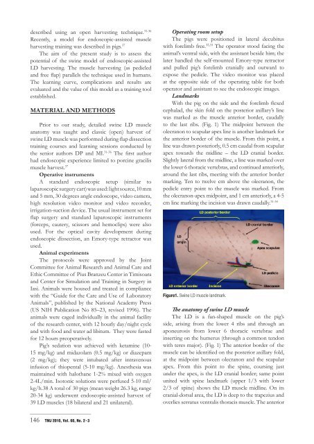
With the pig on the side <strong>and</strong> the forelimb flexed<br />
cephalad, the skin fold on the posterior axillary’s line<br />
was marked as the muscle anterior border, caudally<br />
to the last ribs. (Fig. 1) The midpoint between the<br />
olecranon to scapular apex line is another l<strong>and</strong>mark for<br />
the anterior border <strong>of</strong> the muscle. From this point, a<br />
line was drawn posteriorly, 0.5 cm caudal from scapular<br />
apex towards the midline – the LD cranial border.<br />
Slightly lateral from the midline, a line was marked over<br />
the lower 6 thoracic vertebras, <strong>and</strong> continued anteriorly,<br />
around the last ribs, meeting with the anterior border<br />
marking. Ten to twelve cm above the olecranon, the<br />
pedicle entry point to the muscle was marked. From<br />
the olecranon-apex midpoint, <strong>and</strong> 1 cm anteriorly, a 4-5<br />
cm line marking the incision was drawn caudally. 31-34<br />
Figure1. Swine LD muscle l<strong>and</strong>mark.<br />
The anatomy <strong>of</strong> swine LD muscle<br />
The LD is a fan-shaped muscle on the pig’s<br />
side, arising from the lower 4 ribs <strong>and</strong> through an<br />
aponeurosis from lower 6 thoracic vertebrae <strong>and</strong><br />
inserting on the humerus (through a common tendon<br />
with teres major). (Fig. 1) The anterior border <strong>of</strong> the<br />
muscle can be identified on the posterior axillary fold,<br />
at the midpoint between olecranon <strong>and</strong> the scapular<br />
apex. From this point to the spine, coursing just<br />
under the apex, is the LD cranial border; same point<br />
united with spine l<strong>and</strong>mark (upper 1/3 with lower<br />
2/3 <strong>of</strong> spine) shows the LD muscle midline. On its<br />
cranial-dorsal area, the LD is deep to the trapezius <strong>and</strong><br />
overlies serratus ventralis thoracis muscle. The anterior


