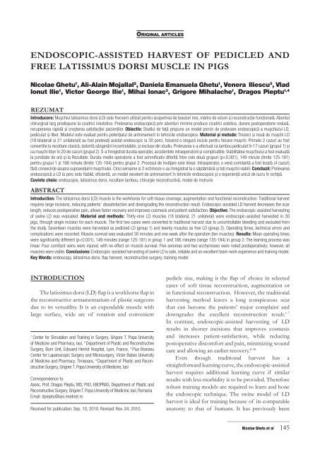
endoscopic-assisted harvest of pedicled and free latissimus dorsi ...
endoscopic-assisted harvest of pedicled and free latissimus dorsi ...
endoscopic-assisted harvest of pedicled and free latissimus dorsi ...

You also want an ePaper? Increase the reach of your titles
YUMPU automatically turns print PDFs into web optimized ePapers that Google loves.
INTRODUCTION<br />
The <strong>latissimus</strong> <strong>dorsi</strong> (LD) flap is a workhorse flap in<br />
the reconstructive armamentarium <strong>of</strong> plastic surgeons<br />
due to its versatility. It is an expendable muscle with<br />
large surface, wide arc <strong>of</strong> rotation <strong>and</strong> convenient<br />
ORIGINAL ARTICLES<br />
ENDOSCOPIC-ASSISTED HARVEST OF PEDICLED AND<br />
FREE LATISSIMUS DORSI MUSCLE IN PIGS<br />
Nicolae Ghetu 1 , Ali-Alain Mojallal 2 , Daniela Emanuela Ghetu 1 , Venera Iliescu 3 , Vlad<br />
Ionut Ilie 1 , Victor George Ilie 1 , Mihai Ionac 3 , Grigore Mihalache 1 , Dragos Pieptu 1,4<br />
REZUMAT<br />
Introducere: Muşchiul <strong>latissimus</strong> <strong>dorsi</strong> (LD) este frecvent utilizat pentru acoperirea de ţesuturi moi, mărire de volum şi reconstrucţie funcţională. Abordul<br />
chirurgical larg predispune la cicatrici inestetice. Prelevarea <strong>endoscopic</strong>ă prin aborduri minime produce cicatrici estetice, durere postoperatorie redusă,<br />
recuperarea rapidă şi creşterea satisfacţiei pacienţilor. Obiectiv: Studiul de faţă propune un model porcin de prelevare <strong>endoscopic</strong>ă a muşchiului LD,<br />
pediculat şi liber. Modelul este evaluat pentru potenţialul de antrenament in tehnicile <strong>endoscopic</strong>e. Material și metode: Treizeci şi nouă de muşchi LD<br />
(18 bilaterali şi 21 unilaterali) au fost prelevaţi asistat-<strong>endoscopic</strong> la 30 porci, folosind o singură incizie pentru fiecare muşchi. Primele 2 cazuri au fost<br />
convertite la recoltare clasică, datorită sângerării incontrolabile, şi excluse din studiu. Prelevarea s-a efectuat ca lambou pediculat în 17 cazuri (grupul 1) şi<br />
ca muşchi liber în 20 de cazuri (grupul 2). S-a înregistrat durata operaţiei, accidentele intraoperatorii şi complicaţiile. Viabilitatea muşchiului a fost evaluată<br />
la jumătate de oră şi la Rezultate: Durata medie operatorie a fost semnificativ diferită între cele două grupuri (p
described using an open <strong>harvest</strong>ing technique. 31-36<br />
Recently, a model for <strong>endoscopic</strong>-<strong>assisted</strong> muscle<br />
<strong>harvest</strong>ing training was described in pigs. 37<br />
The aim <strong>of</strong> the present study is to assess the<br />
potential <strong>of</strong> the swine model <strong>of</strong> <strong>endoscopic</strong>-<strong>assisted</strong><br />
LD <strong>harvest</strong>ing. The muscle <strong>harvest</strong>ing (as <strong>pedicled</strong><br />
<strong>and</strong> <strong>free</strong> flap) parallels the technique used in humans.<br />
The learning curve, complications <strong>and</strong> results are<br />
evaluated <strong>and</strong> the value <strong>of</strong> this model as a training tool<br />
established.<br />
MATERIAL AND METHODS<br />
Prior to our study, detailed swine LD muscle<br />
anatomy was taught <strong>and</strong> classic (open) <strong>harvest</strong> <strong>of</strong><br />
swine LD muscle was performed during flap dissection<br />
training courses <strong>and</strong> learning sessions conducted by<br />
the senior authors DP <strong>and</strong> MI. 31-36 The first author<br />
had <strong>endoscopic</strong> experience limited to porcine gracilis<br />
muscle <strong>harvest</strong>. 37<br />
Operative instruments<br />
A st<strong>and</strong>ard <strong>endoscopic</strong> setup (similar to<br />
laparoscopic surgery cart) was used: light source, 10 mm<br />
<strong>and</strong> 5 mm, 30 degrees angle endoscope, video camera,<br />
high resolution video monitor <strong>and</strong> video recorder,<br />
irrigation-suction device. The usual instrument set for<br />
flap surgery <strong>and</strong> st<strong>and</strong>ard laparoscopic instruments<br />
(forceps, cautery, scissors <strong>and</strong> hemoclips) were also<br />
used. For the optical cavity development during<br />
<strong>endoscopic</strong> dissection, an Emory-type retractor was<br />
used.<br />
Animal experiments<br />
The protocols were approved by the Joint<br />
Committee for Animal Research <strong>and</strong> Animal Care <strong>and</strong><br />
Ethic Committee <strong>of</strong> Pius Branzeu Center in Timisoara<br />
<strong>and</strong> Center for Simulation <strong>and</strong> Training in Surgery in<br />
Iasi. Animals were housed <strong>and</strong> treated in compliance<br />
with the “Guide for the Care <strong>and</strong> Use <strong>of</strong> Laboratory<br />
Animals”, published by the National Academy Press<br />
(US NIH Publication No 85–23, revised 1996). The<br />
animals were caged individually in the animal facility<br />
<strong>of</strong> the research center, with 12 hourly day/night cycle<br />
<strong>and</strong> with food <strong>and</strong> water ad libitum. They were fasted<br />
for 12 hours preoperatively.<br />
Pig’s sedation was achieved with ketamine (10-<br />
15 mg/kg) <strong>and</strong> midazolam (0.5 mg/kg) or diazepam<br />
(2 mg/kg); they were intubated after intravenous<br />
infusion <strong>of</strong> thiopental (5-10 mg/kg). Anesthesia was<br />
maintained with halothane 1-2% mixed with oxygen<br />
2-4L/min. Isotonic solutions were perfused 5-10 ml/<br />
kg/h.38 A total <strong>of</strong> 30 pigs (mean weight 26.3 kg, range<br />
20-34 kg) underwent <strong>endoscopic</strong>-<strong>assisted</strong> <strong>harvest</strong> <strong>of</strong><br />
39 LD muscles (18 bilateral <strong>and</strong> 21 unilateral).<br />
_____________________________<br />
146 TMJ 2010, Vol. 60, No. 2 - 3<br />
Operating room setup<br />
The pigs were positioned in lateral decubitus<br />
with forelimb <strong>free</strong>. 32,33 The operator stood facing the<br />
animal’s ventral side, with the assistant beside him; the<br />
later h<strong>and</strong>led the self-mounted Emory-type retractor<br />
<strong>and</strong> pulled pig’s forelimb cranially <strong>and</strong> outward to<br />
expose the pedicle. The video monitor was placed<br />
at the opposite side <strong>of</strong> the operating table for both<br />
operator <strong>and</strong> assistant to see the <strong>endoscopic</strong> images.<br />
L<strong>and</strong>marks<br />
With the pig on the side <strong>and</strong> the forelimb flexed<br />
cephalad, the skin fold on the posterior axillary’s line<br />
was marked as the muscle anterior border, caudally<br />
to the last ribs. (Fig. 1) The midpoint between the<br />
olecranon to scapular apex line is another l<strong>and</strong>mark for<br />
the anterior border <strong>of</strong> the muscle. From this point, a<br />
line was drawn posteriorly, 0.5 cm caudal from scapular<br />
apex towards the midline – the LD cranial border.<br />
Slightly lateral from the midline, a line was marked over<br />
the lower 6 thoracic vertebras, <strong>and</strong> continued anteriorly,<br />
around the last ribs, meeting with the anterior border<br />
marking. Ten to twelve cm above the olecranon, the<br />
pedicle entry point to the muscle was marked. From<br />
the olecranon-apex midpoint, <strong>and</strong> 1 cm anteriorly, a 4-5<br />
cm line marking the incision was drawn caudally. 31-34<br />
Figure1. Swine LD muscle l<strong>and</strong>mark.<br />
The anatomy <strong>of</strong> swine LD muscle<br />
The LD is a fan-shaped muscle on the pig’s<br />
side, arising from the lower 4 ribs <strong>and</strong> through an<br />
aponeurosis from lower 6 thoracic vertebrae <strong>and</strong><br />
inserting on the humerus (through a common tendon<br />
with teres major). (Fig. 1) The anterior border <strong>of</strong> the<br />
muscle can be identified on the posterior axillary fold,<br />
at the midpoint between olecranon <strong>and</strong> the scapular<br />
apex. From this point to the spine, coursing just<br />
under the apex, is the LD cranial border; same point<br />
united with spine l<strong>and</strong>mark (upper 1/3 with lower<br />
2/3 <strong>of</strong> spine) shows the LD muscle midline. On its<br />
cranial-dorsal area, the LD is deep to the trapezius <strong>and</strong><br />
overlies serratus ventralis thoracis muscle. The anterior
order has loose connections to pectoralis pr<strong>of</strong>undus<br />
ascendens muscle.<br />
The LD is a Mathes <strong>and</strong> Nahai type V muscle, with<br />
a dominant pedicle <strong>and</strong> multiple segmental pedicles<br />
(perforators from intercostals arteries – IAP). The<br />
dominant pedicle consists <strong>of</strong> the thoraco-dorsal artery<br />
(from axillary artery via subscapular artery) <strong>and</strong> paired<br />
venae comitantes (proximally <strong>of</strong>ten united to become<br />
single subscapular vein). Cranially, the muscle curls<br />
on itself in a mild gutter-shape aspect that holds the<br />
vessels. Pedicle runs under teres major muscle, on the<br />
fascia underneath LD. The artery, 1.2-1.5 mm diameter,<br />
enters the muscle 6 cm distal to origin, or 10-12 cm<br />
above the olecranon. Along the cephalic border <strong>of</strong> LD,<br />
branches the arterial supply for teres major muscle. LD<br />
motor innervation is provided by thoraco-dorsal nerve,<br />
situated slightly cranial to the vascular pedicle. 31-34<br />
Harvesting technique<br />
After anesthesia administration, pig’s side <strong>and</strong><br />
forelimb were prepped <strong>and</strong> draped. The skin was<br />
incised 4-5 cm <strong>and</strong> dissection proceeded through<br />
subcutaneous fat layer <strong>and</strong> panniculus carnosus. The<br />
anterior border <strong>of</strong> LD lies under the subpannicular<br />
thin layer <strong>of</strong> translucent fat.<br />
From incision site, dissection proceeds in the<br />
usual open (classic) fashion, both above <strong>and</strong> under the<br />
muscle. (Fig. 2) Once direct vision became limited, the<br />
dissection the proceeds under <strong>endoscopic</strong> assistance,<br />
monitored carefully on the screen. Supramuscular<br />
dense attachments needed electric cautery dissection;<br />
beneath muscle, loose connections allowed easier <strong>and</strong><br />
faster dissection. Small perforators <strong>and</strong> side-branches<br />
(to teres major <strong>and</strong> serratus) were cauterized or clipped.<br />
Figure 2. LD caudal border identified after incision. “OPEN” marks<br />
classic dissection area.<br />
Using Emory-type retractors, the apex <strong>of</strong> the<br />
scapula was visualized, slightly cranial to LD border.<br />
Systematic clockwise or anticlockwise dissection<br />
helped maintain the correct plane <strong>and</strong> dissection <strong>of</strong><br />
the LD, from under the trapezius <strong>and</strong> teres major <strong>and</strong><br />
above the serratus muscle. Near the muscle vertebral<br />
border, the IAPs must be carefully cauterized or<br />
clipped to prevent bleeding; when bleeding occurred,<br />
dissection was resumed after 5 minutes gentle pressure<br />
<strong>and</strong> proper vessel hemoclips ligation. Irrigation-suction<br />
can also be useful for bleeding vessel identification.<br />
After the LD dissection, the muscle origins were<br />
addressed. From underneath the muscle, using a hookshape<br />
cautery, the vertebral aponeurosis <strong>of</strong> LD was<br />
cut. Caudally, the muscle was detached from the ribs,<br />
<strong>and</strong> the anterior margin liberated in a caudal to cranial<br />
direction from thin connections to the pectoralis<br />
muscles. If this sequence is not followed, the muscle<br />
will fall towards the spine, making the vertebral origin<br />
section a difficult task.<br />
Muscle was delivered through the incision, still<br />
attached by pedicle <strong>and</strong> tendon. (Figs. 3, 4) Adequate<br />
arc <strong>of</strong> rotation was provided by 2-3 cm open proximal<br />
mobilization <strong>of</strong> tendon <strong>and</strong> pedicle. The donor site<br />
was inspected for bleeding. Muscle was replaced,<br />
bleeding <strong>and</strong> muscle viability checked again 30 minutes<br />
later, donor site closed using 3/0 absorbable stitches<br />
<strong>and</strong> pigs were returned to animal facility (if follow-up<br />
intended) or euthanized.<br />
Figure 3. Pedicled LD delivered through incision. LD outer surface shown.<br />
Figure 4. Pedicled LD delivered through incision. LD undersurface shown.<br />
_____________________________<br />
Nicolae Ghetu et al 147
Table I: <strong>endoscopic</strong>-<strong>assisted</strong> LD muscles <strong>harvest</strong>ed<br />
Table 1. Endoscopic-<strong>assisted</strong> LD muscles <strong>harvest</strong>ed.<br />
Pig no. Weight<br />
Group 1<br />
Group 2<br />
Case<br />
(kg) no.<br />
time (min) complication<br />
1 34 1 R 205 IAP injury Conversion Muscle viable<br />
2 L 188 IAP injury Conversion “<br />
2 25 3 R 181 VC injury Yes Seroma “<br />
3 32 4 R 168 VC injury Yes “<br />
4 24 5 R 165 Yes Seroma “<br />
5 22 6 R 175 “<br />
7 L 158 “<br />
6 26 8 R 160 “<br />
9 L 152 “<br />
7 31 10 R 142 “<br />
11 L 145 “<br />
For the first 19 cases, the technique described<br />
reproduces the <strong>endoscopic</strong>-<strong>assisted</strong> <strong>harvest</strong> <strong>of</strong> LD<br />
to be used as a <strong>pedicled</strong> muscle (i.e. <strong>pedicled</strong> LD for<br />
breast reconstruction).<br />
For distinction with the subsequent cases, first 19<br />
cases will be included in group 1. (Table 1)<br />
_____________________________<br />
148 TMJ 2010, Vol. 60, No. 2 - 3<br />
Side Operating<br />
Intraoperative<br />
1<br />
Follow-up Complications Results<br />
8 29 12 R 135 “<br />
13 L 136 “<br />
9 27 14 R 142 “<br />
15 L 140 “<br />
10 25 16 R 143 “<br />
17 L 130 “<br />
11 30 18 R 136 “<br />
19 L 125 “<br />
12 25 20 L 184 VC injury Yes Ecchymosis “<br />
13 22 21 R 181 “<br />
22 L 180 “<br />
14 28 23 R 184 VC injury Yes Seroma “<br />
15 24 24 L 175 “<br />
16 21 25 R 181 “<br />
17 27 26 L 176 “<br />
18 29 27 L 172 “<br />
19 23 28 R 165 “<br />
20 24 29 R 164 Yes Seroma “<br />
21 30 30 L 170 “<br />
22 31 32 L 156 Yes Ecchymosis “<br />
23 32 31 R 163 “<br />
24 23 33 R 160 “<br />
25 27 34 L 163 Yes “<br />
26 29 35 R 159 “<br />
27 21 36 R 162 Yes Seroma “<br />
28 20 37 R 150 “<br />
29 23 38 R 140 Yes “<br />
30 25 39 R 135 “<br />
Legend: IAP: intercostals artery perforator, VC: vena comitans, R: right, L: left<br />
Starting with case #20, for the next 20 cases<br />
(group 2), the LD muscle was <strong>harvest</strong>ed as a <strong>free</strong> flap.<br />
Muscle dissection was performed in a similar fashion<br />
<strong>and</strong> continued proximally.<br />
The forelimb was pulled cranially <strong>and</strong> upward<br />
<strong>and</strong> the submuscular plane cranial to the incision was
visualized. Open dissection for 3-4 cm (limited by the<br />
incision direction) preceded the <strong>endoscopic</strong> dissection<br />
<strong>of</strong> the pedicle - artery, vein/paired veins <strong>and</strong> nerve.<br />
The pedicle was skeletonized up to the origin from the<br />
axillary vessels. (Fig. 5) The tendon was isolated from<br />
teres major tendon <strong>and</strong> cut. Half an hour later, the<br />
muscle was inspected for viability. For one week followup,<br />
LD was sutured to the serratus fascia in a position to<br />
prevent pedicle kinking, compression or torsion. After<br />
checking the donor site for bleeding, the incision was<br />
closed by separate absorbable 3/0 stitches. Pigs were<br />
returned to animal facility. If no follow-up intended,<br />
pedicle was clipped near its origin <strong>and</strong> cut, the muscle<br />
was delivered through the incision <strong>and</strong> measured.<br />
Donor site was inspected for bleeding, closed with<br />
absorbable stitches <strong>and</strong> pigs were euthanized.<br />
Figure 5. LD pedicle dissected. Endoscopic view.<br />
Operating times were recorded from incision<br />
to skin closure but the 30 minutes observation<br />
time was not included. Complications <strong>and</strong> muscle<br />
anthropometric measurements were documented.<br />
Neither drains nor dressings were used; antibiotics <strong>and</strong><br />
analgesics were administered for 3 days.<br />
Follow-up<br />
Vital signs, complications, ambulation <strong>and</strong> feeding<br />
habits were inspected on a daily bases. One week<br />
later, under general anesthesia, the incision site was<br />
reopened <strong>and</strong> the muscle viability was checked <strong>and</strong> the<br />
complications were noted.<br />
Statistics<br />
Student t-test was used to analyze the operating<br />
times between the groups 1 <strong>and</strong> 2. P
comitant vein when inserting the retractor (2 cases);<br />
in group 2, one comitant vein injury during pedicle<br />
dissection (two cases). The bleeding was controlled<br />
<strong>and</strong> injured vein clipped. The cases with VC injury<br />
were deliberately included in follow-up group.<br />
First day postoperatively, all animals resumed<br />
ambulation <strong>and</strong> feeding habits, minimal functional<br />
impairment was noticed for 1-2 days postoperatively.<br />
Near the origin <strong>of</strong> LD muscle, small ecchymosis (two<br />
cases) was noticed first day postoperatively that slightly<br />
enlarged for the next 2 days; however, when donorsite<br />
reopened at 1 week, there is no fresh bleeding at<br />
inspection <strong>and</strong> no hematomas. (Fig. 7)<br />
In five cases, non-infected seromas were found<br />
<strong>and</strong> evacuated. All muscles were viable after 30 minutes<br />
observation <strong>and</strong> after 1 week follow-up.<br />
Muscles lengths ranged from 13/10 cm to 16/13<br />
cm on the back table. (Fig. 8)<br />
Figure 7. Ecchymosis at 1 week after LD <strong>endoscopic</strong>-<strong>assisted</strong> <strong>harvest</strong>.<br />
Figure 8. Endoscopic-<strong>assisted</strong> <strong>harvest</strong>ed LD muscle <strong>free</strong> flap.<br />
DISCUSSION<br />
The LD is a workhorse for selected reconstructive<br />
surgeries due to its versatility. It is successfully used,<br />
either as a <strong>pedicled</strong> or <strong>free</strong> flap, in breast, chest wall,<br />
spinal, head <strong>and</strong> neck, lower limb reconstruction <strong>and</strong><br />
scalp resurfacing. Large incisions make the muscle<br />
_____________________________<br />
150 TMJ 2010, Vol. 60, No. 2 - 3<br />
<strong>harvest</strong>ing straightforward but leaves conspicuous<br />
scars, decreasing patient satisfaction in the face <strong>of</strong><br />
otherwise excellent reconstructive results. 1-7<br />
Since the early ‘90s, <strong>endoscopic</strong>-<strong>assisted</strong><br />
<strong>harvest</strong>ing <strong>of</strong> LD muscle achieved shorter incisions<br />
with less postoperative discomfort or pain, earlier<br />
recovery from surgery <strong>and</strong> less expensive wound care;<br />
edema, ecchymosis, seromas <strong>and</strong> infection rates were<br />
not decreased in all studies. 8-30 The advantages have<br />
encouraged surgeons to use the <strong>endoscopic</strong> method<br />
but the reports are still scarce <strong>and</strong> have not gained the<br />
same popularity within plastic surgery as it has in other<br />
surgical specialties. The main hurdle is the additional<br />
learning curve for <strong>endoscopic</strong> techniques; training<br />
models <strong>and</strong> fewer opportunities when compared to<br />
laparoscopy or arthroscopy. 39,40<br />
The swine model <strong>of</strong> muscle myocutaneous LD was<br />
described initially as an excellent model for research<br />
in physiological, pathological <strong>and</strong> pharmacological<br />
experiments. Later the similarities to human anatomy,<br />
favorable position (quickly <strong>and</strong> easily elevated) <strong>and</strong><br />
large, accessible vascular pedicle made it an excellent<br />
model for training <strong>of</strong> the surgical skills. This too has<br />
been our experience. 31-37<br />
The aim <strong>of</strong> this study was to evaluate the<br />
<strong>endoscopic</strong>-<strong>assisted</strong> LD <strong>harvest</strong>ing model in swine.<br />
Two consecutive animal groups underwent the LD<br />
<strong>harvest</strong>ing as <strong>pedicled</strong> <strong>and</strong> <strong>free</strong> flap, respectively.<br />
For all cases, each muscle was <strong>harvest</strong>ed through<br />
single skin incision <strong>of</strong> 4-5 cm, adequate to comfortably<br />
accommodate the instruments (endoscope, retractor,<br />
forceps <strong>and</strong> electrocautery/scissor) <strong>and</strong> to retrieve the<br />
muscle. 41 As Lin et al. advocate, if muscle size is smaller<br />
than 20 cm, an additional incision is unnecessary. 17<br />
Adherent fibr<strong>of</strong>atty tissue overlaying the muscle is<br />
cut first, using electrocautery; if the undersurface is<br />
dissected first, muscle contraction during outersurface<br />
dissection would be too strong, increasing the chances<br />
for tissue injury.<br />
Anatomical <strong>and</strong> technical constraints need to be<br />
overcome for successful operation. Rigid <strong>endoscopic</strong><br />
instruments accommodate poorly to the rigid <strong>and</strong><br />
convex chest wall. 12 Even if the instrument length<br />
is adequate for the optical cavity length, the straight<br />
instruments are not adapted to the three-dimensional<br />
cavity requirements. Increased difficulty is encountered<br />
in the areas most distant from the incision. 40,41 For<br />
the first two cases, bleeding from intercostal artery<br />
perforators was ineffectively addressed, also due to<br />
inexperience in using irrigation-suction device; cases<br />
were converted to classic <strong>harvest</strong> <strong>and</strong> excluded from<br />
the study. For later cases, the use <strong>of</strong> long curved<br />
electrocautery achieved better hemostasis <strong>and</strong> a 30
degree angled scope <strong>assisted</strong> vision over the thorax<br />
convexity, where bleeding occurred. Systematic<br />
steps to <strong>free</strong> the muscle must be followed: vertebral<br />
aponeurosis, costal insertion <strong>and</strong> anterior margin;<br />
otherwise muscle mass retracted towards the spine will<br />
impair aponeurosis sectioning.<br />
The longitudinal incision allowed access to distant<br />
sites, but limited the access proximal to the pedicle.<br />
Compared to humans, swine forelimb mobility is<br />
anatomically limited, with impossible access to the<br />
axilla without increasing the length <strong>of</strong> the incision.<br />
(Fig. 9) Therefore, to maintain the skin incision to the<br />
original size, group 2 underwent <strong>endoscopic</strong> dissection<br />
<strong>of</strong> the thoraco-dorsal pedicle <strong>and</strong> muscle tendon. This<br />
is different from reports in humans, where pedicle<br />
dissection <strong>and</strong> tendon release are performed under<br />
direct visual control. 8-30<br />
Figure 9. Large incision for classic LD <strong>harvest</strong>.<br />
The assistant is critical throughout the entire<br />
operation. The same operating team developed the<br />
gracilis <strong>endoscopic</strong> model, with the first author as<br />
operator <strong>and</strong> four assistants r<strong>and</strong>omly taking turns,<br />
one for each case. Emory-type self-mounted retractor<br />
was single-h<strong>and</strong>edly maneuvered. Forceful retraction<br />
was necessary, particularly near the muscle origin,<br />
due to thick inelastic thoracic skin that increased the<br />
tissue load on the retractor. The aponeurosis division<br />
allowed progressive retractor withdrawal as dissection<br />
advanced around the costal origin <strong>and</strong> the anterior<br />
margin, reducing assistant fatigue. When group 2 was<br />
initiated, assistant’s load increased, his <strong>free</strong> h<strong>and</strong> had<br />
to constantly adapt forelimb position, in abduction<br />
<strong>and</strong> cranial extension, opening the axilla virtual space<br />
<strong>and</strong> allowing <strong>endoscopic</strong> dissection <strong>of</strong> the pedicle <strong>and</strong><br />
muscle tendon. Therefore surgeon-assistant, h<strong>and</strong>-eye<br />
<strong>and</strong> left to right h<strong>and</strong> coordination, learned during<br />
previous model training, were very helpful.<br />
Group 2 differs from group 1 by the <strong>endoscopic</strong><br />
dissection <strong>and</strong> section <strong>of</strong> pedicle <strong>and</strong> muscle tendon<br />
(<strong>free</strong> LD vs. <strong>pedicled</strong> LD). The performance in group<br />
2 relies heavily on the experience accumulated during<br />
group 1 phase. For both groups, the correlation <strong>of</strong><br />
operation time improvement to the number <strong>of</strong> cases<br />
performed is approximated by linear regression, <strong>and</strong><br />
the process is rather monophasic. (Fig. 6) Conversely,<br />
learning process <strong>of</strong> previous swine gracilis <strong>endoscopic</strong><br />
model had biphasic pattern, with a transition from<br />
technique familiarization to skill mastering phase.<br />
LD has no steep learning curve during the first cases:<br />
familiarization phase for LD <strong>endoscopic</strong> <strong>harvest</strong>ing<br />
was achieved with previous model published<br />
(gracilis). 37 The slightly steeper slope for group 1<br />
shows that <strong>pedicled</strong> LD is faster, simpler <strong>and</strong> easier to<br />
learn than <strong>free</strong> flap LD. Remarkably, group 2 maintain<br />
similar linear trend, in spite <strong>of</strong> increased difficulty <strong>and</strong><br />
increased operating time.<br />
Intraoperative accidents related to inadvertent<br />
injury <strong>of</strong> one comitant vein, in early cases from each<br />
group. For the first two cases, the pedicle position was<br />
disregarded when operator introduced the retractor;<br />
subsequent better visual control was assured. The last<br />
two cases represent injury <strong>of</strong> one comitant vein, due<br />
to inadequate evaluation <strong>of</strong> tissue elasticity. Team<br />
brainstorming pointed out the pitfalls leading to the vein<br />
injuries, <strong>and</strong> techniques introduced to prevent this. As a<br />
result, further vein injuries were avoided. One comitant<br />
vein proved sufficient for LD outflow, irrespective <strong>of</strong><br />
proximal or distal level <strong>of</strong> comitant vein injury.<br />
With regard to the ecchymosis occurring in<br />
two cases, no cause-effect could be established: no<br />
accompanying seromas or hematomas were noted<br />
<strong>and</strong> the comitant vein injury was non-contributory.<br />
The initial ecchymosis appeared over the severed<br />
IAP <strong>and</strong> spread as intercostal artery angiosome-type<br />
pattern. Maximum intensity, along with small area<br />
<strong>of</strong> skin necrosis, overlay the IAP. Porcine fixed-skin<br />
has rudimentary panniculus carnosus <strong>and</strong> cutaneous<br />
vascularization is tributary to the fasciocutaneous<br />
perforators. 42 Therefore, we suspect the ecchymosis<br />
occurrence tributary to skin vascularization pattern<br />
rather than a complication or technical error. Seromas<br />
were expected in the context <strong>of</strong> wide dissection <strong>and</strong><br />
no drainage was used.<br />
Our study was not conceived to compare the<br />
classic <strong>and</strong> <strong>endoscopic</strong> <strong>harvest</strong> in terms <strong>of</strong> technique<br />
superiority; several clinical reports address this issue<br />
competently <strong>and</strong> advocate the use <strong>of</strong> <strong>endoscopic</strong><br />
<strong>harvest</strong> for LD for known advantages. 8-30 Yet, the<br />
operating times improve with each case, comparable<br />
to operating times reported in clinical cases. Incision<br />
length is considerably shorter <strong>and</strong> the results are good.<br />
(Fig. 10) The pre-existing <strong>endoscopic</strong> facility made the<br />
operation cost-effective.<br />
_____________________________<br />
Nicolae Ghetu et al 151
Figure 10. Comparison between incisions <strong>of</strong> classic <strong>and</strong> <strong>endoscopic</strong> LD<br />
<strong>harvest</strong>.<br />
CONCLUSIONS<br />
Endoscopic-<strong>assisted</strong> <strong>harvest</strong>ing <strong>of</strong> the swine LD<br />
muscle, as a <strong>pedicled</strong> or <strong>free</strong> flap, is a safe, reliable <strong>and</strong><br />
cost-effective technique. Previous familiarization with<br />
<strong>endoscopic</strong> techniques makes the learning process<br />
faster. The cohesive operating team yields steady<br />
refinements <strong>of</strong> the operating skills, overcoming the<br />
technical <strong>and</strong> anatomical constraints. This <strong>endoscopic</strong><strong>assisted</strong><br />
<strong>harvest</strong>ing <strong>of</strong> the swine LD muscle model is<br />
an excellent learning model <strong>and</strong> will hopefully benefit<br />
future clinical practice.<br />
REFERENCES<br />
1. Hammond DC. Latissimus <strong>dorsi</strong> flap breast reconstruction. Clin Plast<br />
Surg 2007;34(1):75-82.<br />
2. Urschel HC Jr. Pol<strong>and</strong> syndrome. Semin Thorac Cardiovasc Surg<br />
2009;21(1):89-94.<br />
3. Arnold DJ, Wax MK; Microvascular Committee <strong>of</strong> the American<br />
Academy <strong>of</strong> Otolaryngology-Head <strong>and</strong> Neck Surgery. Pediatric<br />
microvascular reconstruction: a report from the Microvascular<br />
Committee. Otolaryngol Head Neck Surg 2007;136(5):848-51.<br />
4. Organek AJ, Klebuc MJ, Zuker RM. Indications <strong>and</strong> outcomes <strong>of</strong> <strong>free</strong><br />
tissue transfer to the lower extremity in children: review. J Reconstr<br />
Microsurg 2006;22(3):173-81.<br />
5. Guettler JH, Basamania CJ. Muscle transfers involving the shoulder. J<br />
Surg Orthop Adv 2006;15(1):27-37.<br />
6. Moelleken BR, Mathes SA, Chang N. Latissimus <strong>dorsi</strong> musclemusculocutaneous<br />
flap in chest-wall reconstruction. Surg Clin North<br />
Am 1989;69(5):977-90.<br />
7. Sherman R. S<strong>of</strong>t-tissue coverage for the elbow. H<strong>and</strong> Clin 1997;13(2):291-<br />
302.<br />
8. Blidişel A, Măciuceanu B, Jiga L, et al. Endoscopy-<strong>assisted</strong> <strong>harvest</strong>ing<br />
<strong>and</strong> <strong>free</strong> <strong>latissimus</strong> <strong>dorsi</strong> muscle flap transfer in reconstructive<br />
microsurgery. Chirurgia (Bucur) 2008;103(1):67-72.<br />
9. Borschel GH, Izenberg PH, Cederna PS. Endoscopically <strong>assisted</strong><br />
reconstruction <strong>of</strong> male <strong>and</strong> female Pol<strong>and</strong> syndrome. Plast Reconstr<br />
Surg 2002;109(5):1536-43.<br />
10. Cho BC, Lee JH, Ramasastry SS, Baik BS. Free <strong>latissimus</strong> <strong>dorsi</strong><br />
muscle transfer using an <strong>endoscopic</strong> technique. Ann Plast Surg<br />
1997;38(6):586-93.<br />
11. Eaves FF, Nahai F, Bostwick J, et al. Early clinical experience in<br />
<strong>endoscopic</strong>-<strong>assisted</strong> muscle flap <strong>harvest</strong> (Discussion). Ann. Plast Surg<br />
1994;33(5):469-72.<br />
12. Fine NA, Orgill DP, Pribaz JJ. Early clinical experience in <strong>endoscopic</strong><strong>assisted</strong><br />
muscle flap <strong>harvest</strong>. Ann Plast Surg 1994;33(5):465-9.<br />
13. Friedl<strong>and</strong>er L, Sundin J. Minimally invasive <strong>harvest</strong>ing <strong>of</strong> the <strong>latissimus</strong><br />
<strong>dorsi</strong>. Plast Reconstr Surg 1994;94(6):881-4.<br />
14. Gröner R, Veishauser M, Brunner C, et al. Endoscopic <strong>harvest</strong>ing <strong>of</strong><br />
the <strong>latissimus</strong> <strong>dorsi</strong> muscle flap. Eur J Plast Surg 1997;20:4-6.<br />
_____________________________<br />
152 TMJ 2010, Vol. 60, No. 2 - 3<br />
15. Jones GE, Eaves FF 3rd. Latissimus <strong>dorsi</strong> <strong>harvest</strong> for <strong>free</strong> <strong>and</strong> <strong>pedicled</strong><br />
tissue transfer. In: Bostwick J 3rd, Eaves FF 3rd, Nahai F, editors.<br />
Endoscopic plastic surgery. Missouri: Quality Medical Publishing<br />
Inc.; 1995. p. 512-25.<br />
16. Karp NS, Bass LS, Kasabian AK, et al. Balloon <strong>assisted</strong> <strong>endoscopic</strong><br />
<strong>harvest</strong> <strong>of</strong> the <strong>latissimus</strong> <strong>dorsi</strong> muscle. Plast Reconstr Surg<br />
1997;100(5):1161-7.<br />
17. Lin CH, Wei FC, Levin LS, et al. Donor-site morbidity comparison<br />
between <strong>endoscopic</strong>ally <strong>assisted</strong> <strong>and</strong> traditional <strong>harvest</strong> <strong>of</strong> <strong>free</strong><br />
<strong>latissimus</strong> <strong>dorsi</strong> muscle flap. Plast Reconstr Surg 1999;104(4):1070-7.<br />
18. Losken A, Schaefer TG, Carlson GW, et al. Immediate <strong>endoscopic</strong><br />
<strong>latissimus</strong> <strong>dorsi</strong> flap: risk or benefit in reconstructing partial<br />
mastectomy defects. Ann Plast Surg 2004;53(1):1-5.<br />
19. Martinez-Ferro M, Fraire C, Saldaña L, et al. Complete video<strong>endoscopic</strong><br />
<strong>harvest</strong> <strong>and</strong> transposition <strong>of</strong> <strong>latissimus</strong> <strong>dorsi</strong> muscle for the treatment<br />
<strong>of</strong> Pol<strong>and</strong> syndrome: a first report. J Laparoendosc Adv Surg Tech<br />
A 2007;17(1):108-13.<br />
20. Masuoka T, Fujikawa M, Yamamoto H, et al. Breast reconstruction after<br />
mastectomy without additional scarring: application <strong>of</strong> <strong>endoscopic</strong><br />
<strong>latissimus</strong> <strong>dorsi</strong> muscle <strong>harvest</strong>. Ann Plast Surg 1998;40(2):123-7.<br />
21. Miller MJ, Robb GL. Endoscopic technique for <strong>free</strong> flap <strong>harvest</strong>ing.<br />
Clin Plast Surg 1995;22(4):755-73.<br />
22. Missana MC, Pomel C. Endoscopic <strong>latissimus</strong> <strong>dorsi</strong> flap <strong>harvest</strong>ing. Am<br />
J Surg 2007;194(2):164-9.<br />
23. Mojallal A, Shipkov C, Braye F. Breast reconstruction in Pol<strong>and</strong><br />
anomaly with <strong>endoscopic</strong>ally-<strong>assisted</strong> <strong>latissimus</strong> <strong>dorsi</strong> muscle flap<br />
<strong>and</strong> autologous fat tissue transfer: a case report <strong>and</strong> review <strong>of</strong> the<br />
literature. Folia Med (Plovdiv). 2008;50(1):63-9.<br />
24. Nakajima H, Fujiwara I, Mizuta N, et al. Clinical outcomes <strong>of</strong> video<strong>assisted</strong><br />
skin-sparing partial mastectomy for breast cancer <strong>and</strong><br />
immediate reconstruction with <strong>latissimus</strong> <strong>dorsi</strong> muscle flap as breastconserving<br />
therapy. World J Surg 2010;34(9):2197-203.<br />
25. Pomel C, Missana MC, Atallah D, et al. Endoscopic muscular <strong>latissimus</strong><br />
<strong>dorsi</strong> flap <strong>harvest</strong>ing for immediate breast reconstruction after skin<br />
sparing mastectomy. Eur J Surg Oncol 2003;29(2):127-31.<br />
26. Pomel C, Missana MC, Lasser P. Endoscopic <strong>harvest</strong>ing <strong>of</strong> the<br />
<strong>latissimus</strong> <strong>dorsi</strong> flap in breast reconstructive surgery. Feasibility study<br />
<strong>and</strong> review <strong>of</strong> the literature. Ann Chir 2002;127(5):337-42.<br />
27. Ramakrishnan V, Southern S. Endoscopic reconstructive plastic<br />
surgery-a review <strong>of</strong> the state <strong>of</strong> art. Min Invas Ther & Allied Technol<br />
1997;5/6:448-57.<br />
28. Southern S, Ramakrishnan V. Re: Breast reconstruction after mastectomy<br />
without additional scarring: application <strong>of</strong> <strong>endoscopic</strong> <strong>latissimus</strong> <strong>dorsi</strong><br />
muscle <strong>harvest</strong>. Ann Plast Surg 1998;41(3):332.<br />
29. Van Buskirk ER, Rehnke RD, Montgomery RL, et al. Endoscopic<br />
<strong>harvest</strong> <strong>of</strong> the <strong>latissimus</strong> <strong>dorsi</strong> muscle using the balloon dissection<br />
technique. Plast Reconstr Surg 1997;99(3):899-903.<br />
30. Vasconez LO. Endoscopic latissmus <strong>dorsi</strong> flap <strong>harvest</strong>ing. Am J Surg<br />
2007;194(2):170-1.<br />
31. Millican PG, Poole MD. A pig model for investigation <strong>of</strong> muscle <strong>and</strong><br />
myocutaneous flaps. Br J Plast Surg 1985;38(3):364-8.<br />
32. Kerrigan CL, Zelt RG, Thomson JG, et al. The pig as an experimental<br />
animal in plastic surgery research for the study <strong>of</strong> skin flaps,<br />
myocutaneous flaps <strong>and</strong> fasciocutaneous flaps. Lab Anim Sci<br />
1986;36(4):408-12.<br />
33. Haughey BH, Panje WR. A porcine model for multiple musculocutaneous<br />
flaps. Laryngoscope 1989;99(2):204-12.<br />
34. Mayer B. Free microsurgical <strong>latissimus</strong> <strong>dorsi</strong> muscular flaps in swine.<br />
In-vitro model for the experimental continuous development<br />
<strong>of</strong> microsurgical reconstruction in the head-<strong>and</strong>-neck area.<br />
Laryngorhinootologie 1991;70(5):272-4. (Article in German)<br />
35. Morris SF, Pang CY, Zhong A, et al. Assessment <strong>of</strong> ischemia-induced<br />
reperfusion injury in the pig <strong>latissimus</strong> <strong>dorsi</strong> myocutaneous flap<br />
model. Plast Reconstr Surg 1993;92(6):1162-72.<br />
36. Gawad AE, Goodenough J, Jiga L, et al. The 8th flap dissection course<br />
on living tissue. Ann Plast Surg 2010;64(4):368.<br />
37. Ghetu N, Iliescu VM, Ghetu DE, et al. Endoscopic-<strong>assisted</strong> <strong>harvest</strong><br />
<strong>of</strong> gracilis muscle in pigs. A model to learn <strong>and</strong> practice <strong>endoscopic</strong>
techniques. Jurnalul de chirurgie. In press.<br />
38. Stanford School <strong>of</strong> Medicine, Comparative medicine. Veterinary<br />
Guidelines for Anesthetics: Pigs. http://med.stanford.edu/<br />
compmed/animal_care/pig.html, accessed last time 09.11.2010.<br />
39. Bostwick J III. Minimally invasive plastic surgery: An overview. In:<br />
Bostwick J III, Eaves FF III, Nahai F, editors. Endoscopic plastic<br />
surgery. St. Louis: Quality Medical Publishing, Inc.; 1995. p. 3-8.<br />
40. Eaves FF III. The Optical Cavity. In: Bostwick J III, Eaves FF III,<br />
Nahai F, editors. Endoscopic plastic surgery. St. Louis: Quality<br />
Medical Publishing, Inc.; 1995. p. 9-22.<br />
41. Eaves FF III, Price CI. Equipment <strong>and</strong> Instrumentation. In: Bostwick<br />
J III, Eaves FF III, Nahai F, editors. Endoscopic plastic surgery. St.<br />
Louis: Quality Medical Publishing, Inc.; 1995. p. 23-58.<br />
42. Murphy RX Jr, Sonntag BV. The axillary tree as a source <strong>of</strong><br />
musculocutaneous <strong>and</strong> fasciocutaneous flaps in a fixed-skin porcine<br />
model. Ann Plast Surg. 1998 May;40(5):473-7.<br />
_____________________________<br />
Nicolae Ghetu et al 153