the use of tissue expansion in the treatment of burn sequelas – our ...
the use of tissue expansion in the treatment of burn sequelas – our ...
the use of tissue expansion in the treatment of burn sequelas – our ...
You also want an ePaper? Increase the reach of your titles
YUMPU automatically turns print PDFs into web optimized ePapers that Google loves.
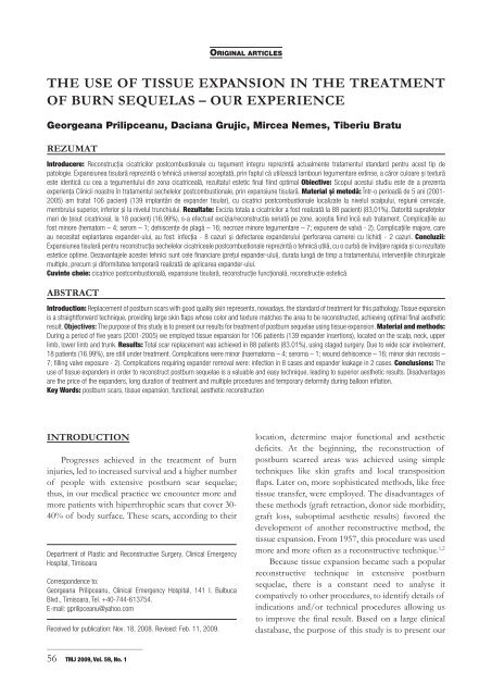
THE USE OF TISSUE EXPANSION IN THE TREATMENT<br />
OF BURN SEQUELAS <strong>–</strong> OUR EXPERIENCE<br />
Georgeana Prilipceanu, Daciana Grujic, Mircea Nemes, Tiberiu Bratu<br />
REZUMAT<br />
Introducere: Reconstrucția cicatricilor postcombustionale cu tegument <strong>in</strong>tegru reprez<strong>in</strong>tă actualmente tratamentul standard pentru acest tip de<br />
patologie. Expansiunea tisulară reprez<strong>in</strong>tă o tehnică universal acceptată, pr<strong>in</strong> faptul că utilizează lamb<strong>our</strong>i tegumentare ext<strong>in</strong>se, a căror culoare și textură<br />
este identică cu cea a tegumentului d<strong>in</strong> zona cicatriceală, rezultatul estetic f<strong>in</strong>al fi<strong>in</strong>d optimal Obiective: Scopul acestui studiu este de a prezenta<br />
experiența Cl<strong>in</strong>icii noastre în tratamentul sechelelor postcombustionale, pr<strong>in</strong> expansiune tisulară. Material și metodă: Într-o perioadă de 5 ani (2001-<br />
2005) am tratat 106 pacienți (139 implantări de expander tisular), cu cicatrici postcombustionale localizate la nivelul scalpului, regiunii cervicale,<br />
membrului superior, <strong>in</strong>ferior și la nivelul trunchiului. Rezultate: Excizia totala a cicatricilor a fost realizată la 88 pacienți (83,01%). Datorită suprafețelor<br />
mari de țesut cicatriceal, la 18 pacienți (16,99%), s-a efectuat excizia/reconstrucția seriată pe zone, aceștia fi<strong>in</strong>d încă sub tratament. Complicațiile au<br />
fost m<strong>in</strong>ore (hematom <strong>–</strong> 4; serom <strong>–</strong> 1; dehiscențe de plagă <strong>–</strong> 16; necroze m<strong>in</strong>ore tegumentare <strong>–</strong> 7; expunere de valvă - 2). Complicațiile majore, care<br />
au necesitat explantarea expander-ului, au fost: <strong>in</strong>fecția - 8 cazuri și defectarea expanderului (perforarea camerei cu lichid) - 2 cazuri. Concluzii:<br />
Expansiunea tisulară pentru reconstrucția sechelelor cicatriceale postcombustionale reprez<strong>in</strong>tă o tehnică utilă, cu o curbă de învățare rapida și cu rezultate<br />
estetice optime. Dezavantajele acestei tehnici sunt cele f<strong>in</strong>anciare (prețul expander-ului), durata lungă de timp a tratamentului, <strong>in</strong>tervențiile chirurgicale<br />
multiple, precum și diformitatea temporară realizată de aplicarea expander-ului.<br />
Cuv<strong>in</strong>te cheie: cicatrice postcombustională, expansiune tisulară, reconstrucție funcțională, reconstrucție estetică<br />
ABSTRACT<br />
Introduction: Replacement <strong>of</strong> post<strong>burn</strong> scars with good quality sk<strong>in</strong> represents, nowadays, <strong>the</strong> standard <strong>of</strong> <strong>treatment</strong> for this pathology. Tissue <strong>expansion</strong><br />
is a straightforward technique, provid<strong>in</strong>g large sk<strong>in</strong> flaps whose color and texture matches <strong>the</strong> area to be reconstructed, achiev<strong>in</strong>g optimal f<strong>in</strong>al aes<strong>the</strong>tic<br />
result. Objectives: The purpose <strong>of</strong> this study is to present <strong>our</strong> results for <strong>treatment</strong> <strong>of</strong> post<strong>burn</strong> sequelae us<strong>in</strong>g <strong>tissue</strong> <strong>expansion</strong>. Material and methods:<br />
Dur<strong>in</strong>g a period <strong>of</strong> five years (2001-2005) we employed <strong>tissue</strong> <strong>expansion</strong> for 106 patients (139 expander <strong>in</strong>sertions), located on <strong>the</strong> scalp, neck, upper<br />
limb, lower limb and trunk. Results: Total scar replacement was achieved <strong>in</strong> 88 patients (83.01%), us<strong>in</strong>g staged surgery. Due to wide scar <strong>in</strong>volvement,<br />
18 patients (16.99%), are still under <strong>treatment</strong>. Complications were m<strong>in</strong>or (haematoma <strong>–</strong> 4; seroma <strong>–</strong> 1; wound dehiscence <strong>–</strong> 16; m<strong>in</strong>or sk<strong>in</strong> necrosis <strong>–</strong><br />
7; fill<strong>in</strong>g valve exposure - 2). Complications requir<strong>in</strong>g expander removal were: <strong>in</strong>fection <strong>in</strong> 8 cases and expander leakage <strong>in</strong> 2 cases. Conclusions: The<br />
<strong>use</strong> <strong>of</strong> <strong>tissue</strong> expanders <strong>in</strong> order to reconstruct post<strong>burn</strong> sequelae is a valuable and easy technique, lead<strong>in</strong>g to superior aes<strong>the</strong>tic results. Disadvantages<br />
are <strong>the</strong> price <strong>of</strong> <strong>the</strong> expanders, long duration <strong>of</strong> <strong>treatment</strong> and multiple procedures and temporary deformity dur<strong>in</strong>g balloon <strong>in</strong>flation.<br />
Key Words: post<strong>burn</strong> scars, <strong>tissue</strong> <strong>expansion</strong>, functional, aes<strong>the</strong>tic reconstruction<br />
INTRODUCTION<br />
Progresses achieved <strong>in</strong> <strong>the</strong> <strong>treatment</strong> <strong>of</strong> <strong>burn</strong><br />
<strong>in</strong>juries, led to <strong>in</strong>creased survival and a higher number<br />
<strong>of</strong> people with extensive post<strong>burn</strong> scar sequelae;<br />
thus, <strong>in</strong> <strong>our</strong> medical practice we encounter more and<br />
more patients with hiperthrophic scars that cover 30-<br />
40% <strong>of</strong> body surface. These scars, accord<strong>in</strong>g to <strong>the</strong>ir<br />
Department <strong>of</strong> Plastic and Reconstructive Surgery, Cl<strong>in</strong>ical Emergency<br />
Hospital, Timisoara<br />
Correspondence to:<br />
Georgeana Prilipceanu, Cl<strong>in</strong>ical Emergency Hospital, 141 I. Bulbuca<br />
Blvd., Timisoara, Tel. +40-744-613754.<br />
E-mail: gprilipceanu@yahoo.com<br />
Received for publication: Nov. 18, 2008. Revised: Feb. 11, 2009.<br />
_____________________________<br />
56 TMJ 2009, Vol. 59, No. 1<br />
ORIGINAL ARTICLES<br />
location, determ<strong>in</strong>e major functional and aes<strong>the</strong>tic<br />
deficits. At <strong>the</strong> beg<strong>in</strong>n<strong>in</strong>g, <strong>the</strong> reconstruction <strong>of</strong><br />
post<strong>burn</strong> scarred areas was achieved us<strong>in</strong>g simple<br />
techniques like sk<strong>in</strong> grafts and local transposition<br />
flaps. Later on, more sophisticated methods, like free<br />
<strong>tissue</strong> transfer, were employed. The disadvantages <strong>of</strong><br />
<strong>the</strong>se methods (graft retraction, donor side morbidity,<br />
graft loss, suboptimal aes<strong>the</strong>tic results) favored <strong>the</strong><br />
development <strong>of</strong> ano<strong>the</strong>r reconstructive method, <strong>the</strong><br />
<strong>tissue</strong> <strong>expansion</strong>. From 1957, this procedure was <strong>use</strong>d<br />
more and more <strong>of</strong>ten as a reconstructive technique. 1,2<br />
Beca<strong>use</strong> <strong>tissue</strong> <strong>expansion</strong> became such a popular<br />
reconstructive technique <strong>in</strong> extensive post<strong>burn</strong><br />
sequelae, <strong>the</strong>re is a constant need to analyse it<br />
compatively to o<strong>the</strong>r procedures, to identify details <strong>of</strong><br />
<strong>in</strong>dications and/or technical procedures allow<strong>in</strong>g us<br />
to improve <strong>the</strong> f<strong>in</strong>al result. Based on a large cl<strong>in</strong>ical<br />
dastabase, <strong>the</strong> purpose <strong>of</strong> this study is to present <strong>our</strong>
esults <strong>in</strong> <strong>tissue</strong> <strong>expansion</strong> for post<strong>burn</strong> sequelae and<br />
to compare it with similar data <strong>in</strong> <strong>the</strong> literature.<br />
MATERIAL AND METHODS<br />
Dur<strong>in</strong>g a five years period (2001-2005), <strong>in</strong> <strong>the</strong><br />
Department <strong>of</strong> Plastic and Reconstructive Surgery <strong>of</strong><br />
<strong>the</strong> Cl<strong>in</strong>ical Emergency County Hospital Timisoara we<br />
have treated 170 patients with scar sequelae <strong>of</strong> various<br />
etiologies, us<strong>in</strong>g <strong>tissue</strong> <strong>expansion</strong> technique. From<br />
<strong>the</strong>se, 106 were admitted with post<strong>burn</strong> sequelae.<br />
Most <strong>of</strong>ten, patients were <strong>burn</strong>ed by fire. (Fig. 1).<br />
The patients’ age was between 4 and 45 years and we<br />
observed a higher rate <strong>of</strong> female patients (70 females<br />
versus 36 males, respectively 66.03% versus 33.96%).<br />
(Figs. 2, 3) There were one or more affected areas per<br />
patient, namely 130 locations: scalp <strong>in</strong> 25/130 locations<br />
(19.23%); nose, ear and face <strong>in</strong> 11/130 locations<br />
(8.46%); neck <strong>in</strong> 26/130 locations (20%), upper limb<br />
<strong>in</strong> 19/130 locations (14.61%), lower limb <strong>in</strong> 15/130<br />
locations (11.54%), anterior trunk <strong>in</strong> 17/130 locations<br />
(13.08 %), and posterior trunk <strong>in</strong> 17/130 locations<br />
(13.08%), some <strong>of</strong> <strong>the</strong> patients present<strong>in</strong>g multiple<br />
locations or requir<strong>in</strong>g multiple or serial <strong>expansion</strong>s <strong>of</strong><br />
<strong>the</strong> same body region. (Fig. 4)<br />
Figure 1. The etiology <strong>of</strong> post<strong>burn</strong> scarr<strong>in</strong>g.<br />
Figure 2. Age repartition with<strong>in</strong> <strong>the</strong> patient group taken <strong>in</strong>to this study.<br />
Expanders produced by Mentor ® , Allergan ® ,<br />
McGhan ® , Eurosilicone ® , Silimed ® , <strong>of</strong> various shapes<br />
and sizes were <strong>use</strong>d (rectangular for <strong>the</strong> scalp, neck,<br />
trunk, upper and lower limb; round expander mostly<br />
<strong>use</strong>d for <strong>the</strong> trunk and kidney-shaped expanders <strong>use</strong>d<br />
mostly for scalp and neck region).<br />
Figure 3. Male/female repatition with<strong>in</strong> patient group taken <strong>in</strong>to this<br />
study.<br />
Figure 4. Localization <strong>of</strong> <strong>the</strong> post<strong>burn</strong> contracture.<br />
When perform<strong>in</strong>g <strong>the</strong> data analysis, we encountered<br />
a variety <strong>of</strong> cl<strong>in</strong>ical situations: (i) patients hav<strong>in</strong>g serial<br />
<strong>expansion</strong> <strong>in</strong> same/different body areas; patients with<br />
simultaneous <strong>expansion</strong> <strong>of</strong> several regions; (ii) cases<br />
<strong>in</strong> which, <strong>in</strong> one operative session, <strong>the</strong> same expander<br />
was removed from an area and <strong>in</strong>serted <strong>in</strong>to ano<strong>the</strong>r<br />
location; (iii) patients that have <strong>the</strong> expander <strong>in</strong>serted<br />
but not removed (<strong>in</strong> c<strong>our</strong>se <strong>of</strong> <strong>in</strong>flation); (iv) patients<br />
that underwent surgery for complications <strong>of</strong> <strong>tissue</strong><br />
<strong>expansion</strong>.<br />
In regard with <strong>the</strong>se f<strong>in</strong>d<strong>in</strong>gs, <strong>the</strong>re is a need to<br />
clarify some notions:<br />
1. Expander-related surgery is <strong>the</strong> central<br />
procedure <strong>of</strong> this method, and refers to expander<br />
<strong>in</strong>sertion or removal after complete <strong>tissue</strong> <strong>expansion</strong>;<br />
it is considered as a separate procedure even if it was<br />
accompanied by o<strong>the</strong>r procedures dur<strong>in</strong>g <strong>the</strong> same<br />
operation.<br />
2. There were 139 expander <strong>in</strong>sertions and 121<br />
expander removals, a total <strong>of</strong> 260 expander-related<br />
procedures. O<strong>the</strong>r <strong>in</strong>terventions were considered<br />
collection evacuation, secondary suture, expander<br />
removal before completion <strong>of</strong> <strong>tissue</strong> <strong>expansion</strong> (43<br />
procedures).<br />
3. A number <strong>of</strong> 18/106 patients underwent<br />
expander <strong>in</strong>sertion and are now <strong>in</strong> c<strong>our</strong>se <strong>of</strong><br />
<strong>in</strong>flation.<br />
_____________________________<br />
Georgeana Prilipceanu et al 57
The expander size was chosen accord<strong>in</strong>g to <strong>the</strong><br />
dimensions <strong>of</strong> <strong>the</strong> defect to be reconstructed and to<br />
<strong>the</strong> characteristics <strong>of</strong> <strong>the</strong> affected zone (smaller <strong>in</strong><br />
<strong>the</strong> scalp, neck, face, nose and upper limb and larger<br />
for trunk and lower limb (thigh and upper 1/3 <strong>of</strong><br />
calf). The volumes <strong>of</strong> <strong>the</strong> expanders volumes varied<br />
between 70-400cc. (Table 1) Preoperatively we checked<br />
up aga<strong>in</strong> <strong>the</strong> measurements taken before order<strong>in</strong>g <strong>the</strong><br />
expanders and do <strong>the</strong> mark<strong>in</strong>gs.<br />
Table 1. The size and types <strong>of</strong> <strong>the</strong> expanders <strong>use</strong>d <strong>in</strong> <strong>the</strong> study, <strong>in</strong><br />
relation with <strong>the</strong> affected body areas.<br />
Region<br />
Expanders<br />
size and type<br />
Rectangular<br />
(100-400 cc)<br />
Croissant<br />
(70-150cc)<br />
Round<br />
(160-180cc)<br />
Scalp<br />
Face/<br />
Nose/<br />
Ear<br />
_____________________________<br />
58 TMJ 2009, Vol. 59, No. 1<br />
Neck<br />
region<br />
Upper<br />
and<br />
lower<br />
limb<br />
17 11 20 34 30<br />
8 - 6 - -<br />
- - - - 4<br />
Anterior<br />
and<br />
posterior<br />
trunk<br />
Operative technique<br />
Under general anes<strong>the</strong>sia, an <strong>in</strong>cision was made<br />
<strong>in</strong> <strong>the</strong> vic<strong>in</strong>ity <strong>of</strong> <strong>the</strong> defect (0.5 cm); its length wass<br />
correlated with <strong>the</strong> affected area and <strong>the</strong> expander’s<br />
size, <strong>the</strong> orientation was perpendicular on <strong>the</strong> long<br />
axis <strong>of</strong> <strong>the</strong> defect <strong>in</strong> 98/139 cases and parallel to<br />
it <strong>in</strong> 41/139 cases. Dissection was cont<strong>in</strong>ued until<br />
<strong>the</strong> suprafascial plane - anterior and/or posterior<br />
trunk/upper and lower limb, or under <strong>the</strong> galeea at<br />
scalp level. Two pockets were dissected, one for <strong>the</strong><br />
expander and one for <strong>the</strong> fill<strong>in</strong>g valve. After plac<strong>in</strong>g<br />
<strong>the</strong> expanders and <strong>the</strong> dra<strong>in</strong>age tubes, <strong>the</strong> wound was<br />
sutured <strong>in</strong> two layers. At <strong>the</strong> end <strong>of</strong> <strong>the</strong> procedure,<br />
<strong>the</strong> expander was filled with sal<strong>in</strong>e (about 10-20% <strong>of</strong><br />
expander’s volume).<br />
Postoperatively, antibiotics were adm<strong>in</strong>istered<br />
for 5-7 days and analgetics for 2-3 days, as needed.<br />
In most <strong>of</strong> <strong>the</strong> cases, <strong>tissue</strong> <strong>expansion</strong> started after<br />
two weeks. In 10/139 cases expander <strong>in</strong>flation was<br />
delayed for several days, until <strong>the</strong> wound was totally<br />
healed.<br />
The expander <strong>in</strong>flations were made as outpatient<br />
procedures. The amount <strong>of</strong> sal<strong>in</strong>e to be <strong>in</strong>troduced<br />
<strong>in</strong> each session depends upon <strong>the</strong> anatomical region,<br />
expander’s capacity and patient’s tolerance. The average<br />
time <strong>of</strong> <strong>tissue</strong> <strong>expansion</strong> was 3 months, at a frequency<br />
<strong>of</strong> one <strong>in</strong>flation/week. In 92/139 cases (66.19%) <strong>the</strong><br />
over<strong>in</strong>flation <strong>of</strong> <strong>the</strong> expanders with 20% excess <strong>of</strong> <strong>the</strong><br />
total volume was achieved.<br />
RESULTS<br />
The second stage <strong>of</strong> <strong>the</strong> surgical procedure<br />
(expander removal and scar replacement: 121 cases)<br />
was performed at 3 months after <strong>the</strong> first <strong>in</strong>tervention<br />
<strong>in</strong> 85 cases (70.25%) that evolved without any<br />
complication.<br />
Complications <strong>of</strong> <strong>tissue</strong> <strong>expansion</strong> were encountered<br />
<strong>in</strong> 40/139 cases (28.78%). M<strong>in</strong>or complications<br />
were considered those that allowed cont<strong>in</strong>uation <strong>of</strong><br />
<strong>tissue</strong> <strong>expansion</strong>, while major complications required<br />
expander removal before completion <strong>of</strong> <strong>treatment</strong>.<br />
In <strong>the</strong> first category we encountered haematoma,<br />
seroma, wound dehiscence and fill valve exposure - <strong>in</strong><br />
30/139 cases (21.58%). From <strong>the</strong>se, 26 were noticed<br />
<strong>in</strong> patients that completed <strong>treatment</strong> and 4 <strong>in</strong> <strong>the</strong><br />
lot <strong>of</strong> 18 patients still under <strong>treatment</strong>. The second<br />
category comprised severe <strong>in</strong>fection and expander<br />
leakage due to manufacture defect - <strong>in</strong> 10/139 cases<br />
(7.19%). (Table 2) In <strong>the</strong> 26 cases that presented<br />
m<strong>in</strong>or complications, but f<strong>in</strong>ished <strong>tissue</strong> <strong>expansion</strong>,<br />
<strong>the</strong> second procedure was delayed for 3 weeks, wait<strong>in</strong>g<br />
for complete wound heal<strong>in</strong>g after necrectomy or<br />
evacuation <strong>of</strong> collections. Wound dehiscence was<br />
encountered mostly <strong>in</strong> cases <strong>of</strong> reconstruction <strong>in</strong><br />
scalp and neck region. The <strong>in</strong>fection (8/139 cases,<br />
5.76%) was secondary to wound dehiscence and<br />
sk<strong>in</strong> necrosis. In <strong>the</strong>se cases <strong>the</strong> expanders had to be<br />
removed with<strong>in</strong> a timeframe <strong>of</strong> 3 weeks to 3 months<br />
from <strong>the</strong> implantation and <strong>the</strong> surgical procedure<br />
was repeated after <strong>the</strong> total heal<strong>in</strong>g <strong>of</strong> <strong>the</strong> wound.<br />
Table 2. Complications encountered with<strong>in</strong> <strong>the</strong> patient group.<br />
Type <strong>of</strong> complication Number<br />
<strong>of</strong> cases<br />
Haematomas 4 3.77<br />
Seromas 1 0.94<br />
Percentage<br />
(%)<br />
Wound dehiscence 16 15.09<br />
Necrosis 7 6.60<br />
Infections 8 7.54<br />
Valve exposure 2 1.88<br />
Expander leakage 2 1.88<br />
After expander removal and scar excision, <strong>the</strong><br />
result<strong>in</strong>g defect was covered with <strong>the</strong> expanded <strong>tissue</strong>,<br />
which had similar characteristics <strong>of</strong> color and texture<br />
with <strong>the</strong> nearby sk<strong>in</strong>. Complete excision <strong>of</strong> <strong>the</strong> scars<br />
was achieved <strong>in</strong> 88/106 patients (83.01%), and it was<br />
done <strong>in</strong> 1-4 <strong>expansion</strong> stages. In 70/106 patients total
scar removal was done <strong>in</strong> a s<strong>in</strong>gle stage <strong>of</strong> <strong>expansion</strong>,<br />
<strong>in</strong> 13/106 patients <strong>the</strong>re were necessary 2 stages, 3/106<br />
patients needed 3 stages, and 2/106 patients needed 4<br />
stages <strong>of</strong> <strong>tissue</strong> <strong>expansion</strong>. There are 18/106 patients<br />
(16.98%) still <strong>in</strong> <strong>treatment</strong>. Some patients were treated<br />
by us<strong>in</strong>g 2 expanders simultaneously; <strong>in</strong> one patient we<br />
<strong>use</strong>d 4 expanders <strong>in</strong> <strong>the</strong> same time (one for cervical<br />
area, one for <strong>the</strong> anterior trunk and 2 for <strong>the</strong> lower<br />
limb). (Table 3)<br />
Table 3. Number <strong>of</strong> patients with def<strong>in</strong>itive scar excision and<br />
reconstruction versus those still <strong>in</strong> c<strong>our</strong>se <strong>of</strong> <strong>treatment</strong>, accord<strong>in</strong>g to <strong>the</strong><br />
<strong>expansion</strong> stage.<br />
Total number<br />
<strong>of</strong> patients <strong>in</strong><br />
<strong>the</strong> study:<br />
106 patients<br />
Patients with<br />
complete scar<br />
removal<br />
88 patients<br />
(83.01%)<br />
Patients <strong>in</strong><br />
c<strong>our</strong>se <strong>of</strong><br />
<strong>treatment</strong><br />
18 patients<br />
(16,98%)<br />
DISCUSSIONS<br />
1 st stage <strong>of</strong><br />
<strong>expansion</strong><br />
2 nd stage <strong>of</strong><br />
<strong>expansion</strong><br />
3 rd stage <strong>of</strong><br />
<strong>expansion</strong><br />
4 th stage <strong>of</strong><br />
<strong>expansion</strong><br />
1 st stage <strong>of</strong><br />
<strong>expansion</strong><br />
2 nd stage <strong>of</strong><br />
<strong>expansion</strong><br />
3 rd stage <strong>of</strong><br />
<strong>expansion</strong><br />
70 patients<br />
13 patients<br />
3 patients<br />
2 patients<br />
12 patients<br />
4 patients<br />
2 patients<br />
Spontaneous heal<strong>in</strong>g <strong>of</strong> <strong>in</strong>termediate deep <strong>burn</strong>s<br />
and sk<strong>in</strong> graft coverage <strong>of</strong> deep <strong>burn</strong>s leaves beh<strong>in</strong>d<br />
areas <strong>of</strong> postcombustional scars that raise o<strong>the</strong>r<br />
issues: <strong>the</strong>y tend to shr<strong>in</strong>k dur<strong>in</strong>g <strong>the</strong>ir evolution and<br />
frequently become unstable, thus hav<strong>in</strong>g a high risk <strong>of</strong><br />
ulceration, leav<strong>in</strong>g beh<strong>in</strong>d functional impairment and<br />
unpleasant appearance.<br />
In <strong>the</strong> past, <strong>the</strong> <strong>treatment</strong> <strong>of</strong> large area post<strong>burn</strong><br />
sequelae consisted ma<strong>in</strong>ly <strong>in</strong> <strong>in</strong>cision/excision<br />
followed by sk<strong>in</strong> grafts or transposition <strong>of</strong> local sk<strong>in</strong><br />
flaps. Frequently encountered problems were sk<strong>in</strong><br />
graft retraction with functional deficit and unpleasant<br />
cosmetic aspects, or <strong>in</strong>sufficient local res<strong>our</strong>ces for<br />
cover<strong>in</strong>g <strong>the</strong> rema<strong>in</strong><strong>in</strong>g sk<strong>in</strong> defects after scar excision.<br />
Use <strong>of</strong> free <strong>tissue</strong> transfer is a tedious procedure,<br />
with more or less donor site availability and morbidity<br />
and higher risk <strong>of</strong> flap necrosis. In <strong>the</strong> last years, <strong>tissue</strong><br />
<strong>expansion</strong> technique has improved <strong>the</strong> postoperative<br />
results <strong>in</strong> <strong>the</strong>se cases.<br />
One <strong>of</strong> <strong>the</strong> ma<strong>in</strong> advantages <strong>of</strong> <strong>tissue</strong> <strong>expansion</strong> is<br />
that it allows full <strong>use</strong> <strong>of</strong> <strong>the</strong> tegument <strong>of</strong> any region. 4<br />
A<br />
B<br />
Figure 5. Male patient, 35 y.o., post-<strong>burn</strong> alopecia (140 cm 2 ), treated<br />
by <strong>tissue</strong> <strong>expansion</strong> us<strong>in</strong>g one croissant-shaped and one rectangularshaped<br />
expanders: (A) preoperative plann<strong>in</strong>g <strong>of</strong> scar excision; (B) six<br />
months after def<strong>in</strong>itive reconstruction.<br />
The rate <strong>of</strong> major <strong>in</strong>fections <strong>in</strong> <strong>our</strong> series<br />
(requir<strong>in</strong>g expander removal before completion <strong>of</strong><br />
<strong>tissue</strong> <strong>expansion</strong>) was 5.76% (8/139 cases), slightly<br />
<strong>in</strong>creased compared to similar data reported <strong>in</strong><br />
literature, 3.1%. 5-8 We also encountered two cases<br />
<strong>of</strong> expander malfunction with consequent leakage,<br />
lead<strong>in</strong>g to expander removal and replacement.<br />
The rate <strong>of</strong> occurence <strong>of</strong> m<strong>in</strong>or complications <strong>in</strong><br />
<strong>our</strong> series was 21.58% (30/139 cases), very close to<br />
data <strong>in</strong> <strong>the</strong> medical literature quot<strong>in</strong>g a rate <strong>of</strong> m<strong>in</strong>or<br />
complications <strong>of</strong> 20%. 9 We consider that <strong>the</strong> relatively<br />
low rate <strong>of</strong> complications is due to <strong>the</strong> <strong>in</strong>creased<br />
experience accumulated over time and to proper<br />
patient selection.<br />
Special issues have appeared <strong>in</strong> <strong>the</strong> reconstruction<br />
<strong>of</strong> <strong>the</strong> neck, where <strong>the</strong> tegument is very th<strong>in</strong>, has great<br />
elasticity and <strong>the</strong>re is no “real” solid plane underneath<br />
<strong>the</strong> expander. In large defects, <strong>the</strong> <strong>expansion</strong> was done<br />
<strong>in</strong> two stages us<strong>in</strong>g <strong>in</strong>creas<strong>in</strong>g expander volumes,<br />
<strong>in</strong> order to achieve f<strong>in</strong>ally defect coverage. At this<br />
level, most frequently, we encountered, marg<strong>in</strong>al flap<br />
necrosis and wound dehiscence, and this was <strong>the</strong><br />
reason for delay<strong>in</strong>g <strong>the</strong> <strong>treatment</strong> about three weeks.<br />
_____________________________<br />
Georgeana Prilipceanu et al 59
A<br />
B<br />
Figure 6. Female patient, 27 y.o., post-<strong>burn</strong> scars (230 cm 2 ) affect<strong>in</strong>g<br />
<strong>the</strong> right thigh, treated by <strong>tissue</strong> <strong>expansion</strong> us<strong>in</strong>g 2 rectangular <strong>tissue</strong><br />
expanders: (A) preoperative plann<strong>in</strong>g; (B) at 2 weeks after def<strong>in</strong>itive<br />
reconstruction.<br />
Similar data from <strong>the</strong> literature emphasize that most<br />
frequent complications appear dur<strong>in</strong>g <strong>tissue</strong> <strong>expansion</strong><br />
<strong>in</strong> <strong>the</strong> cervical region. 10 At this level it is <strong>in</strong>dicated to<br />
<strong>use</strong> free <strong>tissue</strong> transfer or expanded supraclavicular<br />
flaps, especially when <strong>the</strong> scars are extended to <strong>the</strong><br />
anterior chest area or <strong>in</strong> <strong>the</strong> supraclavicular region. 11<br />
On <strong>the</strong> scalp an additional challenge is to<br />
reconstruct <strong>the</strong> hairl<strong>in</strong>e and <strong>the</strong> hair-bear<strong>in</strong>g sk<strong>in</strong>. After<br />
deep <strong>burn</strong>s, <strong>the</strong> destruction <strong>of</strong> <strong>the</strong> hair bulbs leads to<br />
post<strong>burn</strong> alopecia. Sk<strong>in</strong> grafts <strong>use</strong>d <strong>in</strong> <strong>the</strong> emergency<br />
<strong>treatment</strong> <strong>of</strong> scalp <strong>burn</strong>s leaves beh<strong>in</strong>d a sequelar<br />
alopecia which frequently becomes unstable. In those<br />
cases where <strong>the</strong> sk<strong>in</strong> defect is over 50cm 2 , <strong>tissue</strong><br />
<strong>expansion</strong> seems to be <strong>the</strong> only surgical solution. 12,13<br />
Tissue <strong>expansion</strong> at this level was achieved successfully<br />
<strong>in</strong> <strong>our</strong> series <strong>in</strong> 25 out <strong>of</strong> 106 patients, us<strong>in</strong>g rectangular<br />
and reniform <strong>tissue</strong> expanders; <strong>in</strong> 2 cases <strong>the</strong> procedure<br />
was accompanied by <strong>the</strong> reconstruction <strong>of</strong> underly<strong>in</strong>g<br />
excised bone us<strong>in</strong>g titanium plates. The complications<br />
encountered <strong>in</strong> this area were wound dehiscence and<br />
m<strong>in</strong>imal marg<strong>in</strong>al necrosis <strong>of</strong> <strong>the</strong> expanded flaps. This<br />
is most probably due to atrophic or scared <strong>tissue</strong>. 14,15<br />
CONCLUSIONS<br />
Tissue <strong>expansion</strong> is a <strong>use</strong>ful method for<br />
reconstruction <strong>of</strong> any region where we have at least<br />
tegument <strong>in</strong>tegrity. The best results depend on <strong>the</strong><br />
selection <strong>of</strong> patients, <strong>the</strong>ir ability to understand <strong>the</strong><br />
procedure and on surgeon’s experience.<br />
_____________________________<br />
60 TMJ 2009, Vol. 59, No. 1<br />
A<br />
B<br />
C<br />
Figure 7. Female patient, 30 y.o., post-<strong>burn</strong> scars (450 cm 2 ) affect<strong>in</strong>g<br />
<strong>the</strong> hip region, treated by us<strong>in</strong>g two <strong>tissue</strong> rectangular <strong>tissue</strong> expanders:<br />
(A) preoperative appearance; (B) f<strong>in</strong>al <strong>tissue</strong> <strong>expansion</strong> at 3 months; (C)<br />
one week after def<strong>in</strong>itive reconstruction.
REFERENCES<br />
1. Argenta LC, Watanabe MJ, Grabb WC. The <strong>use</strong> <strong>of</strong> <strong>tissue</strong> <strong>expansion</strong> <strong>in</strong><br />
head and neck reconstruction. Ann Plast Surg 1983;11:31-7.<br />
2. Neuman CG. The <strong>expansion</strong> <strong>of</strong> an area <strong>of</strong> sk<strong>in</strong> by progressive distension<br />
<strong>of</strong> subcutaneous ballon. Plast Reconstr Surg 1957;19:124-9.<br />
3. Bratu T, Isac F. Expansiune tisulara dirijata. Mirton: Timisoara, 1995,<br />
p. 43-77.<br />
4. Norstrom RE, Dev<strong>in</strong> JW. Scalp stretch<strong>in</strong>g with a <strong>tissue</strong> expander for<br />
closure scalp defects. Plast Reconstr Surg.1985;75:578-81.<br />
5. Argenta LC. Tissue <strong>expansion</strong>. In Aston Grabb and Smith`s Plastic<br />
Surgery, Lipp<strong>in</strong>cot Raven, 1997, p. 93-105.<br />
6. Gurlek A, Alaybeyoglu N, Canser YD, et al. Aes<strong>the</strong>tic reconstruction <strong>of</strong><br />
large scalp defects by sequential <strong>tissue</strong> <strong>expansion</strong> without <strong>in</strong>terval.<br />
Aest Plast Surg. 2004;28:245-50.<br />
7. Thomas C, Mishra P. Cl<strong>in</strong>ical experience <strong>of</strong> over <strong>in</strong>flation <strong>in</strong> <strong>tissue</strong><br />
<strong>expansion</strong> technique. Eur J Plast Surg 2001;24:234-8.<br />
8. Guzel Z, Yagmur A, Ak<strong>in</strong> Y, et al. Aes<strong>the</strong>tic results <strong>of</strong> treatement <strong>of</strong> large<br />
alopecia with total scalp <strong>expansion</strong>. Aesth Plast Surg 2000;24:130-6.<br />
9. Dogra BB, Sharma A, S<strong>in</strong>gh M, et al. Tissue <strong>expansion</strong>: A simple<br />
technique for complex traumatic s<strong>of</strong>t <strong>tissue</strong> defects. Int Surg<br />
2007;92:37-45.<br />
10. Hurvitz KA, Rosen H, Meara JG. Pediatric cervic<strong>of</strong>acial <strong>tissue</strong><br />
<strong>expansion</strong>. Int J Pediatr Otorh<strong>in</strong>olaryngol 2005;69:1509-13.<br />
11. Moroz VY, Yudenich A, Kafarov TG, et al. Reconstruction <strong>of</strong> extensive<br />
post<strong>burn</strong> scar deformities and contractures <strong>of</strong> <strong>the</strong> neck us<strong>in</strong>g<br />
expanded and nonexpanded free <strong>tissue</strong> transfer. Eur J Plast Surg<br />
2001;24:217-20.<br />
12. Sniezek JC, Sabri A, Buekey BB, et al. Reconstruction after <strong>burn</strong>s <strong>of</strong><br />
<strong>the</strong> face and neck. Otolaryngol Head and Neck Surg 2000;8:277-81.<br />
13. Micheau P, Costagliola M, Chavo<strong>in</strong> JP, et al. Le scalp: la greffe m<strong>in</strong>ce de<br />
premiere <strong>in</strong>tention en urgence. Ann Chir Plast Es<strong>the</strong>t 1986;31:351-9.<br />
14. Antonyshyn O, Gruss JS, MacK<strong>in</strong>non SE, et al. Complication <strong>of</strong> s<strong>of</strong>t<br />
<strong>tissue</strong> <strong>expansion</strong>. Br J Plast Surg, 1988;41:239-59.<br />
15. Manders NK, Schenden MJ, Furrey JA. S<strong>of</strong>t <strong>tissue</strong> <strong>expansion</strong>. Concepts<br />
and complications. Plast Reconstr Surgery 1984;71:493-502.<br />
_____________________________<br />
Georgeana Prilipceanu et al 61


