Bilateral Avascular Necrosis of the Trapezoid - Chirurgie de la Main ...
Bilateral Avascular Necrosis of the Trapezoid - Chirurgie de la Main ...
Bilateral Avascular Necrosis of the Trapezoid - Chirurgie de la Main ...

You also want an ePaper? Increase the reach of your titles
YUMPU automatically turns print PDFs into web optimized ePapers that Google loves.
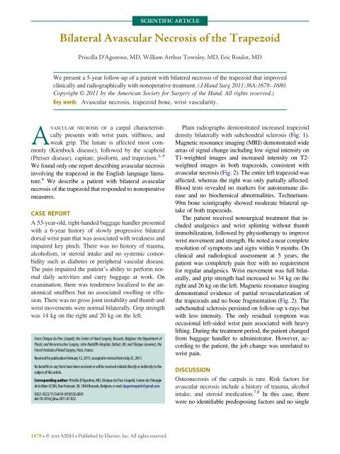
SCIENTIFIC ARTICLE<br />
<strong>Bi<strong>la</strong>teral</strong> <strong>Avascu<strong>la</strong>r</strong> <strong>Necrosis</strong> <strong>of</strong> <strong>the</strong> <strong>Trapezoid</strong><br />
Priscil<strong>la</strong> D’Agostino, MD, William Arthur Townley, MD, Eric Roulot, MD<br />
We present a 5-year follow-up <strong>of</strong> a patient with bi<strong>la</strong>teral necrosis <strong>of</strong> <strong>the</strong> trapezoid that improved<br />
clinically and radiographically with nonoperative treatment. (J Hand Surg 2011;36A:1678–1680.<br />
Copyright © 2011 by <strong>the</strong> American Society for Surgery <strong>of</strong> <strong>the</strong> Hand. All rights reserved.)<br />
Key words <strong>Avascu<strong>la</strong>r</strong> necrosis, trapezoid bone, wrist vascu<strong>la</strong>rity.<br />
AVASCULAR NECROSIS OF a carpal characteristically<br />
presents with wrist pain, stiffness, and<br />
weak grip. The lunate is affected most commonly<br />
(Kienbock disease), followed by <strong>the</strong> scaphoid<br />
(Preiser disease), capitate, pisiform, and trapezium. 1–5<br />
We found only one report <strong>de</strong>scribing avascu<strong>la</strong>r necrosis<br />
involving <strong>the</strong> trapezoid in <strong>the</strong> English <strong>la</strong>nguage literature.<br />
6 We <strong>de</strong>scribe a patient with bi<strong>la</strong>teral avascu<strong>la</strong>r<br />
necrosis <strong>of</strong> <strong>the</strong> trapezoid that respon<strong>de</strong>d to nonoperative<br />
measures.<br />
CASE REPORT<br />
A 53-year-old, right-han<strong>de</strong>d baggage handler presented<br />
with a 6-year history <strong>of</strong> slowly progressive bi<strong>la</strong>teral<br />
dorsal wrist pain that was associated with weakness and<br />
impaired key pinch. There was no history <strong>of</strong> trauma,<br />
alcoholism, or steroid intake and no systemic comorbidity<br />
such as diabetes or peripheral vascu<strong>la</strong>r disease.<br />
The pain impaired <strong>the</strong> patient’s ability to perform normal<br />
daily activities and carry baggage at work. On<br />
examination, <strong>the</strong>re was ten<strong>de</strong>rness localized to <strong>the</strong> anatomical<br />
snuffbox but no associated swelling or effusion.<br />
There was no gross joint instability and thumb and<br />
wrist movements were normal bi<strong>la</strong>terally. Grip strength<br />
was 14 kg on <strong>the</strong> right and 20 kg on <strong>the</strong> left.<br />
From Clinique du Parc Léopold, <strong>the</strong> Center <strong>of</strong> Hand Surgery, Brussels, Belgium; <strong>the</strong> Department <strong>of</strong><br />
P<strong>la</strong>stic and Reconstructive Surgery, John Radcliffe Hospital, Oxford, UK; and Clinique Jouvenet, <strong>the</strong><br />
FrenchInstitute<strong>of</strong>HandSurgery,Paris,France.<br />
Received for publication February 12, 2011; accepted in revised form July 25, 2011.<br />
No benefits in any form have been received or will be received re<strong>la</strong>ted directly or indirectly to <strong>the</strong><br />
subject <strong>of</strong> this article.<br />
Corresponding author: Priscil<strong>la</strong> D’Agostino, MD, Clinique du Parc Léopold, Centre <strong>de</strong> <strong>Chirurgie</strong><br />
<strong>de</strong> <strong>la</strong> <strong>Main</strong> (CCM), Rue Froissart, 38, 1040 Brussels, Belgium; e-mail: dagostinoprish@gmail.com.<br />
0363-5023/11/36A10-0018$36.00/0<br />
doi:10.1016/j.jhsa.2011.07.022<br />
1678 © 2011 ASSH Published by Elsevier, Inc. All rights reserved.<br />
P<strong>la</strong>in radiographs <strong>de</strong>monstrated increased trapezoid<br />
<strong>de</strong>nsity bi<strong>la</strong>terally with subchondral sclerosis (Fig. 1).<br />
Magnetic resonance imaging (MRI) <strong>de</strong>monstrated wi<strong>de</strong><br />
areas <strong>of</strong> signal change including low signal intensity on<br />
T1-weighted images and increased intensity on T2weighted<br />
images in both trapezoids, consistent with<br />
avascu<strong>la</strong>r necrosis (Fig. 2). The entire left trapezoid was<br />
affected, whereas <strong>the</strong> right was only partially affected.<br />
Blood tests revealed no markers for autoimmune disease<br />
and no biochemical abnormalities. Technetium-<br />
99m bone scintigraphy showed mo<strong>de</strong>rate bi<strong>la</strong>teral uptake<br />
<strong>of</strong> both trapezoids.<br />
The patient received nonsurgical treatment that inclu<strong>de</strong>d<br />
analgesics and wrist splinting without thumb<br />
immobilization, followed by physio<strong>the</strong>rapy to improve<br />
wrist movement and strength. He noted a near complete<br />
resolution <strong>of</strong> symptoms and signs within 9 months. On<br />
clinical and radiological assessment at 5 years, <strong>the</strong><br />
patient was completely pain free with no requirement<br />
for regu<strong>la</strong>r analgesics. Wrist movement was full bi<strong>la</strong>terally,<br />
and grip strength had increased to 34 kg on <strong>the</strong><br />
right and 26 kg on <strong>the</strong> left. Magnetic resonance imaging<br />
<strong>de</strong>monstrated evi<strong>de</strong>nce <strong>of</strong> partial revascu<strong>la</strong>rization <strong>of</strong><br />
<strong>the</strong> trapezoids and no bone fragmentation (Fig. 2). The<br />
subchondral sclerosis persisted on follow-up x-rays but<br />
with less intensity. The only residual symptom was<br />
occasional left-si<strong>de</strong>d wrist pain associated with heavy<br />
lifting. During <strong>the</strong> treatment period, <strong>the</strong> patient changed<br />
from baggage handler to administrator. However, according<br />
to <strong>the</strong> patient, <strong>the</strong> job change was unre<strong>la</strong>ted to<br />
wrist pain.<br />
DISCUSSION<br />
Osteonecrosis <strong>of</strong> <strong>the</strong> carpals is rare. Risk factors for<br />
avascu<strong>la</strong>r necrosis inclu<strong>de</strong> a history <strong>of</strong> trauma, alcohol<br />
intake, and steroid medication. 7,8 In this case, <strong>the</strong>re<br />
were no i<strong>de</strong>ntifiable predisposing factors and no single
precipitating event, although as a baggage handler <strong>the</strong><br />
patient may have experienced repetitive microtrauma.<br />
The bi<strong>la</strong>teral occurrence raised <strong>the</strong> possibility <strong>of</strong> a systemic<br />
cause, although none was i<strong>de</strong>ntified. 9–12<br />
AVASCULAR NECROSIS OF THE TRAPEZOID 1679<br />
FIGURE 1: A Right wrist and B left wrist posteroanterior radiographs <strong>de</strong>monstrating sclerosis <strong>of</strong> both trapezoids.<br />
FIGURE 2: A Right wrist and B left wrist T2-weighted coronal MRI showing avascu<strong>la</strong>r necrosis <strong>of</strong> both trapezoids. C Right wrist<br />
and D left wrist T2-weighted Fat saturation coronal MRI showing partial revascu<strong>la</strong>rization 5 years after presentation.<br />
JHS Vol 36A, October 2011<br />
Magnetic resonance imaging is <strong>the</strong> best imaging<br />
technique to diagnose avascu<strong>la</strong>r necrosis. It is more<br />
sensitive than radiographs and bone scintigraphy. 13–19<br />
Necrotic bone generally produces a low or absent signal
1680 AVASCULAR NECROSIS OF THE TRAPEZOID<br />
on T1-weighted images because <strong>of</strong> <strong>the</strong> absence <strong>of</strong> normal<br />
bone marrow elements, whereas signal uptake is<br />
increased on T2-weighted images (bone marrow<br />
e<strong>de</strong>ma). The intravenous injection <strong>of</strong> gadolinium improves<br />
<strong>the</strong> MRI specificity <strong>of</strong> avascu<strong>la</strong>r necrosis diagnosis.<br />
6,13–19 Computed tomography images can help<br />
i<strong>de</strong>ntify secondary changes such as subtle <strong>de</strong>pressions,<br />
col<strong>la</strong>pse, or fragmentation. Sometimes osteoarthritis,<br />
subchondral cysts, and transient osteoporosis may<br />
mimic osteonecrosis on p<strong>la</strong>in films or MRI.<br />
A study by Gelberman and Gross 20 analyzed carpal<br />
vascu<strong>la</strong>r anatomy in cadaveric specimens and i<strong>de</strong>ntified<br />
3 specific patterns, each with a different avascu<strong>la</strong>r necrosis<br />
risk pr<strong>of</strong>ile. The trapezoid and <strong>the</strong> hamate were<br />
c<strong>la</strong>ssified into group 2, a low-risk group characterized<br />
by absence <strong>of</strong> internal anastomoses. <strong>Avascu<strong>la</strong>r</strong> necrosis<br />
<strong>of</strong> <strong>the</strong> hamate is also rare. 21 The trapezoid benefits from<br />
a varied extraosseous blood supply including branches<br />
from <strong>the</strong> dorsal radiocarpal, intercarpal, and basal metacarpal<br />
arches as well as from <strong>the</strong> radial recurrent artery.<br />
Therefore, <strong>the</strong> trapezoid does not <strong>de</strong>pend on a single<br />
vessel but is nourished by a rich network <strong>of</strong> dorsal<br />
(70%) and palmar vessels (30%), which may exp<strong>la</strong>in<br />
why it is affected rarely by avascu<strong>la</strong>r necrosis. 18<br />
The natural history <strong>of</strong> osteonecrosis is unpredictable.<br />
Bone revascu<strong>la</strong>rization may occur or <strong>the</strong> condition may<br />
progress to sclerosis and bone fragmentation. 22,23<br />
Treatment inclu<strong>de</strong>s nonoperative measures as well as<br />
surgical intervention including arthro<strong>de</strong>sis, osteotomy,<br />
and excision arthrop<strong>la</strong>sty. 22,24–26 Some authors have<br />
advocated <strong>the</strong> use <strong>of</strong> vascu<strong>la</strong>rized bone grafts to encourage<br />
bone revascu<strong>la</strong>rization. 6 In addition to having a<br />
protective effect, <strong>the</strong> rich blood supply <strong>of</strong> <strong>the</strong> trapezoid<br />
may also make it more likely to recover. 18,20<br />
REFERENCES<br />
1. Match RM. Nonspecific avascu<strong>la</strong>r necrosis <strong>of</strong> <strong>the</strong> pisiform bone: a<br />
case report. J Hand Surg 1980;5:341–342.<br />
2. Boyer MI, Von Schroe<strong>de</strong>r HP, Axelrod TS. Scaphoid non-union with<br />
avascu<strong>la</strong>r necrosis <strong>of</strong> <strong>the</strong> proximal pole. Treatment with a vascu<strong>la</strong>rized<br />
bone graft from <strong>the</strong> dorsum <strong>of</strong> <strong>the</strong> distal radius. J Hand Surg<br />
1998;23B:686–690.<br />
3. Garcia-Lopez A, Cardoso Z, Ortega L. <strong>Avascu<strong>la</strong>r</strong> necrosis <strong>of</strong> trapezium<br />
bone. J Hand Surg 2002;27A:704–706.<br />
4. Whiting J, Rotman MB. Scaphocapitolunate arthro<strong>de</strong>sis for idiopathic<br />
avascu<strong>la</strong>r necrosis <strong>of</strong> <strong>the</strong> capitate: a case report. J Hand Surg<br />
2002;27A:692–696.<br />
JHS Vol 36A, October 2011<br />
5. Ka<strong>la</strong>inov DM, Cohen MS, Hendrix RW, Sweet S, Culp RW, Osterman<br />
AL. Preiser’s disease: i<strong>de</strong>ntification <strong>of</strong> two patterns. J Hand<br />
Surg 2003;28A:767–768.<br />
6. Sturzenegger M, Mencarelli F. <strong>Avascu<strong>la</strong>r</strong> necrosis <strong>of</strong> <strong>the</strong> trapezoid<br />
bone. J Hand Surg 1998;23B:550–551.<br />
7. Stein AH. Dorsal dislocation <strong>of</strong> <strong>the</strong> lesser multangu<strong>la</strong>r bone. J Bone<br />
Joint Surg 1971;53A:377–379.<br />
8. Dunkerton M, Singer M. Dislocation <strong>of</strong> <strong>the</strong> in<strong>de</strong>x metacarpal and<br />
trapezoid bones. J Hand Surg 1985;10B:377–378.<br />
9. Agus B. <strong>Bi<strong>la</strong>teral</strong> aseptic necrosis <strong>of</strong> <strong>the</strong> lunate in systematic sclerosis.<br />
Clin Exp Rheumatol 1987;5:155–157.<br />
10. Nagazawa K, Ishii Y, Mayumi T, Tada Y, Ueda A, Yamauchi Y, et<br />
al. <strong>Avascu<strong>la</strong>r</strong> necrosis <strong>of</strong> bone in systematic lupus ery<strong>the</strong>matosus:<br />
possible role <strong>of</strong> haemostatic abnormalities. Ann Rheum Dis 1989;<br />
48:672–676.<br />
11. A<strong>de</strong>lowo OO, Oguntona S. <strong>Avascu<strong>la</strong>r</strong> necrosis <strong>of</strong> femoral head<br />
associated with connective tissue disease in Nigerians: case series.<br />
West Afr J Med 2009;28:262–265.<br />
12. Maes M, Hansen L, Cheyns P. Osteochondral mosaicp<strong>la</strong>sty as a<br />
treatment method for bi<strong>la</strong>teral avascu<strong>la</strong>r necrosis <strong>of</strong> <strong>the</strong> long finger<br />
metacarpal: case report. J Hand Surg 2010;35A:1264–1268.<br />
13. Reinus WR, Conway WF, Totty WG, Gilu<strong>la</strong> LA, Murphy WA,<br />
Siegel BA, et al. Carpal avascu<strong>la</strong>r necrosis: MR imaging. Radiology<br />
1986;160:689–693.<br />
14. Cristiani G, Cer<strong>of</strong>olini E, Squarzina PB, Zanasi S, Leoni A, Romagnoli<br />
R, et al. Evaluation <strong>of</strong> ischaemic necrosis <strong>of</strong> carpal bones by<br />
magnetic resonance imaging. J Hand Surg.1990;15B:249–255.<br />
15. Trumble TE. <strong>Avascu<strong>la</strong>r</strong> necrosis after scaphoid fracture: a corre<strong>la</strong>tion<br />
<strong>of</strong> magnetic resonance imaging and histology. J Hand Surg<br />
1990;15A:557–564.<br />
16. Perlik PC, Guilford WB. Magnetic resonance imaging to assess<br />
vascu<strong>la</strong>rity <strong>of</strong> scaphoid nonunions. J Hand Surg 1991;16A:479–484.<br />
17. Golimbu CN, Firooznia H, Rafii M. <strong>Avascu<strong>la</strong>r</strong> necrosis <strong>of</strong> carpal<br />
bones. Magn Reson Imaging Clin N Am 1995;3:281–303.<br />
18. Freedman DM, Botte MJ, Gelberman RH. Vascu<strong>la</strong>rity <strong>of</strong> <strong>the</strong> carpus.<br />
Clin Orthop 2001;383:47–49.<br />
19. Benis J, Turpin F. The role <strong>of</strong> imaging in <strong>the</strong> assessment <strong>of</strong> vascu<strong>la</strong>rity<br />
at hand and wrist. Chir <strong>Main</strong> 2010;29(Suppl 1):S21–S27.<br />
20. Gelberman RH, Gross MS. The vascu<strong>la</strong>rity <strong>of</strong> <strong>the</strong> wrist. I<strong>de</strong>ntification<br />
<strong>of</strong> arterial patterns at risk. Clin Orthop 1986;207:40–49.<br />
21. Van Demark RE, Parke WW. <strong>Avascu<strong>la</strong>r</strong> necrosis <strong>of</strong> <strong>the</strong> hamate: a<br />
case report with reference to <strong>the</strong> hamate blood supply. J Hand Surg<br />
1992;17A:1086–1090.<br />
22. Schuind F, Es<strong>la</strong>mi S, Ledoux P. Kienböck’s <strong>de</strong>sease. J Bone Joint<br />
Surg 2008;20B:133–139.<br />
23. Mont MA, Zywiel MG, Marker DR, McGrath MS, De<strong>la</strong>nois RE. The<br />
natural history <strong>of</strong> untreated asymptomatic osteonecrosis <strong>of</strong> <strong>the</strong> femoral<br />
head. A systematic literature review. J Bone Joint Surg 2010;<br />
92A:2165–2170.<br />
24. Beredjiklian PK. Kienböck’s <strong>de</strong>sease. J Hand Surg 2009;34A:167–<br />
175.<br />
25. Innes L, Strauch RJ. Systematic review <strong>of</strong> <strong>the</strong> treatment <strong>of</strong> Kienböck’s<br />
disease in its early and <strong>la</strong>te stages. J Hand Surg 2010;35A:<br />
713–717.<br />
26. Lichtman DM, Lesley NE, Simmons SP. The c<strong>la</strong>ssification and<br />
treatment <strong>of</strong> Kienböck’s disease: <strong>the</strong> state <strong>of</strong> <strong>the</strong> art and a look at <strong>the</strong><br />
future. J Hand Surg 2010;35B:549–554.