Synchrotron X-ray Absorption Spectroscopy - Stanford Synchrotron ...
Synchrotron X-ray Absorption Spectroscopy - Stanford Synchrotron ...
Synchrotron X-ray Absorption Spectroscopy - Stanford Synchrotron ...

You also want an ePaper? Increase the reach of your titles
YUMPU automatically turns print PDFs into web optimized ePapers that Google loves.
<strong>Synchrotron</strong> X-<strong>ray</strong> <strong>Absorption</strong><br />
<strong>Spectroscopy</strong><br />
Near-edge Spectra (I)<br />
Graham N. George<br />
Ingrid J. Pickering<br />
I. J. Pickering and G. N. George GEOL 498.3/898.3<br />
Near-edge spectra<br />
Nomenclature<br />
Today…<br />
Selection rules and spectra<br />
What are near-edge spectra sensitive to?<br />
Pseudo Voigt peak fitting analysis<br />
I. J. Pickering and G. N. George GEOL 498.3/898.3<br />
X-<strong>ray</strong> <strong>Absorption</strong> <strong>Spectroscopy</strong><br />
EXAFS oscillations (k 3 -weighted)<br />
Near-edge spectrum<br />
I. J. Pickering and G. N. George GEOL 498.3/898.3
Nomenclature<br />
There are a large number of names and acronyms in use –<br />
they all refer to the same thing or are closely related…<br />
Edge Spectra<br />
Near-Edge Spectra<br />
Near-Edge X-<strong>ray</strong> <strong>Absorption</strong> Fine Structure (NEXAFS)<br />
X-<strong>ray</strong> <strong>Absorption</strong> Near-Edge Structure (XANES)<br />
I. J. Pickering and G. N. George GEOL 498.3/898.3<br />
Nomenclature<br />
Sometimes, but not always, “XANES” is used to refer to the region<br />
just above the edge, which is more readily calculable using multiple<br />
scattering theory.<br />
near-edge<br />
} }<br />
XANES<br />
EXAFS<br />
I. J. Pickering and G. N. George GEOL 498.3/898.3<br />
Nomenclature<br />
What is a “White Line”?<br />
The term “white line” refers to an intense absorption in the near-edge. The<br />
nomenclature dates from the days when spectra were recorded on strips of<br />
photographic film, and such intense absorption peaks showed up as a heavily<br />
exposed line on the developed film.<br />
White Line<br />
White Line<br />
photographic film<br />
spectrum<br />
I. J. Pickering and G. N. George GEOL 498.3/898.3
X-<strong>ray</strong> absorption near-edge spectra<br />
Intense features arise due excitation of transitions from the core level<br />
to vacant levels, close to the highest occupied molecular orbital.<br />
hν<br />
2s, l=0<br />
1s,<br />
l=0<br />
vacant orbital<br />
2p, l=1<br />
nucleus<br />
electron<br />
electron-hole<br />
I. J. Pickering and G. N. George GEOL 498.3/898.3<br />
Core level<br />
What is a near-edge spectrum<br />
The photoelectron is excited to a variety of bound states lying<br />
below the threshold energy.<br />
Transitions to<br />
bound states<br />
observed spectrum<br />
Threshold, E 0<br />
I. J. Pickering and G. N. George GEOL 498.3/898.3<br />
µ ( ) = ∑ ψ i H<br />
Near-edge spectra<br />
X-<strong>ray</strong> absorption is given by Fermi’s Golden Rule:<br />
E ψ<br />
f<br />
2<br />
ψ i - the initial state wavefunction<br />
ψ f - the final state wavefunction<br />
H -the interaction<br />
If we wish to quantify spectra, we have two alternatives – evaluate the<br />
integral as completely as possible (molecular orbital approach) or use<br />
multiple scattering theory.<br />
Molecular orbital approach. A chemistry perspective – the X-<strong>ray</strong> excites<br />
transitions between the core level and a molecular orbital. Quantification is<br />
non-trivial, but this approach is highly successful in understanding spectra.<br />
Multiple scattering approach. A physics perspective – the X-<strong>ray</strong> excites a lowenergy<br />
photo-electron which undergoes extensive multiple scattering by nearby<br />
atoms. This success of this approach is limited (to date). It usually cannot<br />
model features due to low-lying bound-state transitions.<br />
I. J. Pickering and G. N. George GEOL 498.3/898.3
σ*<br />
LUMO+1<br />
π*<br />
LUMO<br />
What is a near-edge spectrum?<br />
Molecular orbital approach - transitions to boundstate<br />
molecular orbitals.<br />
S1s →π*<br />
S1s → σ*<br />
I. J. Pickering and G. N. George GEOL 498.3/898.3<br />
Spectral linewidths<br />
Two components contribute to the spectral linewidth – the core-hole<br />
lifetime and the optical resolution.<br />
Core-hole lifetime.<br />
Heisenberg’s uncertainty principal states that:<br />
1<br />
∆E∆t ≥ h<br />
2<br />
Thus, comparing high and low energy edges, we expect the higher<br />
energy edge to have shorter core hole lifetimes (∆t) and<br />
correspondingly broader experimental linewidth (∆E) (assuming that<br />
the spectroscopic resolution is not limiting).<br />
This adds a Lorentzian component to the lineshape.<br />
I. J. Pickering and G. N. George GEOL 498.3/898.3<br />
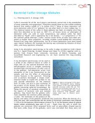
Spectral linewidths<br />
Example – aqueous solution of molybdate [MoO 4 ] 2- measured at the K-edge (1s<br />
excitation) and the L I edge (2s excitation). These are very similar ground<br />
states, and no significant differences in the nature of the near-edge<br />
transitions are expected. The spectra have been offset by 20008.70 eV and<br />
2869.95 eV, respectively. The K edge is has a much shorter core-hole<br />
lifetime than the L I edge, and has corresponding broader linewidths.<br />
L I edge<br />
K edge<br />
I. J. Pickering and G. N. George GEOL 498.3/898.3<br />
S<br />
OH<br />
O
Spectrometer Resolution<br />
Spectral linewidths<br />
In a modern EXAFS beamline this is usually only a function of the<br />
monochromator. Each monochromator material has an inherent energy<br />
resolution - the Darwin width of the crystal.<br />
This adds a Gaussian component to the overall experimental lineshape<br />
function.<br />
The experimental lineshape is expected to be approximated by a convolution<br />
of a Gaussian and a Lorenztian due to monochromator and lifetime broadening,<br />
respectively. This is known as a Voigt lineshape function – in practice it can be<br />
approximated by the sum of a Gaussian and Lorenztian – a pseudo Voigt<br />
lineshape function.<br />
I. J. Pickering and G. N. George GEOL 498.3/898.3<br />
Near-edge Spectra<br />
We can write Fermi’s Golden Rule as:<br />
µ ∝<br />
∑<br />
2<br />
i(<br />
k⋅r<br />
)<br />
i ( e⋅<br />
p)<br />
e ψ f<br />
ψ<br />
If we use a series expansion of<br />
the exponential, and examine<br />
just the first term, we get what<br />
is called the “dipole-allowed”<br />
transitions. These are the most<br />
intense transitions observed, and<br />
can be thought of as being<br />
stimulated by an oscillating<br />
electric field.<br />
ψ i - the initial state wavefunction<br />
ψ f - the final state wavefunction<br />
e - the X-<strong>ray</strong> electric vector<br />
p - the electron momentum vector<br />
k - the X-<strong>ray</strong> forward propagation<br />
vector<br />
r - the transition operator (x, y or z)<br />
in the molecular axis system<br />
I. J. Pickering and G. N. George GEOL 498.3/898.3<br />
Dipole and Quadrupole Transitions<br />
Dipole transitions are described by:<br />
∑<br />
µ D ∝ ψ i ( e⋅<br />
p)<br />
ψ f<br />
These are the most intense transitions observed, and can be thought of as<br />
being stimulated by an oscillating electric field, and have ∆l= ±1.<br />
Including the next term in the series expansion gives “quadrupole<br />
transitions”, which have ∆l= ±2, and these are described by:<br />
∑<br />
µ Q ∝ ψ i ( e⋅<br />
p)(<br />
k ⋅r)<br />
ψ f<br />
Quadrupole transitions are of low intensity and can be thought of as being<br />
stimulated by the electric field gradient, which is significant due to the<br />
short wavelength of the X-radiation being used.<br />
I. J. Pickering and G. N. George GEOL 498.3/898.3<br />
2<br />
2
Transition<br />
Dipole<br />
Quadrupole<br />
Transition<br />
Dipole<br />
Quadrupole<br />
Selection Rules for X-<strong>ray</strong> absorption<br />
near-edge spectra<br />
Selection rule<br />
∆l=±1<br />
∆l=±2<br />
K, L I , M I<br />
ns →n´p<br />
ns →n´d<br />
Strength<br />
Intense<br />
Weak<br />
K-edge<br />
np →n´d<br />
np →n´f<br />
1s →np<br />
1s →nd<br />
L II , L III , M II , M III<br />
LIII-edge 2p →nd<br />
2p →nf<br />
M IV , M V<br />
nd →n´f<br />
nd →n´g<br />
I. J. Pickering and G. N. George GEOL 498.3/898.3<br />
Tungsten L-edges – Selection Rules<br />
XAS L-edge spectra of Na 2 WO 4 –W(VI) is 5d 0 , so we expect strong dipole<br />
allowed transitions to the 5d manifold at the L III and L II edges from the<br />
2p 3/2 and 2p 1/2 , respectively. No such intense transitions are expected at<br />
the L I near-edge (2s excitation).<br />
W L III W L II<br />
W L I<br />
I. J. Pickering and G. N. George GEOL 498.3/898.3<br />
Uranium M-edges – Selection Rules<br />
XAS M-edge spectra of UO 2 (CH 3 CO 2 ) 2 (H 2 O) 2 –U(VI) is 5f 0 , so we expect<br />
strong dipole-allowed transitions to the 5f manifold at the M V and M IV edges<br />
from the 3d 3/2 and 3d 1/2 , respectively. No such intense transitions are<br />
expected at the M III , M II (3p 3/2 and 3p 1/2 excitation, respectively) or M I (3s<br />
excitation) near-edges.<br />
M V<br />
M IV MIII<br />
O O OH2 O U<br />
O<br />
H2O O<br />
O<br />
I. J. Pickering and G. N. George GEOL 498.3/898.3<br />
M II<br />
M I
Dipole and Quadrupole transitions<br />
Cu K-edge spectrum of [Cu(Imidazole) 4](NO 3) 2 is 3d 9 . Spectra arise from 1s excitation,<br />
so we expect strong dipole allowed transitions to orbitals with a lot of 4p character, and<br />
a single weak quadrupole allowed transition to the half-filled 3d level.<br />
Quadrupole 1s→3d transition<br />
x20<br />
Dipole 1s→4p transitions<br />
I. J. Pickering and G. N. George GEOL 498.3/898.3<br />
What do we expect about near-edge<br />
spectra?<br />
• Intense features due to dipole-allowed ∆l=±1 transitions<br />
• Weak features due to quadrupole-allowed ∆l=±2 transitions<br />
• For hard X-<strong>ray</strong> spectra (i.e. E > 1500 eV) the core-hole lies deep<br />
within the atom. One consequence of this is that the final state of an<br />
absorber with atomic number Z approximates to that of Z+1 – i.e. the<br />
next element in the periodic table. This can be important when<br />
comparing splittings measured from UV-visible electronic spectroscopy<br />
with X-<strong>ray</strong> near-edge spectra – e.g splittings of Co 2+ K near-edge<br />
spectra correspond optical spittings observed in the iso-structural<br />
Fe 2+ compound.<br />
I. J. Pickering and G. N. George GEOL 498.3/898.3<br />
What do we expect about near-edge<br />
spectra?<br />
• For hard X-<strong>ray</strong> spectra (i.e. E > 1500 eV) the ejection of a core<br />
electron will cause the outer orbitals to relax to lower energies (e.g.<br />
by about 10 eV for Cu K-edge spectra). This causes a corresponding<br />
shrinkage of the wave function, and thus reduction in the overlap<br />
integrals for molecular orbitals. We therefore expect the spectra to<br />
be very “atomic” in some of their properties.<br />
I. J. Pickering and G. N. George GEOL 498.3/898.3
Influence of core hole on electronic structure<br />
Ejection of metal core-electron causes outer metal orbitals to relax to<br />
lower energies.<br />
4p<br />
3d<br />
1s<br />
metal ligand<br />
Ground state<br />
3p<br />
1s<br />
metal ligand<br />
I. J. Pickering and G. N. George GEOL 498.3/898.3<br />
4p<br />
3d<br />
Final state<br />
What are near-edge spectra sensitive to?<br />
Oxidation State<br />
I. J. Pickering and G. N. George GEOL 498.3/898.3<br />
3p<br />
Pyrococcus furiosus rubredoxin<br />
Fe 2+<br />
Fe 3+<br />
What are near-edge spectra sensitive to?<br />
Nature of the Ligands<br />
Ferric ions with sulfur and oxygen donors<br />
[Fe 3+ (SR) 4 ] -<br />
[Fe 3+ (OR) 4 ] -<br />
I. J. Pickering and G. N. George GEOL 498.3/898.3
What are near-edge spectra sensitive to?<br />
Nature of the Ligands<br />
P 4 O 10 and P 4 S 10 are isostructural, both with P(V) oxidation state<br />
P 4 O 10<br />
P 4 S 10<br />
Covalency of sulfur means that phosphorus appears more reduced than its<br />
formal oxidation state<br />
I. J. Pickering and G. N. George GEOL 498.3/898.3<br />
What are near-edge spectra sensitive to?<br />
Coordination Geometry<br />
Oxygen coordinated ferric ions – octahedral vs. tetrahedral<br />
octahedral<br />
tetrahedral<br />
1s→3d region<br />
I. J. Pickering and G. N. George GEOL 498.3/898.3<br />
Transition metal MO 4 anions<br />
Similar chemical environments give rise to similar spectra<br />
K<br />
K<br />
K<br />
K<br />
L I<br />
K<br />
VO 4 2-<br />
K 2 CrO 4<br />
KMnO 4<br />
K 2 FeO 4<br />
Na 2 WO 4<br />
Na 2 MoO 4<br />
Spectra have been offset to align the lowest energy transition<br />
I. J. Pickering and G. N. George GEOL 498.3/898.3<br />
Edge energy
What are near-edge spectra sensitive to?<br />
Trigonal vs. Digonal cuprous thiolate compounds<br />
Inspection of the chemical literature indicates that Cu(I) prefers two<br />
distinct coordination environments – linear two-coordinate (digonal)<br />
and planar three-coordinate (trigonal) coordination geometries, e.g.<br />
with thiolate ligands:<br />
SR<br />
Cu<br />
SR<br />
-<br />
SR<br />
2-<br />
RS Cu<br />
SR<br />
Cuprous thiolate metalloproteins form a very large group of diverse<br />
function. Both two and three coordinate examples are known.<br />
I. J. Pickering and G. N. George GEOL 498.3/898.3<br />
atom<br />
Isolated atom – degenerate<br />
p-orbital energies<br />
Ligand Field Splitting<br />
p x p y p z<br />
energy<br />
Molecule – ligand-field splitting,<br />
p-orbital degeneracy lifted<br />
I. J. Pickering and G. N. George GEOL 498.3/898.3<br />
What are near-edge spectra sensitive to?<br />
Trigonal vs. Digonal cuprous thiolate compounds<br />
Cu(I) is 3d 10 , so we expect no quadrupole transitions to the 3d<br />
manifold, and the lowest energy features in the near-edge should be<br />
1s→4p transitions. Let us consider the ligand field splitting of the 4p<br />
orbitals.<br />
x<br />
z<br />
digonal<br />
SR<br />
y Cu<br />
SR<br />
p x<br />
p z<br />
p y<br />
RS<br />
trigonal<br />
Cu<br />
SR<br />
SR<br />
I. J. Pickering and G. N. George GEOL 498.3/898.3<br />
p y<br />
p x<br />
p z<br />
RS<br />
p x<br />
Cu<br />
p y<br />
SR<br />
SR<br />
p z<br />
energy<br />
distorted trigonal<br />
p x<br />
pz py
Cu K near-edge spectra of digonal and<br />
trigonal cuprous thiolates<br />
Cu 1s→4p x,y<br />
Cu 1s→4p x<br />
digonal<br />
trigonal<br />
I. J. Pickering and G. N. George GEOL 498.3/898.3<br />
Cu K near-edge spectra of digonal and<br />
trigonal cuprous thiolates<br />
Trigonal vs. Digonal cuprous thiolate compounds<br />
The ~8983 eV peak is diagnostic of digonal Cu(I)<br />
coordination. It can be used as a fingerprint of this kind<br />
of metal coordination.<br />
SR<br />
Cu<br />
SR<br />
RS<br />
SR<br />
Cu<br />
SR<br />
I. J. Pickering and G. N. George GEOL 498.3/898.3<br />
1s→3d transitions of transition metal ions<br />
Octahedral Fe 3+ with oxygen coordination – a small quadrupoleallowed,<br />
dipole-forbidden 1s→3d peak is observed.<br />
The transition has structure is due to the ligand field splitting of the<br />
3d manifold.<br />
1s→3d<br />
1s→3d<br />
I. J. Pickering and G. N. George GEOL 498.3/898.3
Octahedrally coordinated<br />
metal atom<br />
Energy<br />
Ligand Field Splitting<br />
Ligand atom<br />
∆<br />
d 2<br />
z<br />
Those d-orbitals with lobes<br />
directed towards the ligand atoms<br />
will possess higher energies than<br />
those with lobes directed in<br />
between the ligands.<br />
d −<br />
2 2<br />
x y<br />
d xy d d<br />
xz<br />
yz<br />
The energy separation of the orbitals ∆ is known as the ligand field splitting<br />
I. J. Pickering and G. N. George GEOL 498.3/898.3<br />
1s→3d transitions of transition metal ions<br />
The size of the ligand field splitting ∆ (remember this is an excited<br />
state splitting) can tell us about the nature of the metal site.<br />
I. J. Pickering and G. N. George GEOL 498.3/898.3<br />
d −<br />
2 2<br />
x y<br />
d 2<br />
z<br />
d xy d xz d yz<br />
d −<br />
2 2<br />
x y<br />
High-Spin vs. Low-Spin Ferrous<br />
d 2<br />
z<br />
d xy d xz d yz<br />
I. J. Pickering and G. N. George GEOL 498.3/898.3<br />
e g<br />
t2 g<br />
e g<br />
t2 g<br />
∆<br />
Low spin, R ion=0.92 Å<br />
High spin, R ion=0.75 Å<br />
Low-spin Fe 2+ occurs with larger ∆, and gives rise to one peak of<br />
relatively increased intensity.
1s→3d transitons – octahedral vs.<br />
tetrahedral geometry<br />
Centrosymmetric (e.g. octahedral<br />
symmetry) mixing of metal 4p and<br />
3d orbitals forbidden, and the<br />
transition is pure quadrupole.<br />
Non-centrosymmetric (e.g.<br />
tetrahedral symmetry) mixing of<br />
metal 4p and 3d orbitals allowed,<br />
and the transition is quadrupole,<br />
plus dipole-allowed intensity from<br />
admixture of metal 4p levels.<br />
I. J. Pickering and G. N. George GEOL 498.3/898.3<br />
Analysis by peak deconvolution<br />
The experimental spectrum is fitted to a calculated spectrum<br />
comprised of a sum of pseudo Voigt peaks (I V) plus a step function<br />
for the edge (I 0). This is usually done by iteratively minimizing the<br />
sum-of-squares of the differences between calculated and<br />
measured spectra. Each peak should comprise a single transition<br />
(or group of transitions) to a particular bound state (or states).<br />
µ calc(<br />
E) = a0I<br />
0(<br />
E)<br />
+ ∑ aiIVi<br />
( E)<br />
i<br />
I Vi -psudo-Voigt peak i<br />
I 0 -edge function<br />
a 0 - amplitude for edge function<br />
a i - amplitude for peak i<br />
This method allows quantitative analysis of quite subtle changes in<br />
near-edge spectra.<br />
V<br />
I. J. Pickering and G. N. George GEOL 498.3/898.3<br />
I = mI + 1−<br />
G<br />
⎛<br />
I ⎜<br />
G = exp<br />
⎜<br />
⎝<br />
I<br />
L<br />
=<br />
Analysis by peak deconvolution<br />
( m)<br />
I L<br />
( )<br />
[ ( ( ) ) ] ⎟⎟<br />
2<br />
− ln 2 E − E ⎞ m<br />
W + E − E η ξ<br />
[ ( ) ]<br />
[ ( ) ] ( ) 2<br />
2<br />
2<br />
W + E − Em<br />
η<br />
W + E − E η + E − E<br />
m<br />
m<br />
⎠<br />
m<br />
I V - the psudo-Voigt function<br />
I G - Gaussian peak-shape function<br />
I L - Lorentzian peak-shape function<br />
m -mixing factor<br />
E m -peak position<br />
W –half-width of peak<br />
η - peak skew<br />
ξ - ratio of Gaussian to Lorentzian<br />
widths<br />
I. J. Pickering and G. N. George GEOL 498.3/898.3
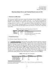
Analysis by peak deconvolution<br />
I. J. Pickering and G. N. George GEOL 498.3/898.3<br />
Sulfur K-edge X-<strong>ray</strong><br />
absorption near-edge<br />
spectra<br />
Sulfur K Near-edge spectra of<br />
biological model compounds.<br />
The spectra are very sensitive<br />
to the chemical form of sulfur<br />
and can be used to “fingerprint”<br />
forms of sulfur present.<br />
SO 4 2-<br />
RSO 3 -<br />
SO 3 2-<br />
RSO 2 -<br />
RS=O<br />
R3S +<br />
RS-Me<br />
RS-H<br />
RS-SR<br />
S8 Fe 4S 4<br />
I. J. Pickering and G. N. George GEOL 498.3/898.3<br />
x2<br />
x2<br />
x2<br />
x2<br />
x2