Post-natal Development in the Linear and Tric Morphometrics of the ...
Post-natal Development in the Linear and Tric Morphometrics of the ...
Post-natal Development in the Linear and Tric Morphometrics of the ...

Create successful ePaper yourself
Turn your PDF publications into a flip-book with our unique Google optimized e-Paper software.
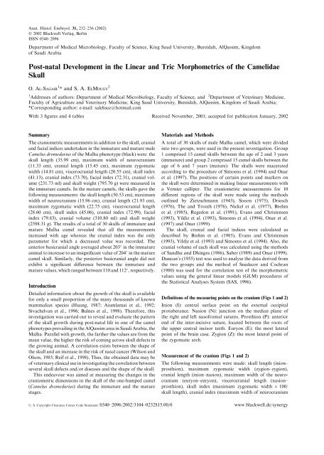
Anat.Histol.Embryol.31, 232–236 (2002)<br />
Ó 2002 Blackwell Verlag, Berl<strong>in</strong><br />
ISSN 0340–2096<br />
Department <strong>of</strong> Medical Microbiology, Faculty <strong>of</strong> Science, K<strong>in</strong>g Saud University, Bureidah, AlQassim, K<strong>in</strong>gdom<br />
<strong>of</strong> Saudi Arabia<br />
<strong>Post</strong>-<strong>natal</strong> <strong>Development</strong> <strong>in</strong> <strong>the</strong> L<strong>in</strong>ear <strong>and</strong> <strong>Tric</strong> <strong>Morphometrics</strong> <strong>of</strong> <strong>the</strong> Camelidae<br />
Skull<br />
O. Al-Sagair 1 * <strong>and</strong> S.A.ElMougy 2<br />
1 2<br />
Addresses <strong>of</strong> authors: Department <strong>of</strong> Medical Microbiology, Faculty <strong>of</strong> Science, <strong>and</strong> Department <strong>of</strong> Veter<strong>in</strong>ary Medic<strong>in</strong>e,<br />
Faculty <strong>of</strong> Agriculture <strong>and</strong> Veter<strong>in</strong>ary Medic<strong>in</strong>e, K<strong>in</strong>g Saud University, Bureidah, AlQassim, K<strong>in</strong>gdom <strong>of</strong> Saudi Arabia;<br />
*Correspond<strong>in</strong>g author: e-mail: salehsce@hotmail.com<br />
With 3 figures <strong>and</strong> 4 tables Received November, 2001; accepted for publication January, 2002<br />
Summary<br />
The craniometric measurements <strong>in</strong> addition to <strong>the</strong> skull, cranial<br />
<strong>and</strong> facial <strong>in</strong>dices undertaken <strong>in</strong> <strong>the</strong> immature <strong>and</strong> mature male<br />
Camelus dromedarius <strong>of</strong> <strong>the</strong> Malha phenotype (black) were: <strong>the</strong><br />
skull length (35.99 cm), maximum width <strong>of</strong> neurocranium<br />
(11.33 cm), cranial length (15.45 cm), maximum zygomatic<br />
width (14.81 cm), viscerocranial length (20.55 cm), skull <strong>in</strong>dex<br />
(41.13), cranial <strong>in</strong>dex (73.70), facial <strong>in</strong>dex (72.31), cranial volume<br />
(231.73 ml) <strong>and</strong> skull weight (795.70 g) were measured <strong>in</strong><br />
<strong>the</strong> immature camels.In <strong>the</strong> mature camels, <strong>the</strong> skulls gave <strong>the</strong><br />
follow<strong>in</strong>g measurements: <strong>the</strong> skull length (50.53 cm), maximum<br />
width <strong>of</strong> neurocranium (15.96 cm), cranial length (21.93 cm),<br />
maximum zygomatic width (22.75 cm), viscerocranial length<br />
(28.60 cm), skull <strong>in</strong>dex (45.06), cranial <strong>in</strong>dex (72.99), facial<br />
<strong>in</strong>dex (79.83), cranial volume (310.80 ml) <strong>and</strong> skull weight<br />
(2598.31 g). The results <strong>of</strong> a total <strong>of</strong> 30 skulls <strong>of</strong> immature <strong>and</strong><br />
mature Malha camel revealed that all <strong>the</strong> measurements<br />
<strong>in</strong>creased with age whereas <strong>the</strong> cranial <strong>in</strong>dex was <strong>the</strong> only<br />
parameter for which a decreased value was recorded.The<br />
anterior basicranial angle averaged about 203 <strong>in</strong> <strong>the</strong> immature<br />
animal to <strong>in</strong>crease to an <strong>in</strong>significant value <strong>of</strong> 204 <strong>in</strong> <strong>the</strong> mature<br />
camel skull.Similarly, <strong>the</strong> posterior basicranial angle did not<br />
exhibit a significant difference between <strong>the</strong> immature <strong>and</strong><br />
mature values, which ranged between 110 <strong>and</strong> 112 , respectively.<br />
Introduction<br />
Detailed <strong>in</strong>formation about <strong>the</strong> growth <strong>of</strong> <strong>the</strong> skull is available<br />
for only a small proportion <strong>of</strong> <strong>the</strong> many thous<strong>and</strong>s <strong>of</strong> known<br />
mammalian species (Huang, 1987; Aiumlamai et al., 1992;<br />
Sivachelvan et al., 1996; Bulnes et al., 1998). Therefore, this<br />
<strong>in</strong>vestigation was carried out to reveal <strong>and</strong> evaluate <strong>the</strong> pattern<br />
<strong>of</strong> <strong>the</strong> skull growth dur<strong>in</strong>g post-<strong>natal</strong> life <strong>in</strong> one <strong>of</strong> <strong>the</strong> camel<br />
phenotypes prevail<strong>in</strong>g <strong>in</strong> <strong>the</strong> AlQassim area <strong>in</strong> Saudi Arabia, <strong>the</strong><br />
Malha.Parallel with growth, <strong>the</strong> far<strong>the</strong>r <strong>the</strong> values are from <strong>the</strong><br />
mean value, <strong>the</strong> higher <strong>the</strong> risk <strong>of</strong> com<strong>in</strong>g across skull defects <strong>in</strong><br />
<strong>the</strong> grow<strong>in</strong>g animal.A correlation exists between <strong>the</strong> shape <strong>of</strong><br />
<strong>the</strong> skull <strong>and</strong> an <strong>in</strong>crease <strong>in</strong> <strong>the</strong> risk <strong>of</strong> nasal cancer (Wilson <strong>and</strong><br />
Olson, 1985; Reif et al., 1998). Thus, <strong>the</strong> obta<strong>in</strong>ed data may be<br />
<strong>of</strong> veter<strong>in</strong>ary cl<strong>in</strong>ical use <strong>in</strong> <strong>in</strong>vestigat<strong>in</strong>g <strong>the</strong> correlation between<br />
several skull defects <strong>and</strong>/or diseases <strong>and</strong> <strong>the</strong> shape <strong>of</strong> <strong>the</strong> skull.<br />
This endeavour was aimed at measur<strong>in</strong>g <strong>the</strong> changes <strong>in</strong> <strong>the</strong><br />
craniometric dimensions <strong>in</strong> <strong>the</strong> skull <strong>of</strong> <strong>the</strong> one-humped camel<br />
(Camelus dromedarius) dur<strong>in</strong>g <strong>the</strong> immature <strong>and</strong> <strong>the</strong> mature<br />
stages.<br />
Materials <strong>and</strong> Methods<br />
A total <strong>of</strong> 30 skulls <strong>of</strong> male Malha camel, which were divided<br />
<strong>in</strong>to two groups, were used <strong>in</strong> <strong>the</strong> present <strong>in</strong>vestigation.Group<br />
1 comprised 15 camel skulls between <strong>the</strong> age <strong>of</strong> 2 <strong>and</strong> 3 years<br />
(immature) <strong>and</strong> group 2 comprised 15 camel skulls between <strong>the</strong><br />
age <strong>of</strong> 6 <strong>and</strong> 7 years (mature).The skulls were macerated<br />
accord<strong>in</strong>g to <strong>the</strong> procedure <strong>of</strong> Simoens et al.(1994) <strong>and</strong> Onar<br />
et al.(1997).The positions <strong>of</strong> certa<strong>in</strong> po<strong>in</strong>ts <strong>and</strong> markers on<br />
<strong>the</strong> skull were determ<strong>in</strong>ed <strong>in</strong> mak<strong>in</strong>g l<strong>in</strong>ear measurements with<br />
a Vernier calliper.The craniometric measurements for 10<br />
different regions <strong>of</strong> <strong>the</strong> skull were made us<strong>in</strong>g <strong>the</strong> methods<br />
outl<strong>in</strong>ed by Zietzschmann (1943), Sisson (1975), Driesch<br />
(1976), The <strong>and</strong> Trouth (1976), Nickel et al.(1977), Brehm<br />
et al.(1985), Regedon et al.(1991), Evans <strong>and</strong> Christensen<br />
(1993), Yildiz et al.(1993), Simoens et al.(1994), Onar et al.<br />
(1997) <strong>and</strong> Onar (1999).<br />
The skull, cranial <strong>and</strong> facial <strong>in</strong>dices were calculated as<br />
described by Brehm et al.(1985), Evans <strong>and</strong> Christensen<br />
(1993), Yildiz et al.(1993) <strong>and</strong> Simoens et al.(1994).Also, <strong>the</strong><br />
cranial volume <strong>of</strong> each skull was calculated us<strong>in</strong>g <strong>the</strong> methods<br />
<strong>of</strong> S<strong>and</strong>hu <strong>and</strong> Dh<strong>in</strong>gra (1986), Saber (1989) <strong>and</strong> Onar (1999).<br />
Duncan’s (1955) test was used to analyse <strong>the</strong> data derived from<br />
<strong>the</strong> two groups <strong>and</strong> <strong>the</strong> method <strong>of</strong> Snedecor <strong>and</strong> Cochran<br />
(1980) was used for <strong>the</strong> correlation test <strong>of</strong> <strong>the</strong> morphometric<br />
values us<strong>in</strong>g <strong>the</strong> general l<strong>in</strong>ear models (GLM) procedures <strong>of</strong><br />
<strong>the</strong> Statistical Analyses System (SAS, 1996).<br />
Def<strong>in</strong>itions <strong>of</strong> <strong>the</strong> measur<strong>in</strong>g po<strong>in</strong>ts on <strong>the</strong> cranium (Figs 1<strong>and</strong> 2)<br />
Inion (I): central surface po<strong>in</strong>t on <strong>the</strong> external occipital<br />
protuberance.Nasion (N): junction on <strong>the</strong> median plane <strong>of</strong><br />
<strong>the</strong> right <strong>and</strong> left nas<strong>of</strong>rontal sutures.Prosthion (P): anterior<br />
end <strong>of</strong> <strong>the</strong> <strong>in</strong>ter-<strong>in</strong>cisive suture, located between <strong>the</strong> roots <strong>of</strong><br />
<strong>the</strong> upper central <strong>in</strong>cisor teeth.Euryon (E): <strong>the</strong> most lateral<br />
po<strong>in</strong>t <strong>of</strong> <strong>the</strong> bra<strong>in</strong> case.Zygion (Z): <strong>the</strong> most lateral po<strong>in</strong>t <strong>of</strong><br />
<strong>the</strong> zygomatic arch.<br />
Measurement <strong>of</strong> <strong>the</strong> cranium (Figs 1<strong>and</strong> 2)<br />
The follow<strong>in</strong>g measurements were made: skull length (<strong>in</strong>ion–<br />
prosthion), maximum zygomatic width (zygion–zygion),<br />
cranial length (<strong>in</strong>ion–nasion), maximum width <strong>of</strong> <strong>the</strong> neurocranium<br />
(euryon–euryon), viscerocranial length (nasion–<br />
prosthion), skull <strong>in</strong>dex (maximum zygomatic width 100/<br />
skull length), cranial <strong>in</strong>dex (maximum width <strong>of</strong> neurocranium<br />
U.S.Copyright Clearance Center Code Statement: 0340–2096/2002/3104–0232$15.00/0 www.blackwell.de/synergy
<strong>Post</strong>-<strong>natal</strong> <strong>Development</strong> <strong>of</strong> <strong>the</strong> Camelidae Skull 233<br />
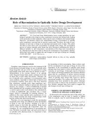
Fig.1. Measurments <strong>of</strong> <strong>the</strong> camel skull (dorsal view): <strong>in</strong>ion (I), nasion<br />
(N), prosthion (P), zygion (Z), skull length (1), cranial length (2)<br />
viscerocranial length (3), maximum width <strong>of</strong> neurocranium (4) <strong>and</strong><br />
maximum zygomatic width (5).<br />
100/cranial length), facial <strong>in</strong>dex (maximum zygomatic width<br />
100/viscerocranial length), cranial volume <strong>and</strong> skull weight.<br />
The measurement <strong>of</strong> <strong>the</strong> cranial volume was commenced by<br />
plugg<strong>in</strong>g <strong>the</strong> foram<strong>in</strong>a open<strong>in</strong>g <strong>in</strong>to <strong>the</strong> cranial cavity with<br />
fresh Plastic<strong>in</strong>e.The cavity was <strong>the</strong>n filled with f<strong>in</strong>e-quality rice<br />
seeds to <strong>the</strong> level <strong>of</strong> <strong>the</strong> distal border <strong>of</strong> <strong>the</strong> foramen magnum.<br />
When <strong>the</strong> cranial cavity was filled with rice seeds via <strong>the</strong><br />
foramen magnum, <strong>the</strong> space immediately cranial to <strong>the</strong><br />
tentorium cerebelli osseum was also filled.The contents were<br />
emptied <strong>and</strong> measured <strong>in</strong> a measur<strong>in</strong>g cyl<strong>in</strong>der calibrated <strong>in</strong><br />
millilitres.<br />
L<strong>in</strong>ear <strong>and</strong> angular dimensions <strong>of</strong> <strong>the</strong> cranial base (Fig 3)<br />
The cranial base, as traditionally def<strong>in</strong>ed <strong>in</strong> growth studies <strong>of</strong><br />
<strong>the</strong> mammalian skull, consists (<strong>in</strong> <strong>the</strong> median plane) <strong>of</strong> a<br />
segment, termed <strong>the</strong> basicranial axis, stretch<strong>in</strong>g from <strong>the</strong><br />
pituitary region to <strong>the</strong> ventral (anterior) marg<strong>in</strong> <strong>of</strong> <strong>the</strong> foramen<br />
magnum, an anterior extension from <strong>the</strong> pituitary region to <strong>the</strong><br />
junction <strong>of</strong> <strong>the</strong> frontal <strong>and</strong> nasal bones, <strong>and</strong> a posterior<br />
extension from <strong>the</strong> ventral to <strong>the</strong> dorsal marg<strong>in</strong> <strong>of</strong> <strong>the</strong> foramen<br />
magnum (Moore, 1981).<br />
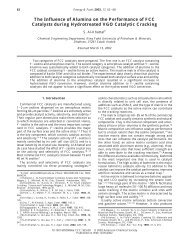
Fig.2. Measurments <strong>of</strong> <strong>the</strong> camel skull (lateral view): <strong>in</strong>ion (I),<br />
prosthion (p), nasion (N), skull length (1), viscerocranial length (2) <strong>and</strong><br />
cranial length (3).<br />
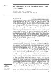
Fig.3. Sagittal sections <strong>of</strong> <strong>the</strong> skull <strong>of</strong> camel to show l<strong>in</strong>ear <strong>and</strong><br />
angular dimensions <strong>of</strong> cranial base.E ¼ endobasion; N ¼ nasion; O ¼<br />
opisthion; PP ¼ pituitarty po<strong>in</strong>t.The anterior <strong>and</strong> posterior basicranial<br />
angles are shown by dashed l<strong>in</strong>es.<br />
Statistical analysis<br />
Changes <strong>in</strong> <strong>the</strong> craniometric measurements with<strong>in</strong> <strong>and</strong> between<br />
age groups were statistically evaluated by <strong>the</strong> least-squares<br />
analysis <strong>of</strong> variance us<strong>in</strong>g <strong>the</strong> GLM procedures <strong>of</strong> <strong>the</strong><br />
statistical analysis system (SAS, 1996).<br />
Results<br />
The craniometric measurements <strong>and</strong> <strong>the</strong> cranial volume<br />
<strong>in</strong>creased significantly with age.The results recorded <strong>in</strong> Table<br />
1 reveal that <strong>the</strong> skull, cranial <strong>and</strong> viscerocranial lengths,<br />
neurocranium <strong>and</strong> <strong>the</strong> maximum zygomatic widths, <strong>in</strong>creased<br />
significantly with age.Although <strong>the</strong> skull <strong>and</strong> facial <strong>in</strong>dices<br />
<strong>in</strong>creased, <strong>the</strong> cranial <strong>in</strong>dex <strong>in</strong>significantly decreased <strong>in</strong> value<br />
with age.<br />
The results <strong>of</strong> <strong>the</strong> correlation analyses <strong>of</strong> <strong>the</strong> features<br />
exam<strong>in</strong>ed <strong>in</strong> <strong>the</strong> immature <strong>and</strong> mature animals are recorded <strong>in</strong><br />
Table 2.<br />
There was a strongly positive correlation among <strong>the</strong> skull,<br />
viscerocranial lengths, cranial <strong>and</strong> <strong>the</strong> maximum zygomatic<br />
width <strong>in</strong> <strong>the</strong> immature <strong>and</strong> mature animals.<br />
The maximum width <strong>of</strong> <strong>the</strong> neurocranium showed a strong<br />
significant positive correlation with all <strong>of</strong> <strong>the</strong> measurements <strong>in</strong><br />
both <strong>the</strong> immature <strong>and</strong> mature groups <strong>of</strong> camels.<br />
The skull <strong>and</strong> facial <strong>in</strong>dices <strong>in</strong> <strong>the</strong> immature <strong>and</strong> mature<br />
camels were found to have a positive correlation with <strong>the</strong><br />
maximum zygomatic width <strong>and</strong> <strong>the</strong> skull, cranial <strong>and</strong> viscerocranial<br />
lengths <strong>in</strong> <strong>the</strong> immature <strong>and</strong> mature animals.The<br />
cranial <strong>in</strong>dex was found to have a negative correlation with all<br />
<strong>of</strong> <strong>the</strong> craniometric measurements with <strong>the</strong> exception <strong>of</strong> <strong>the</strong><br />
viscerocranial length, which was <strong>in</strong>significantly positive.<br />
However, a similar comparison <strong>in</strong> <strong>the</strong> immature <strong>and</strong> mature<br />
groups <strong>of</strong> camels showed that <strong>the</strong> cranial <strong>in</strong>dex had a weak<br />
negative significant correlation with <strong>the</strong> cranial length.<br />
A positive correlation between <strong>the</strong> facial <strong>in</strong>dex <strong>and</strong> <strong>the</strong><br />
maximum width <strong>of</strong> <strong>the</strong> neurocranium was found <strong>in</strong> <strong>the</strong><br />
immature <strong>and</strong> mature camels.<br />
The correlation between <strong>the</strong> skull <strong>and</strong> facial <strong>in</strong>dices <strong>in</strong> both<br />
groups was positive <strong>and</strong> strong.The correlation between <strong>the</strong><br />
cranial <strong>and</strong> facial <strong>in</strong>dices was weakly significant <strong>in</strong> <strong>the</strong><br />
immature <strong>and</strong> mature camels.
234 O. Al-Sagair <strong>and</strong> S. A. ElMougy<br />
Table 2.Correlation analysis <strong>of</strong> <strong>the</strong> skull measurements <strong>of</strong> <strong>the</strong> immature <strong>and</strong> mature Malha (black) camels (Camelus dromedarius)<br />
Skull<br />
length<br />
(cm)<br />
Viscerocranial<br />
length<br />
(cm)<br />
Cranial<br />
length<br />
(cm)<br />
In both <strong>the</strong> immature <strong>and</strong> mature camels, <strong>the</strong> cranial volume<br />
generally <strong>in</strong>dicated a significant positive correlation with <strong>the</strong><br />
skull measurements, but <strong>the</strong>re was a weak negative correlation<br />
between <strong>the</strong> cranial volume <strong>and</strong> <strong>the</strong> skull, cranial <strong>and</strong> facial<br />
<strong>in</strong>dices.In nei<strong>the</strong>r <strong>of</strong> <strong>the</strong> camels, immature <strong>and</strong> mature, was<br />
this significant.<br />
The skull weight showed a negative correlation with <strong>the</strong><br />
cranial <strong>in</strong>dex only <strong>in</strong> <strong>the</strong> immature camel, whereas <strong>in</strong> <strong>the</strong><br />
mature camels <strong>the</strong> skull weight evidenced a positive correlation<br />
with <strong>the</strong> skull <strong>and</strong> facial <strong>in</strong>dices <strong>in</strong> addition to all <strong>of</strong> <strong>the</strong> o<strong>the</strong>r<br />
craniometric measurements.<br />
The results <strong>of</strong> <strong>the</strong> correlation analysis between <strong>the</strong> immature<br />
camels, as recorded <strong>in</strong> Table 3, reveal a strongly positive<br />
correlation between <strong>the</strong> craniometric measurements, with one<br />
exception.The cranial <strong>in</strong>dex had an <strong>in</strong>significant negative<br />
correlation with all craniometric parameters except with <strong>the</strong><br />
facial <strong>in</strong>dex where it had a weak negative significant correlation<br />
<strong>and</strong> an <strong>in</strong>significant positive correlation with <strong>the</strong> viscerocranial<br />
length.<br />
The results <strong>of</strong> <strong>the</strong> correlation analysis between <strong>the</strong> mature<br />
camels, as recorded <strong>in</strong> Table 4, reveal that <strong>the</strong> only strong<br />
significant correlation values were between <strong>the</strong> viscerocranial<br />
length <strong>and</strong> <strong>the</strong> skull length, viscerocranial length <strong>and</strong> <strong>the</strong> facial<br />
Maximum width Maximum<br />
<strong>of</strong> neurocranium zygomatic<br />
(cm) width (cm)<br />
Skull<br />
weight<br />
(g)<br />
Skull<br />
<strong>in</strong>dex<br />
Cranial<br />
<strong>in</strong>dex<br />
Facial<br />
<strong>in</strong>dex<br />
Cranial<br />
volume<br />
(ml)<br />
Skull length (cm) 0.965*** 0.946*** 0.967*** 0.963*** 0.952*** 0.549*** 0.181 0.448* 0.900***<br />
Viscerocranial<br />
length (cm)<br />
0.965*** 0.828*** 0.906*** 0.912*** 0.893*** 0.484** 0.006 0.271 0.856***<br />
Cranial length (cm) 0.946*** 0.828*** 0.948*** 0.926*** 0.932*** 0.577*** 0.395* 0.624*** 0.867***<br />
Maximum width <strong>of</strong><br />
neurocranium (cm)<br />
0.967*** 0.906*** 0.948*** 0.960*** 0.967*** 0.624*** 0.087 0.549** 0.912***<br />
Maximum zygomatic<br />
width (cm)<br />
0.963*** 0.917*** 0.926*** 0.960*** 0.931*** 0.751*** 0.135 0.628*** 0.900***<br />
Skull weight (g) 0.952*** 0.893*** 0.932*** 0.967*** 0.931*** 0.576*** 0.128 0.509** 0.905***<br />
Skull <strong>in</strong>dex 0.549** 0.484* 0.577*** 0.624*** 0.751*** 0.576*** 0.003 0.880*** 0.594***<br />
Cranial <strong>in</strong>dex 0.181 0.006 0.395* 0.087 0.135 0.128 0.003 0.358* 0.083<br />
Facial <strong>in</strong>dex 0.448* 0.271 0.624*** 0.549** 0.628*** 0.509** 0.880*** 0.358* 0.503**<br />
Cranial volume (ml) 0.900*** 0.856*** 0.867*** 0.912*** 0.900*** 0.905*** 0.594*** 0.083 0.503**<br />
*P < 0:05.<br />
**P < 0:01.<br />
***P < 0:001.<br />
Immature Mature<br />
Mean SE Mean SE<br />
Skull length (cm) 35.99 b 0.44 50.53 a 0.44<br />
Maximum width <strong>of</strong> neurocranium(cm) 11.33 b 0.11 15.96 a 0.11<br />
Cranial length (cm) 15.45 b 0.34 21.93 a 0.34<br />
Maximum zygomatic width (cm) 14.81 b 0.24 22.75 a 0.24<br />
Viscerocranial length (cm) 20.55 b 0.42 28.60 a 0.42<br />
Skull <strong>in</strong>dex 41.13 b 0.61 45.06 a 0.61<br />
Cranial <strong>in</strong>dex 73.70 a 1.17 72.99 a 1.17<br />
Facial <strong>in</strong>dex 72.31 b 1.56 79.83 a 1.56<br />
Cranial volume (ml) 231.73 b 4.22 310.80 a 4.22<br />
Skull weight (g) 795.70 b 70.33 2598.31 a 70.33<br />
SE st<strong>and</strong>ard error <strong>of</strong> mean.<br />
P < 0:001 for all parameters.The difference between <strong>the</strong> mean values <strong>of</strong> measurements is statistically<br />
significant.<br />
a;b Means with <strong>the</strong> same letter are not significantly different.<br />
Table 1.Skull measurements <strong>of</strong> <strong>the</strong><br />
immature (n ¼ 30) <strong>and</strong> mature<br />
(n ¼ 30) Malha (black) camels<br />
(Camelus dromedarius)<br />
<strong>in</strong>dex, cranial length <strong>and</strong> cranial <strong>in</strong>dex <strong>and</strong> between <strong>the</strong><br />
maximum zygomatic width <strong>and</strong> skull <strong>in</strong>dex.<br />
In <strong>the</strong> present <strong>in</strong>vestigation, <strong>the</strong> OE angle, <strong>of</strong> <strong>the</strong> camel<br />
skull, did not exceed <strong>the</strong> level <strong>of</strong> 112 <strong>in</strong> <strong>the</strong> mature animal<br />
whereas dur<strong>in</strong>g <strong>the</strong> immature stage it was 110 , which did not<br />
reveal a significant difference.In addition, <strong>the</strong> PP angle<br />
rema<strong>in</strong>ed at an angle <strong>of</strong> 204 <strong>and</strong> did not change with growth.<br />
Discussion<br />
Because <strong>of</strong> <strong>the</strong> absence <strong>of</strong> any available <strong>in</strong>formation on <strong>the</strong><br />
craniometric measurements <strong>of</strong> <strong>the</strong> adult camel, it was a must<br />
to <strong>in</strong>volve <strong>the</strong> immature animal <strong>in</strong> our measurements <strong>and</strong><br />
<strong>the</strong>reafter compare with measurements on <strong>the</strong> mature camels.<br />
An <strong>in</strong>crease <strong>in</strong> <strong>the</strong> skull, cranial <strong>and</strong> viscerocranial lengths,<br />
<strong>the</strong> neurocranium <strong>and</strong> <strong>the</strong> maximum zygomatic widths, <strong>the</strong><br />
cranial volume <strong>and</strong> <strong>the</strong> skull weight, <strong>and</strong> a decrease <strong>in</strong> <strong>the</strong><br />
skull, cranial <strong>and</strong> facial <strong>in</strong>dices was found when <strong>the</strong> immature<br />
camels reached maturity.<br />
In addition, when <strong>the</strong> maximum zygomatic width <strong>and</strong> <strong>the</strong><br />
maximum width <strong>of</strong> <strong>the</strong> neurocranium, were compared with <strong>the</strong><br />
skull, cranial <strong>and</strong> viscerocranial lengths, <strong>the</strong> latter had a higher<br />
<strong>in</strong>crement rate with age.This showed that <strong>the</strong> skull <strong>of</strong> <strong>the</strong>
<strong>Post</strong>-<strong>natal</strong> <strong>Development</strong> <strong>of</strong> <strong>the</strong> Camelidae Skull 235<br />
Table 3.Correlation analysis <strong>of</strong> <strong>the</strong> skull measurements <strong>of</strong> <strong>the</strong> immature Malha (black) camels (Camelus dromedarius)<br />
Skull<br />
length<br />
(cm)<br />
Viscerocranial<br />
length<br />
(cm)<br />
Cranial<br />
length<br />
(cm)<br />
Maximum width<br />
<strong>of</strong> neurocranium<br />
(cm)<br />
Artiodactyla (Camelidae) took on a slightly long-nosed shape<br />
as <strong>the</strong> animals grew older, whereas <strong>in</strong> <strong>the</strong> maximum zygomatic<br />
width <strong>and</strong> <strong>the</strong> maximum width <strong>of</strong> <strong>the</strong> neurocranium, smaller<br />
changes occurred.The change <strong>in</strong> <strong>the</strong> maximum width <strong>of</strong> <strong>the</strong><br />
neurocranium was even smaller with age.This was supported<br />
by <strong>the</strong> fact that <strong>the</strong>re was a low correlation between <strong>the</strong><br />
maximum width <strong>of</strong> <strong>the</strong> neurocranium <strong>and</strong> <strong>the</strong> skull, cranial<br />
<strong>and</strong> viscerocranial lengths.<br />
Both <strong>the</strong> determ<strong>in</strong>ation <strong>of</strong> a strongly positive correlation<br />
between <strong>the</strong> skull, cranial <strong>and</strong> viscerocranial lengths <strong>and</strong><br />
<strong>in</strong>dices, <strong>and</strong> a decrease <strong>in</strong> <strong>in</strong>dices with growth <strong>of</strong> <strong>the</strong> immature<br />
camels reflected <strong>the</strong> development <strong>of</strong> an Artiodactyla-shaped<br />
head.<br />
Camels are considered to belong to <strong>the</strong> mammalian Artiodactyla<br />
order <strong>and</strong> <strong>in</strong> <strong>the</strong> Camelidae group.In <strong>the</strong> present<br />
study, <strong>the</strong> length–width <strong>in</strong>dex was 0.67 <strong>and</strong> 0.44 <strong>in</strong> <strong>the</strong><br />
immature <strong>and</strong> mature camels, respectively.Also, <strong>the</strong> ratio <strong>of</strong><br />
<strong>the</strong> total skull length to <strong>the</strong> maximum length between <strong>the</strong> two<br />
zygomatic arches was 2.43 <strong>in</strong> <strong>the</strong> immature camels <strong>and</strong> 2.22 <strong>in</strong><br />
<strong>the</strong> mature ones, which <strong>in</strong>dicates that as <strong>the</strong> camels grew, <strong>the</strong><br />
Maximum<br />
zygomatic<br />
width (cm)<br />
Skull<br />
weight<br />
(g)<br />
Skull<br />
<strong>in</strong>dex<br />
Cranial<br />
<strong>in</strong>dex<br />
Facial<br />
<strong>in</strong>dex<br />
Cranial<br />
volume<br />
(ml)<br />
Skull length (cm) 0.965*** 0.946*** 0.967*** 0.963*** 0.952*** 0.549** 0.181 0.448* 0.900***<br />
Viscerocranial<br />
length (cm)<br />
0.965*** 0.828*** 0.906*** 0.917*** 0.893*** 0.484** 0.006 0.271 0.856***<br />
Cranial length (cm) 0.946*** 0.828*** 0.948*** 0.926*** 0.932*** 0.577*** 0.395 0.624*** 0.867***<br />
Maximum width <strong>of</strong><br />
neurocranium (cm)<br />
0.967*** 0.906*** 0.948*** 0.960*** 0.967*** 0.624*** 0.087 0.549** 0.912***<br />
Maximum zygomatic<br />
width (cm)<br />
0.963*** 0.917*** 0.926*** 0.960*** 0.931*** 0.751*** 0.135 0.628*** 0.900***<br />
Skull weight (g) 0.952*** 0.893*** 0.932*** 0.967*** 0.931*** 0.576*** 0.128 0.509** 0.905***<br />
Skull <strong>in</strong>dex 0.549** 0.484** 0.577*** 0.624*** 0.751*** 0.576*** 0.003 0.880*** 0.594***<br />
Cranial <strong>in</strong>dex 0.181 0.006 0.395 0.087 0.135 0.128 0.003 0.358* 0.083<br />
Facial <strong>in</strong>dex 0.448* 0.271 0.624*** 0.549** 0.628*** 0.509* 0.880*** 0.358* 0.503**<br />
Cranial volume (ml) 0.900*** 0.856*** 0.867*** 0.912*** 0.900*** 0.905*** 0.594*** 0.083 0.503**<br />
*P < 0:05.<br />
**P < 0:01.<br />
***P < 0:001.<br />
Table 4.Correlation analysis <strong>of</strong> <strong>the</strong> skull measurements <strong>of</strong> <strong>the</strong> mature Malha (black) camels (Camelus dromedarius)<br />
Skull<br />
length<br />
(cm)<br />
Viscerocranial<br />
length<br />
(cm)<br />
Cranial<br />
length<br />
(cm)<br />
Maximum width<br />
<strong>of</strong> neurocranium<br />
(cm)<br />
Maximum<br />
zygomatic<br />
width (cm)<br />
Skull<br />
weight<br />
(g)<br />
Skull<br />
<strong>in</strong>dex<br />
Cranial<br />
<strong>in</strong>dex<br />
Facial<br />
<strong>in</strong>dex<br />
Cranial<br />
volume<br />
(ml)<br />
Skull length (cm) 0.692** 0.165 0.008 0.248 0.292 0.382 0.186 0.469 0.132<br />
Viscerocranial length<br />
(cm)<br />
0.692** 0.598 0.347 0.212 0.033 0.225 0.470 0.758** 0.069<br />
Cranial length (cm) 0.165 0.598 0.483 0.0152 0.369 0.117 0.849*** 0.516 0.052<br />
Maximum width <strong>of</strong><br />
neurocranium (cm)<br />
0.008 0.347 0.483 0.078 0.493 0.078 0.050 0.234 0.005<br />
Maximum zygomatic<br />
width (cm)<br />
0.248 0.212 0.015 0.078 0.171 0.800*** 0.015 0.474 0.051<br />
Skull weight (g) 0.292 0.033 0.369 0.493 0.171 0.348 0.130 0.110 0.336<br />
Skull <strong>in</strong>dex 0.382 0.225 0.117 0.078 0.800*** 0.348 0.104 0.741 0.125<br />
Cranial <strong>in</strong>dex 0.186 0.470 0.849*** 0.050 0.015 0.130 0.104 0.435 0.036<br />
Facial <strong>in</strong>dex 0.469 0.758** 0.516 0.234 0.474 0.110 0.741 0.435 0.118<br />
Cranial volume (ml) 0.132 0.069 0.052 0.005 0.051 0.336 0.125 0.036 0.118<br />
**P < 0:01.<br />
***P < 0:001.<br />
ratio between skull length <strong>and</strong> maximum zygomatic width<br />
decreased.<br />
The cranial volume was found to <strong>in</strong>crease <strong>in</strong> l<strong>in</strong>e with <strong>the</strong><br />
<strong>in</strong>crements <strong>in</strong> o<strong>the</strong>r parameters reflected as a high correlation<br />
<strong>in</strong> <strong>the</strong> immature <strong>and</strong> mature camels with a high correlation<br />
with <strong>the</strong> <strong>in</strong>dices.However, <strong>the</strong> relatively large changes with<br />
age <strong>in</strong> <strong>the</strong>se values compared to those tak<strong>in</strong>g place <strong>in</strong> <strong>the</strong><br />
skull, cranial <strong>and</strong> viscerocranial lengths showed that <strong>the</strong><br />
cranial cavity <strong>in</strong>creased breadthwise, which meant that<br />
<strong>the</strong> development <strong>of</strong> <strong>the</strong> skull was compatible with <strong>the</strong><br />
Artiodactyla type (Camelidae) <strong>and</strong> which is quite opposite to<br />
what happens <strong>in</strong> <strong>the</strong> dolichocephalic-shaped head (Onar,<br />
1999).In <strong>the</strong> dolichocephalic dogs, <strong>the</strong> skull takes on a longnosed<br />
shape as <strong>the</strong> animals grow older, <strong>the</strong> cranial cavity<br />
widened lengthwise with a strongly negative correlation<br />
between <strong>the</strong> skull, cranial <strong>and</strong> viscerocranial lengths <strong>and</strong><br />
<strong>in</strong>dices, <strong>and</strong> a decrease <strong>in</strong> <strong>in</strong>dices with growth <strong>of</strong> <strong>the</strong> immature<br />
dogs.<br />
The growth changes <strong>in</strong> <strong>the</strong> l<strong>in</strong>ear dimensions <strong>of</strong> <strong>the</strong><br />
aforementioned segments <strong>of</strong> <strong>the</strong> cranial base <strong>and</strong> <strong>in</strong> <strong>the</strong> angles
236 O. Al-Sagair <strong>and</strong> S. A. ElMougy<br />
between <strong>the</strong>m record that <strong>the</strong> anterior basicranial angle<br />
averaged about 203 <strong>in</strong> <strong>the</strong> immature animal <strong>in</strong>creased<br />
<strong>in</strong>significantly to a value <strong>of</strong> 204 <strong>in</strong> <strong>the</strong> mature camel skull.<br />
Similarly, <strong>the</strong> posterior basicranial angle did not exhibit a<br />
significant difference between <strong>the</strong> immature <strong>and</strong> mature values,<br />
which ranged between 110 <strong>and</strong> 112 , respectively.These l<strong>in</strong>ear<br />
dimensions have been studied <strong>in</strong> several mammalian species.In<br />
non-primates, <strong>the</strong> angle tends to <strong>in</strong>crease dur<strong>in</strong>g post-<strong>natal</strong><br />
growth to a value well <strong>in</strong> excess <strong>of</strong> 180 .In <strong>the</strong> rat, for<br />
example, <strong>the</strong> anterior basicranial angle at maturity is <strong>of</strong> <strong>the</strong><br />
order <strong>of</strong> 240 (Moore, 1981).In <strong>the</strong> higher primates (Ceboidea,<br />
Cercopi<strong>the</strong>coidea <strong>and</strong> <strong>the</strong> apes amongst <strong>the</strong> Hom<strong>in</strong>oidea) <strong>the</strong><br />
anterior basicranial angle averages about 150 at <strong>the</strong> stage <strong>of</strong><br />
<strong>the</strong> milk dentition <strong>and</strong>, as <strong>in</strong> non-primates, cont<strong>in</strong>ues to<br />
<strong>in</strong>crease throughout <strong>the</strong> growth period but to a much lesser<br />
degree (Zuckerman, 1926; Ashton, 1957; Ashton et al., 1977).<br />
Their result <strong>in</strong>dicated that <strong>the</strong> anterior basicranial angle<br />
rema<strong>in</strong>s below 180 throughout life.This difference is attributable<br />
to <strong>the</strong> much enhanced growth <strong>of</strong> <strong>the</strong> primate bra<strong>in</strong><br />
which is accommodated, anteriorly, by enlargement <strong>of</strong> <strong>the</strong><br />
bra<strong>in</strong>case forwards above <strong>the</strong> facial skeleton.The primate<br />
pattern, as well as <strong>the</strong> recorded results with <strong>the</strong> camel, can be<br />
regarded as a tendency to reta<strong>in</strong> <strong>the</strong> immature shape <strong>and</strong><br />
correlates with <strong>the</strong> observation, as already noted, that <strong>the</strong><br />
bra<strong>in</strong> is large relative to <strong>the</strong> rema<strong>in</strong>der <strong>of</strong> <strong>the</strong> head early <strong>in</strong> <strong>the</strong><br />
growth period.In prosimians, <strong>the</strong> degree <strong>of</strong> cranial flexion is<br />
generally less than <strong>in</strong> anthropoid primates although <strong>the</strong><br />
anterior basicranial angle is still usually somewhat below<br />
180 .It is <strong>of</strong> <strong>in</strong>terest that <strong>in</strong> Daubentonia cranial flexion is<br />
much greater than <strong>in</strong> o<strong>the</strong>r prosimians.Cartmill (1977) has<br />
attributed this to <strong>the</strong> need to streng<strong>the</strong>n <strong>the</strong> skull, by<br />
augment<strong>in</strong>g facial height, aga<strong>in</strong>st <strong>the</strong> bend<strong>in</strong>g forces produced<br />
dur<strong>in</strong>g use <strong>of</strong> <strong>the</strong> <strong>in</strong>cisor teeth (Daubentonia possesses rodentlike<br />
<strong>in</strong>cisors, which it uses <strong>in</strong> a powerful gnaw<strong>in</strong>g action).<br />
The data obta<strong>in</strong>ed <strong>in</strong> this study us<strong>in</strong>g <strong>the</strong> correlations<br />
amongst shape <strong>and</strong> dimension parameters for <strong>the</strong> bra<strong>in</strong> <strong>and</strong><br />
<strong>the</strong> skull <strong>and</strong> <strong>the</strong> changes that occur with age may prove <strong>of</strong><br />
cl<strong>in</strong>ical usefulness to those cl<strong>in</strong>ically associated with <strong>the</strong><br />
camels.<br />
References<br />
Aiumlamai, S., G. Fredriksson, <strong>and</strong> L. Nilsfors, 1992: Real-time<br />
ultrasonography for determ<strong>in</strong><strong>in</strong>g <strong>the</strong> gestational age <strong>of</strong> ewes.Vet.<br />
Rec. 131, 560–562.<br />
Ashton, E.H., 1957: Age changes <strong>in</strong> <strong>the</strong> basicranial axis <strong>of</strong> <strong>the</strong><br />
Authropoidea.Proc.Zool.Soc.Lond.129, 61–74.<br />
Ashton, E.H., R.M.Fl<strong>in</strong>n, <strong>and</strong> W.J.Moore, 1977: The basicranial<br />
axis <strong>in</strong> certa<strong>in</strong> fossil hom<strong>in</strong>oids.J.Zool.176, 577–591.<br />
Brehm, H., K. Loeffler, <strong>and</strong> H. Komeyli, 1985: Skull shape <strong>in</strong> <strong>the</strong> dog.<br />
Anat.Histol.Embryol.14, 324–331.<br />
Bulnes, A.G.de, J.S.Moreno, A.L.Sebastian, <strong>and</strong> A.G.De-Bulness,<br />
1998: Estimation <strong>of</strong> fetal development <strong>in</strong> Manchega dairy ewes by<br />
transrectal ultrasonographic measurements.Small Rum<strong>in</strong>ant Res.<br />
27, 243–250.<br />
Cartmill, M., 1977: Daubentonia, Dactylopsila, woodpeckers <strong>and</strong><br />
Kl<strong>in</strong>orhynchy.In: Prosimian Anatomy, Biochemistry <strong>and</strong> Evolution<br />
(R.D.Mart<strong>in</strong>, G.A.Doyle <strong>and</strong> A.C.Walker, eds).London:<br />
Duckworth.<br />
Driesch, A.V.D., 1976: A Guide to <strong>the</strong> Measurement <strong>of</strong> Animal<br />
Bones from Archaeological Sites.Peabody Museum Bullet<strong>in</strong> 1.<br />
Massachusetts: Harvard University.<br />
Duncan, D.B., 1955: Multiple range <strong>and</strong> multiple F-tests.Biometrics<br />
11, 1–42.<br />
Evans, H.E.<strong>and</strong> G.C.Christensen, 1993: Miller’s Anatomy <strong>of</strong> <strong>the</strong><br />
Dog, 3rd edn.Philadelphia, PA: Saunders Co.<br />
Huang, C.M., 1987: Heritability <strong>and</strong> genetic <strong>and</strong> phenotypic correlation<br />
<strong>of</strong> skull traits <strong>in</strong> <strong>the</strong> rabbit.Bull.Inst.Zool.Acad.S<strong>in</strong>ica 26,<br />
133–141.<br />
Moore, W.J., 1981: The Mammalian Skull.Cambridge University<br />
Press: Cambridge/London/New York.<br />
Nickel, R., A. Schummer, <strong>and</strong> H. Wille, 1977: Passive Bewegungsapparat,<br />
Skelettsystem.In: Lehrbuch der Anatomie der Haustiere<br />
Bd.1, 4.Auflage (R.Nickel, A.Schummer <strong>and</strong> E.Seiferle, eds).<br />
Berl<strong>in</strong>: Verlag Paul Parey, pp.150–151.<br />
Onar, V., 1999: A morphometric study on <strong>the</strong> skull <strong>of</strong> <strong>the</strong> German<br />
Shepherd dog (Alsatian).Anat.Histol.Embryol.28, 253–256.<br />
Onar, V., R. Mutu, <strong>and</strong> K. O. Kahvecioglu, 1997: Morphometric<br />
analysis <strong>of</strong> <strong>the</strong> foramen magnum <strong>in</strong> German Shepherd dogs<br />
(Alsatians).Ann.Anat.179, 563–568.<br />
Regedon, S., A.Rob<strong>in</strong>a, A.Franco, J.M.Vivo, <strong>and</strong> Y.Lignereux,<br />
1991: Radiographic evaluation <strong>and</strong> statistical analysis <strong>of</strong> can<strong>in</strong>e<br />
skull form.Dolichocephaly, Mesocephaly, Brachycephaly.Anat.<br />
Histol.Embryol.20, 129–138.<br />
Reif, J.S., C.Bruns, <strong>and</strong> K.S.Lowe, 1998: Cancer <strong>of</strong> <strong>the</strong> nasal cavity<br />
<strong>and</strong> paranasal s<strong>in</strong>uses <strong>and</strong> exposure to environmental tobacco<br />
smoke <strong>in</strong> pet dogs.Am J Epidemiol.147, 488–492.<br />
Saber, A.S., 1989: Cranial capacity <strong>of</strong> <strong>the</strong> sheep <strong>and</strong> goat.Assiut Vet.<br />
Med.J.21, 1–6.<br />
S<strong>and</strong>hu, P.S.<strong>and</strong> L.D.Dh<strong>in</strong>gra, 1986: Cranial capacity <strong>of</strong> <strong>the</strong> Indian<br />
camel.Indian J Anim.Sci.56, 870–872.<br />
SAS (1996).SAS User’s Guide, SAS Institute, Cary, New York, USA.<br />
Simoens, P., P. Poels, <strong>and</strong> H. Lauwers, 1994: Morphometric analysis<br />
<strong>of</strong> <strong>the</strong> foramen magnum <strong>in</strong> Pek<strong>in</strong>gese dogs.Am.J.Vet.Res.55,<br />
34–39.<br />
Sisson, S., 1975: Carnivore osteology. In: The Anatomy <strong>of</strong> Domestic<br />
Animals, Vol.2, 5th edn (S.Sisson <strong>and</strong> J.D.Grossman, eds).<br />
Philadelphia, PA: Saunders Co, pp.1474–1479.<br />
Sivachelvan, M.N., A.M.Ghali, <strong>and</strong> G.A.Chibuzo, 1996: Foetal age<br />
estimation <strong>in</strong> sheep <strong>and</strong> goats.Small Rum<strong>in</strong>ant Res 19, 69–76.<br />
Snedecor, G.W., <strong>and</strong> W.G.Cochran, 1980: Statistical Methods, 7th<br />
edn.Ames, IA: Iowa State University Press.<br />
The, T.L., <strong>and</strong> C.O.Trouth, 1976: Sexual dimorphism <strong>in</strong> <strong>the</strong> basilar<br />
part <strong>of</strong> <strong>the</strong> occipital bone <strong>of</strong> <strong>the</strong> dog (Canis familiaris).Acta Anat.<br />
95, 565–571.<br />
Wilson, G.P., <strong>and</strong> L.E.Olson, 1985: Can<strong>in</strong>e skull l<strong>in</strong>ear <strong>and</strong> tric<br />
morphometrics.Anat.Histol.Embryol.14, 162–191.<br />
Yildiz, B., A.Serbest, O.Yilmaz, <strong>and</strong> H.Kirbiyik, 1993: The comparison<br />
<strong>of</strong> <strong>the</strong> head measures <strong>of</strong> Turkish <strong>and</strong> German shepherd dog<br />
breeds.J.Fac.Vet.Med.Univ.Uludag 1, 35–39.<br />
Zietzschmann, O., 1943: 1. Das Skeletsystem. Der passive Bewegungsapparat.In:<br />
H<strong>and</strong>buch der Vergleichenden Anatomie der Haustiere,<br />
18.Auflage (W.Ellenberger <strong>and</strong> H.Baum, eds).Berl<strong>in</strong>:<br />
Spr<strong>in</strong>ger Verlag, pp.114–116.<br />
Zuckerman, S., 1926: Growth changes <strong>in</strong> <strong>the</strong> skull <strong>of</strong> <strong>the</strong> baboon,<br />
Papio porcarius.Proc.Zool.Soc.Lond.26, 843–873.