Product brochure 6.19MB - Siemens Healthcare
Product brochure 6.19MB - Siemens Healthcare
Product brochure 6.19MB - Siemens Healthcare

Create successful ePaper yourself
Turn your PDF publications into a flip-book with our unique Google optimized e-Paper software.
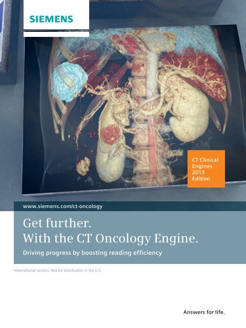
www.siemens.com/ct-oncology<br />
Get further.<br />
With the CT Oncology Engine.<br />
Driving progress by boosting reading efficiency<br />
International version. Not for distribution in the U.S.<br />
CT Clinical<br />
Engines<br />
2013<br />
Edition<br />
Answers for life.
2<br />
What is a CT Clinical Engine?<br />
• A powerful combination of software applications and<br />
scanner features – tailored to meet your clinical challenges<br />
• A solution that helps you get the most from your CT scanner<br />
With a CT Clinical Engine, you can continuously enhance<br />
speed, workflow efficiency, and diagnostic information.
How far can you get<br />
with your CT?<br />
Rising patient expectations, increasing efficiency needs: The best way to meet these<br />
challenges is to get detailed diagnostic information faster.<br />
The CT Oncology Engine delivers the clinical information you need – where and when you<br />
need it. You can easily refer to multi-modality and Dual Energy imaging as well as findings<br />
made at any previous point in time. Automated measurements and comparisons help you<br />
quantify suspicious lesions and provide a sound basis for further medical action. Especially<br />
when it comes to follow-up assessment, you will appreciate having all results from previous<br />
exams at hand so that you can compare them with current findings and combine all available<br />
information into one holistic picture. This way, you can reliably assess tumor progression as<br />
well as your patient’s response to therapy.<br />
Driving progress by boosting reading efficiency.<br />
2013 Edition: Overview<br />
3
4<br />
Rib and spine<br />
assessment redefined –<br />
speed up
2013 Edition: What‘s the top innovation?<br />
and streamline your<br />
diagnostic bone readings.<br />
5
Increased speed and<br />
specificity in bone assessment<br />
syngo.CT Bone Reading *<br />
Is there only one way to assess bones in stacks of<br />
CT images – scrolling back and forth, always trying to<br />
stick to the point of interest? Decide for a new method<br />
that’s as simple as effective: You can see the bones<br />
“rolled” and displayed in a plain and easy-to-view way.<br />
6<br />
Reaching beyond standard image stacks and MPR displays,<br />
syngo.CT Bone Reading offers you revolutionary new visualizations<br />
for completely new diagnostic insights. Complex<br />
anatomies are adapted to your reading needs. You can<br />
speed up and streamline your assessments of ribs and<br />
spine – complex anatomical structures that need to be<br />
assessed in every routine chest and abdominal CT scan.<br />
* patent-registered by <strong>Siemens</strong>
SOMATOM Perspective – the perfect fit for radiological<br />
assessment in oncology with up to 128 slice image<br />
acquisition, a spatial resolution of 0.4 mm, and with<br />
SAFIRE - the raw-data-based iterative reconstruction –<br />
consequently following the ALARA principle.<br />
Oncology imaging is all about reliably finding the tumor<br />
and then staging it; in other words, measuring it and<br />
identifying all its metastases. After initial diagnosis, the<br />
follow-up comparison of all cancerous findings at different<br />
points in time is a large part of ongoing patient monitoring.<br />
2013 Edition: What‘s the top innovation?<br />
In performing these diagnostic steps, fast acquisition<br />
covering large scan areas and eliminating motion artifacts<br />
caused by breathing is essential in order to guarantee<br />
precise and reliable image quality.<br />
In addition, X-ray exposure must be minimized, especially<br />
when numerous follow-up CT scans may need to be performed.<br />
SAFIRE – the raw-data-based iterative reconstruction<br />
– reduces the need for additional doses without compromising<br />
diagnostic information in any way. When combined<br />
with the CT Oncology Engine, it is an ideal way to facilitate<br />
reliable quantification for oncological diagnoses.<br />
7
Quantification of tumor tissue Complete 3D assessment in virtual colonography<br />
Holistic oncological imaging<br />
Advanced HU Statistics – quantification beyond<br />
tumor size<br />
syngo.CT Segmentation<br />
Automated segmentation for measuring suspicious lesions<br />
(RECIST, WHO, and volume) can be performed with a single<br />
click. And even beyond – advanced HU Statistics provides<br />
you quantification of tumor tissue, identifying hypodense<br />
areas that could be an indicator for tumor necrosis and<br />
potential therapy response. This enables you to provide a<br />
set of quantitative information to referring physicians that<br />
allows them to make a well-informed decision.<br />
Trending – tumor response at a glance<br />
syngo.PET&CT Cross-Timepoint Evaluation<br />
Reading follow-up procedures is essential in the assessment<br />
of cancer disease progression. After measurement in<br />
the CT scan, syngo.via automatically calculates tumor<br />
growth rate, tumor burden, and tumor volume doubling<br />
time of findings already available from prior studies.<br />
Trending helps you assess relevant changes at a glance by<br />
providing graphical visualization of changes in tumor<br />
growth over time.<br />
8<br />
3D polyp measurement – perform your entire<br />
assessment in 3D<br />
syngo.CT Colonography and syngo.CT Colonography<br />
Advanced<br />
Why not perform your entire virtual colonography assessment<br />
in 3D? With our exceptional tools, you can now<br />
make polyp size measurements in the 3D endoluminal<br />
view. In addition, the Virtual Dissection feature provides a<br />
planar visualization of the mucosa, enabling you to assess<br />
the colon surface from another perspective. While flying<br />
through the colon, this display is updated continuously so<br />
that the entire length of the colon can be evaluated.
syngo.CT Pulmo 3D syngo.CT Segmentation<br />
Specialized in diagnosis of diseases affecting the lung<br />
parenchyma and airways<br />
syngo.CT Pulmo 3D*<br />
To perform clinical assessments and therapy monitoring of<br />
lung diseases like COPD (chronic obstructive pulmonary<br />
disease), fibrosis, and bronchiectasia, syngo.CT Pulmo 3D<br />
preprocesses unenhanced CT chest scans. In order to facilitate<br />
quantitative assessments, it provides you with automated<br />
lung lobe and airway segmentation as well as precise<br />
calculations and measurements in tables and graphs.<br />
Thanks to the spatial details a native CT chest scan delivers<br />
as well as specific color-coded displays, you will obtain a<br />
fast overview of pathological areas, enabling you to identify<br />
the severity and localization of the disease.<br />
* Optional<br />
2013 Edition: What else is new?<br />
Your benefits at a glance<br />
• Speed up your diagnostic reading – and provide<br />
results earlier<br />
• Assess and quantify with a single click<br />
• Help your referring physician make a timely<br />
adaptation of your patient’s therapeutic regimen<br />
• Potentially increase your patients’ quality of life<br />
9
Second reader support – automatic detection of<br />
lung nodules<br />
syngo.CT Lung CAD<br />
Visualization, comparison, and synchronized navigation of up to eight different time points<br />
syngo.PET&CT Onco Multi-Timepoint<br />
Trending and tumor growth rate at a glance<br />
syngo.PET&CT Cross-Timepoint Evaluation<br />
Get further – with our<br />
* Optional<br />
10<br />
Quantitative oncological assessment through<br />
Dual Energy<br />
syngo.CT DE Virtual Unenhanced*<br />
CT Oncology Engine
Second reader support – automatic detection of colon polyps<br />
syngo.CT Colonography – PEV<br />
Comprehensive PET segmentation and evaluation<br />
syngo.PET Segmentation *<br />
Quantitative dynamic volume perfusion assessment<br />
syngo Volume Perfusion CT Body *<br />
Do you know the full potential of CT Oncology?<br />
and optional applications<br />
11
Global <strong>Siemens</strong> Headquarters<br />
<strong>Siemens</strong> AG<br />
Wittelsbacherplatz 2<br />
80333 Muenchen<br />
Germany<br />
Global <strong>Siemens</strong><br />
<strong>Healthcare</strong> Headquarters<br />
<strong>Siemens</strong> AG<br />
<strong>Healthcare</strong> Sector<br />
Henkestrasse 127<br />
91052 Erlangen<br />
Germany<br />
Phone: +49 9131 84 0<br />
www.siemens.com/healthcare<br />
In the event that upgrades require FDA approval,<br />
<strong>Siemens</strong> cannot predict whether or when the FDA will<br />
issue its approval. Therefore, if regulatory clearance<br />
is obtained and is applicable to this package, it will be<br />
made available according to the terms of this offer.<br />
On account of certain regional limitations of sales<br />
rights and service availability, we cannot guarantee<br />
that all products included in this <strong>brochure</strong> are available<br />
through the <strong>Siemens</strong> sales organization worldwide.<br />
Availability and packaging may vary by country and is<br />
subject to change without prior notice. Some/all of the<br />
features and products described herein may not be<br />
available in the United States.<br />
www.siemens.com/healthcare<br />
Global Business Unit<br />
<strong>Siemens</strong> AG<br />
Medical Solutions<br />
Computed Tomography<br />
& Radiation Oncology<br />
<strong>Siemens</strong>str. 1<br />
DE-91301 Forchheim<br />
Germany<br />
Phone: +49 9191 18 0<br />
Fax: +49 9191 18 9998<br />
Order No. A91CT-13018-23C1-7600 | Printed in Germany<br />
CC CT WS 11120.5 | © 11.2012, <strong>Siemens</strong> AG<br />
The information in this document contains general<br />
technical descriptions of specifications and options as<br />
well as standard and optional features which do not<br />
always have to be present in individual cases.<br />
<strong>Siemens</strong> reserves the right to modify the design,<br />
packaging, specifications, and options described herein<br />
without prior notice. Please contact your local <strong>Siemens</strong><br />
sales representative for the most current information.<br />
Note: Any technical data contained in this document<br />
may vary within defined tolerances. Original images<br />
always lose a certain amount of detail when reproduced.

![WalkAway plus Technical Specifications [41 KB] - Siemens Healthcare](https://img.yumpu.com/51018135/1/190x253/walkaway-plus-technical-specifications-41-kb-siemens-healthcare.jpg?quality=85)