

proceedings - International Tissue Elasticity Conference
proceedings - International Tissue Elasticity Conference
proceedings - International Tissue Elasticity Conference

You also want an ePaper? Increase the reach of your titles
YUMPU automatically turns print PDFs into web optimized ePapers that Google loves.
102<br />
Session MIP–3: Methods for Imaging Elastic <strong>Tissue</strong> Properties – III<br />
Thursday, October 30 1:15P – 3:00P<br />
052 ELASTIC MODULUS RECONSTRUCTION USING A NOVEL FAST FINITE ELEMENT MODEL.<br />
Iman Khalaji 1 , Kaamran Rahemifar 2 , Abbas Samani 1 .<br />
1 University of Western Ontario, London, Ontario, CANADA; 2 Ryerson University, Toronto, Ontario,<br />
CANADA.<br />
Background: Ultrasound (US) elastograms are known to have artifacts due to the stress uniformity<br />
assumption used in strain imaging. To eliminate such artifacts, elastic modulus reconstruction<br />
techniques based on inverse problems have been developed. While accurate, such techniques are<br />
computer time demanding. This drawback would deprive US elastography from its attractive real–time<br />
feature. As an attempt to address this drawback, we introduce a novel elastography reconstruction<br />
technique. This technique is based on an accelerated finite element method (FEM) we have recently<br />
developed [1]. While sufficiently accurate, this technique preserves the attractive real–time feature.<br />
Aims: To assess the accuracy and speed of a novel elastography reconstruction technique using a phantom study.<br />
Methods: The proposed technique is based on the iterative reconstruction technique developed by<br />
Samani et al. [2] where the elastic modulus (E) is iteratively updated using Hooke’s law in conjunction<br />
with the stress field calculated from FE analysis. To accelerate this technique, we have made two<br />
modifications: limiting the number of iterations to one and using a novel accelerated FEM. The novel FEM<br />
is based on the Statistical Shape Model (SSM) concept [3] where every shape, X, in a class of objects can<br />
be represented by the summation of the mean shape Xmean and a linear combination of main modes of<br />
variability P, i.e., X = Xmean + Pb. In our technique, the same concept is used in finding tissue<br />
deformation. This means that for the same class of objects, similar boundary conditions and constitutive<br />
model, the displacement field, U, can be obtained by summing the mean displacement field of the object<br />
class, Umean, and a linear combination of the displacements’ principal modes Q, i.e., U = Umean + Qc. In<br />
this equation, c and Q are a weight factor vector and the displacements principal modes, respectively. The<br />
latter fields can be obtained by performing conventional FE analysis on a sufficient number of objects in<br />
the class. For a new object not included in the class, b can be obtained easily. To find U corresponding to<br />
this object, only c needs to be determined. To establish a relationship between the shape space and Finite<br />
Element space, we employed a four–layer feed–forward back–propagation neural network (NN) that<br />
outputs c for any new shape. To demonstrate this reconstruction method, we used a numerical prostate<br />
phantom, which has a stiff inclusion with an inclusion to background modulus ratio of 5.<br />
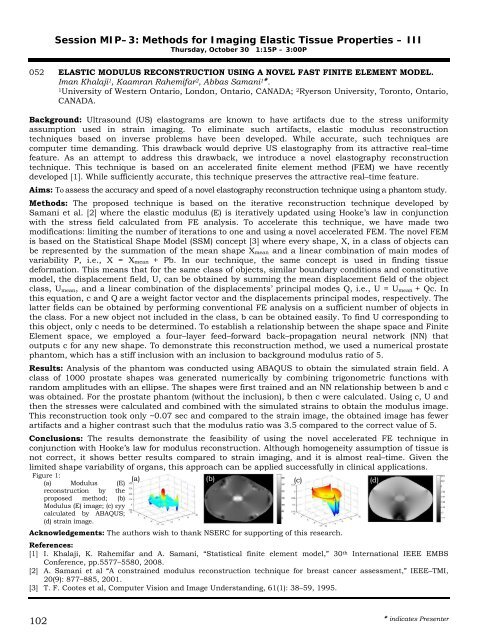
Results: Analysis of the phantom was conducted using ABAQUS to obtain the simulated strain field. A<br />
class of 1000 prostate shapes was generated numerically by combining trigonometric functions with<br />
random amplitudes with an ellipse. The shapes were first trained and an NN relationship between b and c<br />
was obtained. For the prostate phantom (without the inclusion), b then c were calculated. Using c, U and<br />
then the stresses were calculated and combined with the simulated strains to obtain the modulus image.<br />
This reconstruction took only ~0.07 sec and compared to the strain image, the obtained image has fewer<br />
artifacts and a higher contrast such that the modulus ratio was 3.5 compared to the correct value of 5.<br />
Conclusions: The results demonstrate the feasibility of using the novel accelerated FE technique in<br />
conjunction with Hooke’s law for modulus reconstruction. Although homogeneity assumption of tissue is<br />
not correct, it shows better results compared to strain imaging, and it is almost real–time. Given the<br />
limited shape variability of organs, this approach can be applied successfully in clinical applications.<br />
Acknowledgements: The authors wish to thank NSERC for supporting of this research.<br />
References:<br />
[1] I. Khalaji, K. Rahemifar and A. Samani, “Statistical finite element model,” 30th Figure 1:<br />
(a) (b)<br />
(a) Modulus (E)<br />
(c)<br />
(d)<br />
reconstruction by the<br />
proposed method; (b)<br />
Modulus (E) image; (c) εyy<br />
calculated by ABAQUS;<br />
(d) strain image.<br />
<strong>International</strong> IEEE EMBS<br />
<strong>Conference</strong>, pp.5577–5580, 2008.<br />
[2] A. Samani et al “A constrained modulus reconstruction technique for breast cancer assessment,” IEEE–TMI,<br />
20(9): 877–885, 2001.<br />
[3] T. F. Cootes et al, Computer Vision and Image Understanding, 61(1): 38–59, 1995.<br />
indicates Presenter