

proceedings - International Tissue Elasticity Conference
proceedings - International Tissue Elasticity Conference
proceedings - International Tissue Elasticity Conference

You also want an ePaper? Increase the reach of your titles
YUMPU automatically turns print PDFs into web optimized ePapers that Google loves.
087 IMPROVING SHEAR VELOCITY ESTIMATION ACCURACY BY DETECTING POTENTIAL FALSE<br />
PEAKS IN TISSUE DISPLACEMENT RESPONSE.<br />
Liexiang Fan 1 , Paul Freiburger 1 .<br />
1 Siemens Medical Solutions USA, Ultrasound Division, 22010 SE 51 st Street, Issaquah, WA, USA.<br />
Background: It has been proven feasible to detect tissue shear velocity by monitoring the shear wave<br />
arrival times at multiple locations after acoustic radiation force impulse (ARFI) excitation [1]. In [1], it is<br />
also demonstrated that accurate results can be achieved by using the peak of the tissue displacement<br />
response to track the wave front propagation. When we applied this method, we observed the appearance<br />
of two peaks in the displacement response in both phantom and in–vivo data. In some cases, the highest<br />
peak was a false indicator of the wave front propagation. A false peak detected before the true peak<br />
resulted in overestimation of the shear velocity, and a false peak detected after the true peak resulted in<br />
underestimation of the shear velocity. There are many possible causes for a second peak which will not<br />
be addressed in this work. Instead, we developed a method which can identify a false peak. This has<br />
enabled improved shear velocity estimation.<br />
Aims: The aim of this work is to identify the true peak in the tissue displacement response after ARFI<br />
excitation in order to prevent over/under–estimation of shear velocity.<br />
Methods: A set of displacements over time are generated within a region on interest (ROI) after ARFI<br />
excitation. A temporal displacement profile is generated from the median value of the displacements<br />
calculated within the ROI and is used as a reference. The highest peak is assumed to be the reference<br />
peak reflecting the average arriving time for the ROI. For each individual profile, the peak closest to the<br />
reference peak is taken as the true peak when two peaks are observed.<br />
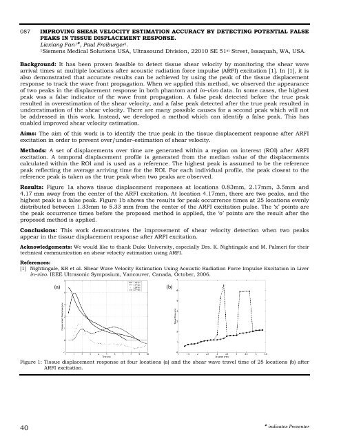
Results: Figure 1a shows tissue displacement responses at locations 0.83mm, 2.17mm, 3.5mm and<br />
4.17 mm away from the center of the ARFI excitation. At location 4.17mm, there are two peaks, and the<br />
highest peak is a false peak. Figure 1b shows the results for peak occurrence times at 25 locations evenly<br />
distributed between 1.33mm to 5.33 mm from the center of the ARFI excitation pulse. The ‘x’ points are<br />
the peak occurrence times before the proposed method is applied, the ‘o’ points are the result after the<br />
proposed method is applied.<br />
Conclusions: This work demonstrates the improvement of shear velocity detection when two peaks<br />
appear in the tissue displacement response after ARFI excitation.<br />
Acknowledgements: We would like to thank Duke University, especially Drs. K. Nightingale and M. Palmeri for their<br />
technical communication on shear velocity estimation using ARFI.<br />
References:<br />
[1] Nightingale, KR et al. Shear Wave Velocity Estimation Using Acoustic Radiation Force Impulse Excitation in Liver<br />
in–vivo. IEEE Ultrasonic Symposium, Vancouver, Canada, October, 2006.<br />
40<br />
(a) (b)<br />
Figure 1: <strong>Tissue</strong> displacement response at four locations (a) and the shear wave travel time of 25 locations (b) after<br />
ARFI excitation.<br />
indicates Presenter