

proceedings - International Tissue Elasticity Conference
proceedings - International Tissue Elasticity Conference
proceedings - International Tissue Elasticity Conference

Create successful ePaper yourself
Turn your PDF publications into a flip-book with our unique Google optimized e-Paper software.
029 FIRST RESULTS IN THE MEASUREMENT OF RAYLEIGH ELASTICITY PARAMETERS IN<br />
PHANTOMS AND IN–VIVO BREAST TISSUE.<br />
EEW Van Houten 1 , MDJ McGarry 1 , JB Weaver 2 , KD Paulsen 3 .<br />
1 University of Canterbury, Private Bag 4800, Christchurch, New Zealand; 2 Dartmouth Hitchcock<br />
Medical Center, Lebanon, NH 03756, USA, 3 Thayer School of Engineering, Dartmouth College,<br />
Hanover, NH 03755, USA.<br />
Background: The attenuating behavior of soft tissue shows evidence of providing clinical and diagnostic<br />
insight into various soft tissue disorders [1–3]. To make the most of this diagnostic capability,<br />
characterization of the damping behavior of tissue should be as accurate as possible. To date, attenuation<br />
in the case of the time–harmonic tissue behavior found in dynamic elastography has captured the damping<br />
behavior through a single parameter that takes the form of an imaginary component of a complex valued<br />
shear modulus. A more generalized damping formulation for the time–harmonic case, known commonly as<br />
Rayleigh or proportional damping, includes an additional parameter that takes the form of an imaginary<br />
component of a complex valued density. The effects of these two different damping mechanisms can be<br />
shown to be independent across homogeneous distributions and mischaracterization of the damping<br />
structure can be shown to lead to artifacts in the reconstructed attenuation profile [4].<br />
Methods: A novel method for reconstructing the Rayleigh damping distribution of within tissue and<br />
tissue–mimicking phantoms from 3D time–harmonic displacement data obtained via MR imaging has<br />
been developed based on a subzone approach to the global inverse problem [5]. This technique has been<br />
used to evaluate the ability to assess Rayleigh damping distribution from measured data and to initiate<br />
an investigation into the potential clinical value of these measurements.<br />
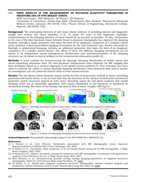
Results: The two figures below illustrate typical results for this reconstruction method in tissue mimicking<br />
phantoms and breast tissue. It can be seen that that the structure of the various reconstructed mechanical<br />
properties match structural aspects in both cases. Resulting values for real shear modulus and overall<br />
damping levels are in reasonable agreement with values established in the literature or measured by<br />
mechanical testing. The value of real density was fixed at that of water, roughly 1000 kg/m 3 .<br />
42<br />
Figure 1: Rayleigh damping reconstruction from a tissue mimicking heterogeneous phantom.<br />
Figure 2: 3 slices of a Rayleigh damping reconstruction from breast tissue.<br />
Acknowledgements: We gratefully acknowledge support from NIH/NIBIB R01–EB004632–02.<br />
References:<br />
[1] L Huwart et al., Liver Fibrosis: Noninvasive Assessment with MR Elastography versus Aspartate<br />
Aminotransferase–to–Platelet Ratio Index, Radiology, 245, 2007.<br />
[2] R Sinkus et al., Viscoelastic shear properties of in vivo breast lesions measured by MR elastography, J Magn<br />
Reson Imaging, 23(2), 2005.<br />
[3] N Salameh et al., Hepatic viscoelastic parameters measured with MR elastography: correlations with quantitative<br />
analysis of liver fibrosis in the rat, J. Magn Reson Imag, 26(4), 2007.<br />
[4] MDJ McGarry et. al., Reconstructing spatially varying rayleigh damping parameters in magnetic resonance<br />
elastography, Sixth Int Conf Ultrason Meas Imag Tiss Elast, 54, 2007.<br />
[5] M Doyley et al., Steady–state magnetic resonance harmonic elastography: contrast detailed analysis and<br />
preliminary clinical evaluation, First Int Conf Ultrason Meas Imag Tiss Elast, 45, 2002.<br />
indicates Presenter