Plasmonic organic solar cell.pdf - 123SeminarsOnly
Plasmonic organic solar cell.pdf - 123SeminarsOnly
Plasmonic organic solar cell.pdf - 123SeminarsOnly

You also want an ePaper? Increase the reach of your titles
YUMPU automatically turns print PDFs into web optimized ePapers that Google loves.
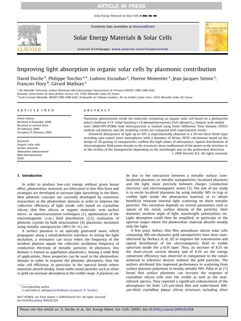
Improving light absorption in <strong>organic</strong> <strong>solar</strong> <strong>cell</strong>s by plasmonic contribution<br />
David Duche a , Philippe Torchio a,<br />
, Ludovic Escoubas a , Florent Monestier a , Jean-Jacques Simon a ,<br />
Franc-ois Flory b ,Gérard Mathian a<br />
a<br />
Aix-Marseille University, Institut Matériaux Microélectronique Nanosciences de Provence-IM2NP, CNRS-UMR 6242,<br />
Domaine Universitaire de Saint-Jérôme, Service 231, 13397 Marseille Cedex 20, France<br />
b<br />
Ecole Centrale Marseille, IM2NP CNRS-UMR 6242, Technopôle de Château-Gombert, 38 rue Frédéric Joliot Curie, 13451 Marseille Cedex 20, France<br />
article info<br />
Article history:<br />
Received 4 December 2008<br />
Received in revised form<br />
20 February 2009<br />
Accepted 25 February 2009<br />
Keywords:<br />
Organic <strong>solar</strong> <strong>cell</strong>s<br />
Surface plasmon<br />
Absorption enhancement<br />
Bulk heterojunction<br />
FDTD<br />
1. Introduction<br />
abstract<br />
In order to produce low-cost energy without green house<br />
effect, photovoltaic materials are fabricated in thin film form and<br />
strategies are developed to increase light harvesting in the films.<br />
New photonic concepts are currently developed by numerous<br />
researchers in the photovoltaic domain in order to improve the<br />
collection efficiency of light inside <strong>cell</strong>s based on crystalline<br />
silicon, thin film silicon, or <strong>organic</strong> materials: new surface<br />
micro- or nanostructuration techniques [1], optimization of the<br />
electromagnetic (e.m.) field distribution [2,3], realization of<br />
photonic crystals by bulk structuration [4], plasmonic excitation<br />
using metallic nanoparticles (NPs) [6–11], etc.<br />
A surface plasmon is an optically generated wave, which<br />
propagates along a metal/dielectric interface. In tuning the light<br />
excitation, a resonance can occur when the frequency of the<br />
incident photons equals the collective oscillation frequency of<br />
conduction electrons of metallic particles. In photonics, this<br />
domain is known as appellation plasmonics. Among a wide field<br />
of applications, these properties can be used in the photovoltaic<br />
domain in order to improve the photonic absorption, thus the<br />
<strong>solar</strong> <strong>cell</strong> efficiency, in particular in the spectral bands where<br />
materials absorb weakly. Some noble metal particles such as silver<br />
or gold can increase absorption in the visible range. A plasmon can<br />
Corresponding author.<br />
E-mail address: philippe.torchio@univ-cezanne.fr (P. Torchio).<br />
0927-0248/$ - see front matter & 2009 Elsevier B.V. All rights reserved.<br />
doi:10.1016/j.solmat.2009.02.028<br />
ARTICLE IN PRESS<br />
Solar Energy Materials & Solar Cells ] (]]]]) ]]]–]]]<br />
Contents lists available at ScienceDirect<br />
Solar Energy Materials & Solar Cells<br />
journal homepage: www.elsevier.com/locate/solmat<br />
<strong>Plasmonic</strong> phenomenon inside the materials composing an <strong>organic</strong> <strong>solar</strong> <strong>cell</strong> based on a photoactive<br />
poly(2-methoxy-5-(2 0 -ethyl-hexyloxy)-1,4-phenylenevinylene):(6,6)-phenyl-C61-butyric-acid-methyl<br />
ester (MEH-PPV:PCBM) bulk heterojunction is studied using Finite Difference Time Domain (FDTD)<br />
method calculations and the modeling results are compared with experimental results.<br />
Enhanced absorptance of light up to 50% is experimentally obtained in a 50-nm-thick blend layer<br />
including spin-coated silver nanospheres with a diameter of 40 nm. FDTD calculations based on the<br />
design of 2D-grating of nanoparticles confirm the high values of absorptance. Spatial distributions of<br />
electromagnetic field power density in the structures show confinement of the power at the interface or<br />
in the vicinity of the nanoparticles depending on the wavelength and on the preferential directions.<br />
& 2009 Elsevier B.V. All rights reserved.<br />
be due to the interaction between a metallic surface (nonlocalized<br />
plasmon) or metallic nanoparticles (localized plasmon)<br />
and the light, more precisely between charges (conduction<br />
electrons) and electromagnetic waves [5]. The aim of our study<br />
is to excite localized plasmons by using metallic NPs to trap or<br />
confine light inside the photoactive material, or to obtain<br />
beneficial resonant internal light scattering on these metallic<br />
particles. This excitation depends on several parameters such as<br />
nature of the metal, surface density of the particles, their<br />
diameter, incident angle of light, wavelength, polarization, etc.<br />
Light absorption could then be amplified, in particular in the<br />
spectral ranges where the photovoltaic material absorbs weakly<br />
only the light.<br />
A few years before, thin film amorphous silicon <strong>solar</strong> <strong>cell</strong>s<br />
containing 100-nm-diameter gold nanoparticles have been manufactured<br />
by Derkacs et al. [6] to engineer the transmission and<br />
spatial distribution of the electromagnetic field in visible<br />
spectrum inside the a-Si:H layer. Thus, an increase of 8.1% on<br />
the short-circuit current density and of 8.3% on the energy<br />
conversion efficiency was observed in comparison to the values<br />
achieved in reference devices without the gold particles. The<br />
authors attributed this improved performance to scattering from<br />
surface plasmon polaritons in nearby metallic NPs. Pillai et al. [7]<br />
found that surface plasmons can increase the response of<br />
crystalline silicon <strong>cell</strong>s over the visible as well as the nearinfrared<br />
spectra. They reported a significant enhancement of the<br />
absorptance for both 1.25-mm-thick film and wafer-based 300mm-thick<br />
crystalline planar silicon structures including silver<br />
Please cite this article as: D. Duche, et al., Sol. Energy Mater. Sol. Cells (2009), doi:10.1016/j.solmat.2009.02.028
2<br />
islands of 12 and 16 nm diameters. The photocurrent response<br />
was increased by about 30% over the whole visible range, and also<br />
in the near-infrared with a factor of 16 at l ¼ 1050 nm. This light<br />
trapping approach, also based on scattering by particles in silicon<br />
devices, clearly allows amplifying the interaction between light<br />
and material. Schaadt et al. [8] reported an engineered enhancement<br />
of optical absorptance and photocurrent in silicon p–n<br />
junction via the excitation of surface plasmon resonances in<br />
spherical Au nanoparticles and suggested their use for improving<br />
performances of photodetectors, imaging arrays and photovoltaics.<br />
Similar studies were carried out with <strong>organic</strong> materials to<br />
enhance light absorptance, subsequently leading to an increase in<br />
the amount of excitons. Stenzel et al. [9] have already demonstrated<br />
in systems ITO/NPs/CuPc/In that incorporating copper or<br />
gold NPs was increasing the photocurrent by a factor of more than<br />
two. This result was then confirmed by Westphalen et al. [10] on<br />
components such as ITO/Ag clusters/ZnPc/Ag in which the exciton<br />
rate was increased resulting in a photocurrent multiplied by a<br />
factor of two. Rand et al. [11] have studied the optical properties of<br />
NPs and have shown that enhancing electromagnetic field by<br />
plasmonic excitation allows to increase conversion efficiency of<br />
tandem <strong>organic</strong> <strong>cell</strong>s by increasing the excitation generation rate.<br />
Their multilayer device ITO/CuPc/PTCBI/silver NPs/CuPc/PTCBI/Ag<br />
consisted of a serial connection of two donor–acceptor heterojunctions<br />
separated by a very thin layer of metallic nanoagregates,<br />
which were used as charge recombination centers.<br />
They also exhibited an increased absorptance by a factor of 2, for<br />
7-nm-thick CuPc single layers including 1-nm-diameter silver NPs<br />
in comparison with single layers without these NPs.<br />
The work presented in this paper aims at studying the<br />
potentiality of this plasmonic technique on <strong>cell</strong>s, of which the<br />
active layer is composed by an interpenetrated donor–acceptor<br />
Ag<br />
1000<br />
nm<br />
0<br />
200nm<br />
1 x 1 µm 2<br />
ARTICLE IN PRESS<br />
50<br />
10<br />
0<br />
1000nm<br />
Fig. 1. Atomic force microscopy (AFM) image of silver nanoparticles spin-coated<br />
on a SiO 2 substrate.<br />
Metallic Nanoparticles<br />
D. Duche et al. / Solar Energy Materials & Solar Cells ] (]]]]) ]]]–]]]<br />
nm<br />
MEH-PPV:PCBM<br />
blend and it also aims at modeling this phenomenon by using a<br />
software based on the Finite Difference Time Domain (FDTD)<br />
method [12]. A bulk heterojunction (BHJ) device has been chosen<br />
because this kind of photoactive material is very promising and,<br />
presently, exhibits the best photovoltaic efficiencies in the field of<br />
<strong>organic</strong> <strong>solar</strong> <strong>cell</strong>s [13].<br />
2. Results and discussion<br />
2.1. Experimental<br />
Photoactive layers were fabricated from an interpenetrated<br />
network of conjugated polymer poly(2-methoxy-5-(2 0 -ethyl-hexyloxy)-1,4-phenylenevinylene)<br />
(MEH-PPV) as electron donor and<br />
fullerene derivative (6,6)-phenyl-C61-butyric-acid-methyl ester<br />
(PCBM) as electron acceptor. These layers were deposited on<br />
silica substrates, after incorporation of silver nanoparticles in<br />
some of the samples.<br />
SiO2 substrates were first sequentially cleaned in an ultrasonic<br />
bath by using acetone and isopropanol, then rinsed with<br />
deionised water, dried in an oven at 120 1C for 30 min and finally<br />
treated with UV-generated ozone. Colloïdal solutions rich in<br />
spherical silver nanoparticles with diameter 40 nm were spincoated<br />
on the silica substrates. These NPs were diluted in 10%wt.<br />
ethylene glycol. The adhesion of NPs on the silica surface was<br />
previously ensured by dip-coating the substrates in an organosilane<br />
(APTMS: 3-aminopropyl trimethoxysilane) in ethanol solution.<br />
This latter step also allows avoiding a too strong aggregation<br />
of the nanoparticles and preventing particle migration in the film.<br />
The parameters of these inhomogeneous metallic deposits have<br />
been adjusted to obtain coalescent deposits as thin as possible,<br />
with a control by atomic force microscopy (AFM) as shown in<br />
Fig. 1. We observe a random array of silver clusters of about<br />
100 nm width and 40 nm height. If the lateral size is higher<br />
than that of one isolated particle, the height is well conserved.<br />
Some substrates, with or without NPs, were then covered by<br />
spin-coating a bulk heterojunction from an anhydrous<br />
chlorobenzene solution of MEH-PPV:PCBM at 1:4 weight ratio<br />
(Fig. 2). The films were spun at 1500 rpm during 1 min. The<br />
thickness of the film was 50 nm, measured by using a mechanical<br />
profilometer.<br />
Spectrophotometric measurements of the reflectance (R) and<br />
the transmittance (T) allows to deduce the values of absorptance<br />
(A) and scattering (S) A+S ¼ 1 R T of these samples in the visible<br />
range (Fig. 3). We clearly observe an improvement of (1 R T)<br />
over a broad spectral range between 375 and 800 nm for stacks<br />
including NPs, with a maximum gain of 50% at 500 nm compared<br />
to those without NPs. In the 375–575 nm spectral domain, the<br />
improvement of (A+S) in the polymer:fullerene heterojunction<br />
including NPs can be mainly attributed to a plasmonic effect<br />
inside the heterojunction while, in the 575–800 nm region, the<br />
improved (A+S) values could be essentially due to the absorptance<br />
and scattering of the NPs themselves.<br />
MEH-PPV:PCBM<br />
Silica substrate Silica substrate<br />
Silica substrate<br />
Fig. 2. Structure of the fabricated samples: (a) silica/silver nanoparticles, (b) silica/MEH-PPV:PCBM 1:4 and (c) silica/silver nanoparticles/MEH-PPV:PCBM 1:4.<br />
Please cite this article as: D. Duche, et al., Sol. Energy Mater. Sol. Cells (2009), doi:10.1016/j.solmat.2009.02.028
A + S = 1 - R - T<br />
0.55<br />
0.50<br />
0.45<br />
0.40<br />
0.35<br />
0.30<br />
0.25<br />
0.20<br />
0.15<br />
0.10<br />
0.05<br />
0.00<br />
300<br />
.<br />
.<br />
2.2. Numerical model<br />
SiO 2 / SilverNPs<br />
SiO 2 / MEH-PPV:PCBM (50 nm)<br />
SiO 2 / SilverNPs/ MEH-PPV:PCBM (50 nm)<br />
375<br />
400 500<br />
575<br />
600<br />
Wavelength(nm)<br />
In order to understand the A+S mechanisms previously shown<br />
in Section 2.1, the absorptance was calculated and the electromagnetic<br />
field was modeled, thanks to a Finite Difference Time<br />
Domain method. The numerical FDTD method [12] allows solving<br />
the Maxwell’s equations versus time on a discrete spatial grid.<br />
Moreover, a discretization of time Dt allows taking into account<br />
the propagation phenomenon of the electromagnetic field. To<br />
perform the calculation of light propagation through a structure,<br />
several numerical parameters have to be considered, e.g., finite<br />
computational domain (x, y, z), the boundary conditions as the<br />
periodicity properties of the structures, spatial grid sizes (Dx, Dy,<br />
Dz), temporal grid Dt and thus, the computation time, relative<br />
permittivity e(r, o) and relative permeability m(r, o) for each<br />
material of the structure, etc.<br />
We modeled previous structures but with a simplified design<br />
constituted of a two-dimensional (2D) array of silver nanospheres<br />
having a 40 nm diameter rather than an inhomogeneous and<br />
coalescent layer. As shown in Fig. 4, the structure had 2D-grating<br />
composed of a squared lattice with a period equal to 75 nm. This<br />
structure was first placed in the air as the surrounding medium,<br />
then embedded in a host matrix of 50-nm-thick MEH-PPV:PCBM<br />
blend. It is important to notice that, for the calculation, each<br />
device was illuminated with plane waves in normal incidence,<br />
while reflectance (R) and transmittance (T) were calculated using<br />
two infinite sensors, which received all the reflected and<br />
ARTICLE IN PRESS<br />
700<br />
800<br />
Fig. 3. Values of A+S ¼ 1 R T deduced from spectrophotometric measurement of<br />
reflectance and transmittance for the devices: silica/silver nanoparticles (solid<br />
line), silica/MEH-PPV:PCBM 1:4 without (dotted line) or with silver nanoparticles<br />
(filled circles line).<br />
Y<br />
Z<br />
40 nm<br />
X<br />
Z<br />
75 nm X<br />
Y<br />
⊗<br />
∞ ∞<br />
hν<br />
40 nm<br />
∞ ∞<br />
Sensor (T)<br />
50 nm-<br />
BHJ<br />
Sensor (R)<br />
Fig. 4. Schematics of the metallic nanospheres in a 2D-grating design, embedded<br />
or not in a bulk heterojunction film, used for FDTD calculations: (a) top view and<br />
(b) side view.<br />
D. Duche et al. / Solar Energy Materials & Solar Cells ] (]]]]) ]]]–]]] 3<br />
2<br />
1.5<br />
1<br />
0.5<br />
transmitted energies (Fig. 4b), including in particular the nonspecular<br />
reflected part. If it exists, the scattered light would be<br />
included here in the non-specular reflectance. It means that the<br />
absorptance in the device is then obtained by 1 R T. Sensors<br />
were considered sufficiently far from the device to avoid nearfield<br />
effects. The temporal grid was Dt ¼ 17 10 19 s and the<br />
spatial grid was Dx ¼ Dy ¼ Dz ¼ 0.9 nm. Computations have been<br />
performed using monochromatic excitations at wavelengths<br />
ranging between 350 and 800 nm. The polarization state TE or<br />
TM of the light has no influence because computations were done<br />
in normal incidence. Nevertheless, in order to have a better<br />
understanding of the phenomena, we decided to keep only the E x<br />
component along the x-axis of the electrical field of the incident<br />
light and to cancel the E y component along the y-axis.<br />
Furthermore, the dispersion curves of the complex refractive<br />
indices of the materials have to be taken into account. Real and<br />
imaginary parts of the complex refractive index, n and k, of MEH-<br />
PPV:PCBM, deduced from experimental ellipsometric measurements<br />
performed by SOPRA S.A. company (Fig. 5), have been<br />
introduced in the computation for each wavelength. The energy<br />
gap of the considered bulk heterojunction is found to be equal to<br />
2.16 eV (l ¼ 575 nm). Refractive index dispersion curves of silver<br />
in thin film form were obtained by fitting the experimental<br />
ellipsometric data found in the literature [14]. A fit of metallic<br />
material parameters by using only a Drude or Drude–Lorentz<br />
model is highly recommended in our FDTD software. The Drude<br />
model given by Eq. (1) was then used as a fit law for the complex<br />
dielectric constant of silver:<br />
mðoÞ ¼ inf þ D=ð ao2 (1)<br />
The obtained constants were einf ¼ 5, D ¼ 2181.58,<br />
a ¼ 1.11 10 17 and b ¼ 5.79 10 9 .<br />
Comparison between fitted data and measured data for optical<br />
constants n and k of silver are shown in Fig. 6. The differences are<br />
due to the fact that law (1) has to be validated by the two parts of<br />
the complex dielectric constants, i.e. both n and k.<br />
2.3. Numerical results<br />
n (MEH-PPV:PCBM)<br />
k (MEH-PPV:PCBM)<br />
iboÞ ¼ðn þ ikÞ 2<br />
E g = 2.16 eV<br />
0<br />
300 400 500<br />
575<br />
600 700 800<br />
Wavelength (nm)<br />
Fig. 5. Dispersion curves obtained by spectroscopic ellipsometry representing the<br />
real (full line) and the imaginary (dotted line) parts of the complex refractive index<br />
of the MEH-PPV:PCBM bulk heterojunction.<br />
We have computed by the FDTD method the reflectance (R),<br />
the transmittance (T) and the deduced absorptance (A) from the<br />
equation 1 R T. R, T and A are presented in Fig. 7 versus<br />
wavelength for the metallic 2D-structure depicted in Fig. 4 and<br />
placed in the air. A surface plasmon effect at the surface of the<br />
Please cite this article as: D. Duche, et al., Sol. Energy Mater. Sol. Cells (2009), doi:10.1016/j.solmat.2009.02.028
4<br />
n<br />
1.8<br />
1.6<br />
1.4<br />
1.2<br />
1.0<br />
0.8<br />
0.6<br />
0.4<br />
0.2<br />
nanospheres appears, which results in a strong decrease of the<br />
transmission and then an increase of the absorptance, at<br />
wavelength 360 nm. Nevertheless, a maximum appears also on<br />
the R curve at l ¼ 375 nm. We can also attribute this small peak to<br />
the surface plasmon, which could cause a decoupling of light in<br />
the plane of periodicity of the NPs grating and an increase of R, as<br />
it was noticed in [15] in similar coupling devices using photonic<br />
crystals. Because our 2D structure exhibits a dual periodicity of<br />
the dielectric constant, it can be considered as a 2D photonic<br />
crystal, but this appellation is usually used for all dielectric<br />
structures.<br />
The diameter of the nanospheres is very small compared to the<br />
wavelengths of the incident waves. Thus, the Mie Theory [16] can be<br />
used to describe the interaction of the incident energy with<br />
the localized plasmons due to the metallic nanoparticles. This<br />
model consists of the rigorous resolution of Maxwell’s equation by<br />
taking both the electromagnetic field inside the NPs and the field<br />
scattered by the NPs into account. The first-order Mie Theory leads<br />
to the expression of the extinction coefficient s(o), also called<br />
‘‘absorption-scattering cross-section’’, of a set of metallic NPs [17]<br />
sðoÞ ¼<br />
9NoV 3=2<br />
s<br />
c<br />
k data<br />
k fit<br />
n data<br />
n fit<br />
0.4 0.5<br />
0.6<br />
Wavelength (μm)<br />
0.7 0.8<br />
Fig. 6. Measured data from [12] compared with our fitted data with a Drude model<br />
(used as input in our numerical method) for optical constants of silver.<br />
A, R, T (%)<br />
100<br />
90<br />
80<br />
70<br />
60<br />
50<br />
40<br />
30<br />
20<br />
10<br />
360<br />
0<br />
350<br />
450<br />
550 650<br />
Wavelength (nm)<br />
2ðoÞ<br />
½ 1ðoÞþ2 sðoÞŠ 2 þ 2 2ðoÞ !<br />
A(Ag)<br />
R(Ag)<br />
T(Ag)<br />
Fig. 7. Calculated values of reflectance (&), transmittance (J) and absorptance<br />
(1 R T) (’) versus wavelength for a square matrix of silver nanoparticles placed<br />
in air. Plasmon resonance at l ¼ 360 nm.<br />
ARTICLE IN PRESS<br />
D. Duche et al. / Solar Energy Materials & Solar Cells ] (]]]]) ]]]–]]]<br />
750<br />
9<br />
8<br />
7<br />
6<br />
5<br />
4<br />
3<br />
2<br />
1<br />
0<br />
k<br />
(2)<br />
where o ¼ 2p/l is the pulsation of the incident electromagnetic<br />
field, c the speed of light in vacuum, es thedielectricconstantofthe<br />
surrounding medium, V thevolumeofoneNP,N the volumic density<br />
of NPs, and e1 and e2 are, respectively, the real part and the<br />
imaginary part of the dielectric constant em of the metal<br />
(e m ¼ e 1+ie 2). It is important to notice that both scattering out of<br />
the NPs and absorption inside the NPs are included in s(o).<br />
According to Eq. (2), s(o) is maximal when<br />
[(e1(o)+2es(o)) 2 +e2 2 (o)] is equal to zero. This condition is called<br />
the Mie resonance.<br />
Thus, assuming that e1 2 be2 2 for frequencies in the Mie<br />
resonance region (typically true for Ag), Mie Theory can predict<br />
the plasmon resonance wavelength of a layer integrating NPs by<br />
using the following formula:<br />
1ðoÞ ¼ 2 sðoÞ (3)<br />
According to Eq. (3) and silver optical constants, we calculate<br />
and obtain the plasmon resonance wavelength at l ¼ 357 nm.<br />
A very close value of that resonance wavelength is observed in<br />
Fig. 7 (l ¼ 360 nm).<br />
In order to highlight and explain the experimental plasmon<br />
effect by numerical calculation, we have calculated values of<br />
(1–R T), i.e. absorptance, versus the wavelength for a squared 2Dgrating<br />
silver nanoparticles placed in the air, for the MEH-<br />
PPV:PCBM interpenetrating network alone, and for silver nanoparticles<br />
embedded in the network (Fig. 8). Even if the Ag<br />
distribution is slightly different in our experiments and in our<br />
calculations, we observe that the main behaviour of the three<br />
calculated curves is similar to experiments curves (see Fig. 3). The<br />
behaviour of the absorptance curve concerning the MEH-<br />
PPV:PCBM alone is in accordance with those of its extinction<br />
coefficient k (Fig. 5), like the value of the energy gap at 575 nm.<br />
The enhancement of the absorptance for samples consisting in a<br />
blend including NPs in comparison with those without NPs is also<br />
clearly observed on a large spectral range (350 nmolo800 nm).<br />
This enhancement is attributed to the localized plasmon effect<br />
due to the metallic NPs. We can also notice that the plasmon<br />
resonance wavelength is redshifted by 95 nm to l ¼ 455 nm in<br />
comparison with the resonance at l ¼ 360 nm of the NPs placed in<br />
air. This result fulfils the Mie Theory of Eq. (2), which shows that<br />
the plasmon resonance depends on the ‘‘difference’’ between the<br />
real part of the relative permittivity of the metal and the relative<br />
permittivity of the host material surrounding the nanospheres<br />
(either air or active blend). The beneficial effect is mainly<br />
interesting in the long-wavelength range 500 nmolo800 nm, in<br />
a spectral region where the MEH-PPV:PCBM absorbs weakly or<br />
A (%)<br />
60<br />
50<br />
40<br />
30<br />
20<br />
10<br />
0<br />
350<br />
400<br />
450<br />
455 575<br />
500<br />
550<br />
600<br />
A(Ag+MEH-PPV:PCBM)<br />
A(MEH-PPV:PCBM)<br />
A(Ag)<br />
Wavelength (nm)<br />
Fig. 8. Calculated values of absorptance A ¼ (1 R T) versus wavelength for<br />
devices: silver nanoparticles in air (’), MEH-PPV:PCBM alone (K), and silver<br />
nanoparticles embedded in MEH-PPV:PCBM (m) showing a resonance at l ¼ 455<br />
nm.<br />
Please cite this article as: D. Duche, et al., Sol. Energy Mater. Sol. Cells (2009), doi:10.1016/j.solmat.2009.02.028<br />
650<br />
800<br />
750<br />
800
Y (µm)<br />
0.03<br />
0.02<br />
0.01<br />
0.00<br />
-0.01<br />
-0.02<br />
-0.03<br />
75 nm<br />
NP NP<br />
NP NP<br />
-0.03 -0.02-0.010.000.010.020.03<br />
X (µm)<br />
very weakly, while the metal has a very low level of absorptance.<br />
In this spectral region, the absorptance gain in the <strong>solar</strong> <strong>cell</strong> can be<br />
very strong. In the other part of the spectrum, 350 nmolo500<br />
nm, the absorptance in the metal is not negligible, as shown on<br />
the curve of the NPs in air, and the absorptance of the plasmon<br />
structures may not be profitable to the final device performance.<br />
However, the <strong>solar</strong> spectral intensity at lo350–400 nm is rather<br />
weak and does not critically impact the performances as<br />
compared with longer wavelengths.<br />
Therefore, it would be interesting to separate the contribution of<br />
the absorptance due to the metallic nanospheres from that of the<br />
bulk heterojunction. Our numerical FDTD method is able to calculate<br />
and to map the distribution of the power density (P) carried by the<br />
electromagnetic plane wave in our plasmonic structure. This value of<br />
P power flow is calculated over one period in steady state and can be<br />
thought of as a type of time average. P can also represent the<br />
modulus of the Poynting vector. Fig. 9(a) shows the spatial<br />
distribution of the power density of the e.m. field in the<br />
nanoparticles plane {x, y} and in the dielectric vicinity of an array<br />
of 4 silver NPs at the excitation wavelength 450 nm in normal<br />
incidence (direction z). This wavelength is very close to the<br />
maximum absorptance peak of the NPs embedded in the BHJ<br />
(455 nm). We locally observe a very strong increase of the power<br />
density at the interface between the blend and the NPs (‘‘bright<br />
areas’’), a non-negligeable level inside the NPs, and also a mean level<br />
of P in the blend surrounding the NPs, which occurs preferentially<br />
along the x-axis in the plane of the squared lattice (which is in<br />
accordance with the E x incident electrical field orientation). At the<br />
interface between both media, in the internal side of NP, the distance<br />
where P decreases exponentially down to P/e is in agreement with<br />
the value of silver skin thickness (between 2.5 and 3 nm for<br />
ARTICLE IN PRESS<br />
20 nm 17.5 nm<br />
1.97397<br />
P 0.03<br />
0.02<br />
0.01<br />
0.00<br />
-0.01<br />
-0.02<br />
NP NP<br />
2.20483<br />
P<br />
-0.03<br />
NP NP<br />
0.0<br />
0.0<br />
Y (µm)<br />
wavelengths ranging between 450 and 600 nm). Results confirm<br />
that silver is able to absorb a part of the incident light at l ¼ 450 nm.<br />
At the excitation wavelength of 600 nm, corresponding to a low-level<br />
absorptance of BHJ with NPs, the power density is decreased inside<br />
the NPs and beneficially enhanced in the interface and in the<br />
dielectric medium between NPs along the x-axis (Fig. 9(b)). The<br />
maximal P-values are enhanced from 1.97 at l ¼ 450 nm to 2.2 at<br />
l ¼ 600 nm. The P-values are referred to the incident power density<br />
value which equals 1. Along this x-axis,wecannoticeintheFig. 9(b)<br />
that the power enhancement is produced significantly in the<br />
interstice up to the midpoint between 2 NPs, i.e. 17.5 nm. Other P<br />
distributions carried out across the z-axis transversal to the 2D-array<br />
are presented in Figs. 10(a) and (b). Fig. 10(a), computed at<br />
l ¼ 450 nm, confirms the very strong increase of P at the interface<br />
between the NPs and the environment (with values up to 5.68). At<br />
l ¼ 600 nm, the P enhancement is mainly confined in the medium<br />
between the NPs (Fig. 10(b)), thus improving the absorptance of light<br />
in the BHJ film. In figures (a) and (b), we observe that the increase in<br />
P is prolonged in the whole embedded medium along the z-axis up<br />
to about 10 nm out of the NP (corresponding to the limit of the BHJ<br />
thickness in our design) towards the light source. Power density<br />
enhancements happening along preferential directions, are very<br />
promising for multilayer <strong>organic</strong> <strong>solar</strong> <strong>cell</strong>s applications. Indeed, it is<br />
observed in our computations that the interaction length between<br />
light and matter can be significantly increased.<br />
3. Conclusion<br />
-0.03 -0.02-0.010.000.010.020.03<br />
Fig. 9. Spatial distribution of the power density P of the electromagnetic field in the nanoparticles plane {x, y} and in the dielectric vicinity of an array of 4 silver NPs at the<br />
incident wavelength of: (a) 450 nm and (b) 600 nm.<br />
Y (µm)<br />
0.02<br />
0.01<br />
0.00<br />
-0.01<br />
-0.02<br />
D. Duche et al. / Solar Energy Materials & Solar Cells ] (]]]]) ]]]–]]] 5<br />
X µm)<br />
5.68066<br />
4.31154<br />
P 0.02<br />
0.01<br />
P<br />
NP NP<br />
0.00<br />
NP NP<br />
-0.03<br />
-0.02<br />
-0.01<br />
0.00<br />
0.01<br />
X (µm)<br />
0.02<br />
0.03<br />
0.0<br />
40 (nm)<br />
10 (nm)<br />
hv<br />
-0.03<br />
-0.02<br />
-0.01<br />
0.00<br />
0.01<br />
Fig. 10. Spatial distribution of the power density P of the electromagnetic field in the orthogonal plane {x, z} of the NPs and in the dielectric vicinity of an array of 2 silver<br />
NPs at the incident wavelength of: (a) 450 nm and (b) 600 nm.<br />
Y (µm)<br />
-0.01<br />
-0.02<br />
X (µm)<br />
0.02<br />
0.03<br />
We investigate the absorption enhancement induced by silver<br />
nanoparticles within a dielectric layer. Experimental results were<br />
Please cite this article as: D. Duche, et al., Sol. Energy Mater. Sol. Cells (2009), doi:10.1016/j.solmat.2009.02.028<br />
0.0
6<br />
obtained by using a spin-coating method to spread the colloidal<br />
nanoparticles on wide areas of organosilane-coated silica substrates.<br />
Enhanced absorptance up to 50% was obtained for a bulk heterojunction<br />
layer including these nanospheres. Power density of the<br />
electromagnetic field computations confirms that an increased<br />
absorptance and increased concentration of the power are possible<br />
in the vicinity of a two-dimensional array of nanoparticles embedded<br />
in the BHJ. Preferential distributions of the electromagnetic field in<br />
the longitudinal and transversal planes with respect to the nanoparticles<br />
2D-grating are shown. Both effects, the increase of the local<br />
field strength and the internal light scattering, can contribute to<br />
stronger absorption. The investigation has to be completed by optical<br />
measurements in order to separate the contribution to the global<br />
absorption from possible external scattering. The best positioning of<br />
these nanoparticles in a multilayer <strong>organic</strong> <strong>solar</strong> <strong>cell</strong> has to be<br />
analyzed in order to maintain the strong benefit of the localized<br />
surface plasmons on absorption without degrading the electrical<br />
properties, and thus to obtain a significant photovoltaic power<br />
efficiency improvement.<br />
Acknowledgements<br />
The authors acknowledge C. Defranoux from SOPRA S.A.<br />
company (Bois Colombes-France) for spectroscopic ellipsometry<br />
measurements of MEH-PPV:PCBM, and Agence Nationale de la<br />
Recherche (ANR) for support under the photovoltaic program<br />
SPARCS (ANR-07-PSPV-006-02).<br />
References<br />
[1] F. Flory, L. Escoubas, B. Lazarides, Artificial anisotropy and polarizing films,<br />
Appl. Opt. 41 (16) (2002) 3332–3335.<br />
[2] F. Monestier, Ph. Torchio, J.J. Simon, L. Escoubas, M. Cathelinaud, Demonstration<br />
of a software for automatic optimization of the electromagnetic<br />
ARTICLE IN PRESS<br />
D. Duche et al. / Solar Energy Materials & Solar Cells ] (]]]]) ]]]–]]]<br />
field in <strong>organic</strong> <strong>solar</strong> <strong>cell</strong>s, Nonlinear Opt. Quantum Opt. 37 (2007)<br />
159–168.<br />
[3] F. Monestier, J.J. Simon, Ph. Torchio, L. Escoubas, F. Flory, S. Bailly, R. de<br />
Bettignies, S. Guillerez, C. Defranoux, Modeling the short circuit current<br />
density of polymer <strong>solar</strong> <strong>cell</strong>s based on P3HT:PCBM blend, Sol. Energ. Mater.<br />
Sol. Cells 91 (2007) 405–410.<br />
[4] D. Duche, L. Escoubas, J.J. Simon, Ph. Torchio, W. Vervisch, F. Flory, Slow blochmodes<br />
for enhancing the absorption of light in thin films for photovoltaic<br />
<strong>cell</strong>s, Appl. Phys. Lett. 92 (2008) 193310.1–193310.3.<br />
[5] W.L. Barnes, A. Dereux, T.W. Ebbesen, Surface plasmon subwavelength optics,<br />
Nature (London) 424 (2003) 824–830.<br />
[6] D. Derkacs, S.H. Lim, P. Matheu, W. Mar, E.T. Yu, Improved performance<br />
of amorphous silicon <strong>solar</strong> <strong>cell</strong>s via scattering from surface plasmon<br />
polaritons in nearby metallic nanoparticles, Appl. Phys. Lett. 89 (2006)<br />
093103.1–093103.3.<br />
[7] S. Pillai, K.R. Catchpole, T. Trupke, M.A. Green, Surface plasmon enhanced<br />
silicon <strong>solar</strong> <strong>cell</strong>s, J. Appl. Phys. 101 (2007) 093105.1–093105.8.<br />
[8] D.M. Schaadt, B. Feng, E.T. Yu, Enhanced semiconductor optical absorption via<br />
surface plasmon excitation in metal nanoparticles, Appl. Phys. Lett. 86 (2005)<br />
063106.1–063106.3.<br />
[9] O. Stenzel, A. Stendhal, K. Voigtsberger, C. von Borczyskowski, Enhancement<br />
of the photovoltaïc conversion efficiency of copper phthalocyanine thin film<br />
devices by incorporation of metal clusters, Sol. Energy Mater. Sol. Cells 37 (1995)<br />
337–348.<br />
[10] W. Westphalen, U. Kreibig, J. Rostalski, H. Lüth, D. Meissner, Metal<br />
cluster enhanced <strong>organic</strong> <strong>solar</strong> <strong>cell</strong>s, Sol. Energy Mater. Sol. Cells 61 (2000)<br />
97–105.<br />
[11] B.P. Rand, P. Peumans, S.R. Forrest, Long-range absorption enhancement in<br />
<strong>organic</strong> tandem thin-film <strong>solar</strong> <strong>cell</strong>s containing silver nanoclusters, J. Appl.<br />
Phys. 96 (12) (2004) 7519–7526.<br />
[12] A. Tavlove, Computational Electrodynamics: The Finite-Difference Time-<br />
Domain Method, Artech House, Norwood, MA, 1995.<br />
[13] J.Y. Kim, K. Lee, N.E. Coates, D. Moses, T. Nguyen, M. Dante, J. Heeger, Efficient<br />
Tandem polymer <strong>solar</strong> <strong>cell</strong>s fabricated by all-solution processing, Science 317<br />
(2007) 222–225.<br />
[14] E.D. Palik, Handbook of Optical Constants of Solids, Academic Press, New York,<br />
1985, p. 12.<br />
[15] C. Seassal, Y. Park, A. Fave, E. Drouard, E. Fourmond, A. Kaminski, M. Lemiti, X.<br />
Letartre, P. Viktorovitch, Photonic crystal assisted ultra-thin silicon<br />
photovoltaic <strong>solar</strong> <strong>cell</strong>, in: SPIE Proceedings of the Photonics Europe Conference,<br />
2008.<br />
[16] G. Mie, Beiträge zur optik trüber medien, Speziell Kolloidaler Metallösungen,<br />
Leipzig, Ann. Phys. 330 (1908) 377–445.<br />
[17] U. Kreibig, M. Vollmer, Optical Properties of Metal Clusters, Springer, Berlin,<br />
1995.<br />
Please cite this article as: D. Duche, et al., Sol. Energy Mater. Sol. Cells (2009), doi:10.1016/j.solmat.2009.02.028