Bioorganic & Medicinal Chemistry Letters
Bioorganic & Medicinal Chemistry Letters
Bioorganic & Medicinal Chemistry Letters

You also want an ePaper? Increase the reach of your titles
YUMPU automatically turns print PDFs into web optimized ePapers that Google loves.
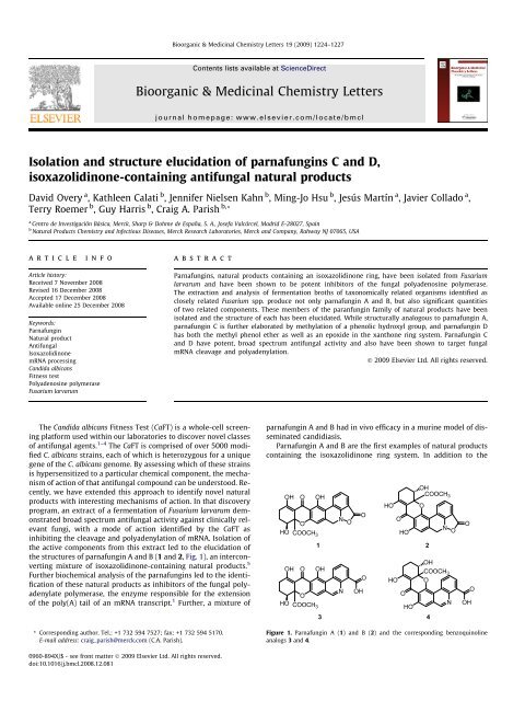
Isolation and structure elucidation of parnafungins C and D,<br />
isoxazolidinone-containing antifungal natural products<br />
David Overy a , Kathleen Calati b , Jennifer Nielsen Kahn b , Ming-Jo Hsu b , Jesús Martín a , Javier Collado a ,<br />
Terry Roemer b , Guy Harris b , Craig A. Parish b, *<br />
a Centro de Investigación Básica, Merck, Sharp & Dohme de España, S. A., Josefa Valcárcel, Madrid E-28027, Spain<br />
b Natural Products <strong>Chemistry</strong> and Infectious Diseases, Merck Research Laboratories, Merck and Company, Rahway NJ 07065, USA<br />
article info<br />
Article history:<br />
Received 7 November 2008<br />
Revised 16 December 2008<br />
Accepted 17 December 2008<br />
Available online 25 December 2008<br />
Keywords:<br />
Parnafungin<br />
Natural product<br />
Antifungal<br />
Isoxazolidinone<br />
mRNA processing<br />
Candida albicans<br />
Fitness test<br />
Polyadenosine polymerase<br />
Fusarium larvarum<br />
abstract<br />
The Candida albicans Fitness Test (CaFT) is a whole-cell screening<br />
platform used within our laboratories to discover novel classes<br />
of antifungal agents. 1–4 The CaFT is comprised of over 5000 modified<br />
C. albicans strains, each of which is heterozygous for a unique<br />
gene of the C. albicans genome. By assessing which of these strains<br />
is hypersensitized to a particular chemical component, the mechanism<br />
of action of that antifungal compound can be understood. Recently,<br />
we have extended this approach to identify novel natural<br />
products with interesting mechanisms of action. In that discovery<br />
program, an extract of a fermentation of Fusarium larvarum demonstrated<br />
broad spectrum antifungal activity against clinically relevant<br />
fungi, with a mode of action identified by the CaFT as<br />
inhibiting the cleavage and polyadenylation of mRNA. Isolation of<br />
the active components from this extract led to the elucidation of<br />
the structures of parnafungin A and B (1 and 2, Fig. 1), an interconverting<br />
mixture of isoxazolidinone-containing natural products. 5<br />
Further biochemical analysis of the parnafungins led to the identification<br />
of these natural products as inhibitors of the fungal polyadenylate<br />
polymerase, the enzyme responsible for the extension<br />
of the poly(A) tail of an mRNA transcript. 1 Further, a mixture of<br />
* Corresponding author. Tel.: +1 732 594 7527; fax: +1 732 594 5170.<br />
E-mail address: craig_parish@merck.com (C.A. Parish).<br />
0960-894X/$ - see front matter Ó 2009 Elsevier Ltd. All rights reserved.<br />
doi:10.1016/j.bmcl.2008.12.081<br />
<strong>Bioorganic</strong> & <strong>Medicinal</strong> <strong>Chemistry</strong> <strong>Letters</strong> 19 (2009) 1224–1227<br />
Contents lists available at ScienceDirect<br />
<strong>Bioorganic</strong> & <strong>Medicinal</strong> <strong>Chemistry</strong> <strong>Letters</strong><br />
journal homepage: www.elsevier.com/locate/bmcl<br />
Parnafungins, natural products containing an isoxazolidinone ring, have been isolated from Fusarium<br />
larvarum and have been shown to be potent inhibitors of the fungal polyadenosine polymerase.<br />
The extraction and analysis of fermentation broths of taxonomically related organisms identified as<br />
closely related Fusarium spp. produce not only parnafungin A and B, but also significant quantities<br />
of two related components. These members of the paranfungin family of natural products have been<br />
isolated and the structure of each has been elucidated. While structurally analogous to parnafungin A,<br />
parnafungin C is further elaborated by methylation of a phenolic hydroxyl group, and parnafungin D<br />
has both the methyl phenol ether as well as an epoxide in the xanthone ring system. Parnafungin C<br />
and D have potent, broad spectrum antifungal activity and also have been shown to target fungal<br />
mRNA cleavage and polyadenylation.<br />
Ó 2009 Elsevier Ltd. All rights reserved.<br />
parnafungin A and B had in vivo efficacy in a murine model of disseminated<br />
candidiasis.<br />
Parnafungin A and B are the first examples of natural products<br />
containing the isoxazolidinone ring system. In addition to the<br />
OH O OH<br />
OH<br />
COOCH3 HO O<br />
O<br />
HO COOCH3 N<br />
O<br />
O<br />
O<br />
HO<br />
N<br />
O<br />
1 2<br />
OH O OH<br />
O HO<br />
OH<br />
COOCH3 O<br />
O<br />
N OH<br />
O<br />
HO COOCH3 HO<br />
N<br />
3 4<br />
Figure 1. Parnafungin A (1) and B (2) and the corresponding benzoquinoline<br />
analogs 3 and 4.<br />
O<br />
OH<br />
O
isoxazolidinone, these compounds also contain a xanthone ring<br />
system, closely related to the ergochrome subunit that dimerizes<br />
to form various secalonic acids. 6,7 Ring-opening of the xanthone<br />
by a retro-Michael reaction leads to the interconversion of 1 and<br />
2, along with epimerization of the quaternary carbon bearing the<br />
methyl carboxylate. Further, the chemistry of these natural products<br />
is complicated by the instability of the isoxazolidinone ring.<br />
Hydrolysis of the isoxazolidinone ring, which is accompanied by<br />
the elimination of a molecule of water, generates the inactive benzoquinoline<br />
analogs 3 and 4.<br />
Parnafungin A and B were initially discovered from two fungal<br />
strains that resembled F. larvarum Fuckel (Ascomycota, Hypocreales)<br />
and were isolated from lichens obtained from the province<br />
of Madrid, Spain. 1,5 Subsequent to the discovery of parnafungin A<br />
and B, additional fungal species resembling F. larvarum, which included<br />
strains isolated from plants, plant litter and lichens, were<br />
identified as parnafungin A and B producers. From this taxonomic<br />
study, 8 it was concluded that the F. larvarum complex could be resolved<br />
into at least six or, possibly, seven different species. However,<br />
the examination of additional related strains is necessary<br />
before definitive taxa can be described. After fermentation of these<br />
strains, an acetone extract of each sample was analyzed by reversed<br />
phase HPLC with diode array and mass spectrometric detection<br />
(HPLC-DAD-MS) in order to confirm the production of 1 and 2<br />
(MW 451). In all cases, the production of 1 and 2 were confirmed<br />
and, further, the CaFT profiles of these extracts matched that observed<br />
for purified parnafungins A and B.<br />
While probing the taxonomic relationship of these parnafungin<br />
producing strains, an acetone extract from strain F-155,597 was<br />
identified by HPLC-DAD-MS analysis as producing substantial<br />
quantities of two analogs along with lesser quantities of parnafungins<br />
A and B. 8 These new analogs shared similar absorbance spectra<br />
to that obtained for parnafungin A and B (kmax 350 nm), but<br />
differed in retention time and molecular weight (MW 465 and<br />
479). Additional metabolites having similar absorbance spectra to<br />
that of the inactive benzoquinoline analogs (kmax 450 nm) were<br />
also present in this sample with corresponding molecular weights<br />
(MW 465 and 479). Following the discovery of these additional<br />
parnafungins, single ion monitoring was able to confirm that several<br />
other strains from the F. larvarum complex also produced these<br />
compounds, but in less pronounced quantities than that obtained<br />
from strain F-155,597. Here, we describe the isolation, structure<br />
elucidation and comparative antifungal activities of the two additional<br />
parnafungins identified from strain F-155,597, designated<br />
here as parnafungin C (5) and parnafungin D (6).<br />
In order to determine the structure of parnafungin C and D, a<br />
large scale fermentation (1 L) of F-155,597 was prepared and extracted<br />
with one volume of EtOAc. After adsorbing this extract onto<br />
silica gel by removal of the solvent and then loading it on a silica<br />
cartridge, the column was eluted successively with 30%, 50%, and<br />
80% EtOAc in hexanes, followed by 30% methanol in EtOAc.<br />
HPLC-DAD-MS analysis (C18) indicated that the two new parnafungin<br />
analogs were present in the 50% and 80% EtOAc in hexane<br />
fractions, with the latter cut containing predominantly 5 and 6.<br />
The combined 80% EtOAc fractions were concentrated to dryness<br />
and further purified by preparative reversed phase C18 HPLC. This<br />
fractionation step provided purified components for full chemical<br />
characterization and structure elucidation. High resolution mass<br />
spec analysis of these samples was consistent with the molecular<br />
formulas of C24H19NO9 (466.1131, calcd for M+H 466.1138) and<br />
C24H17NO10 (480.0924, calcd for M+H 480.0931) for parnafungin<br />
C and parnafungin D, respectively.<br />
In our earlier work, 5 the structures of parnafungins A and B<br />
were determined after methylation of the mixture of those natural<br />
products with ethereal diazomethane, thereby stabilizing the components<br />
from interconverting. The mono-methylated products<br />
D. Overy et al. / Bioorg. Med. Chem. Lett. 19 (2009) 1224–1227 1225<br />
OH O<br />
8<br />
OCH3 12 11 7<br />
10<br />
O OCH 3<br />
A 15a<br />
15 O 6<br />
HO COOCH3 16 17<br />
N<br />
O 1<br />
4<br />
O O A<br />
O<br />
HO COOCH3 5<br />
6<br />
OH<br />
O OCH 3<br />
O<br />
HO COOCH3 7<br />
N<br />
OH<br />
O<br />
N O<br />
were purified and the structures of the methylated derivatives<br />
were resolved by NMR spectroscopy and X-ray crystallography.<br />
In that case, methylation occurred at the C12 position providing<br />
the corresponding methyl enol ethers. Methylation at the enol position<br />
prevented the retro-Michael opening of the A ring (Fig. 2),<br />
thereby blocking the equilibration between parnafungin A and parnafungin<br />
B as well as the epimerization at C15a.<br />
NMR spectroscopy was used to determine the structure of parnafungin<br />
C and parnafungin D. It was readily apparent from an<br />
analysis of the 1 H and 13 C 1D NMR spectra that there were two<br />
methyl groups present in parnafungin C as compared with only<br />
one in parnafungins A and B (Table 1). One methyl singlet (d<br />
3.57 ppm) corresponded to the methyl carboxylate at the quaternary<br />
chiral center (C15a). The additional methyl singlet resonance<br />
in paranfungin C (5) atd 3.86 ppm was distinct from that in the<br />
OH<br />
O OCH 3<br />
O<br />
HO COOCH3 Figure 2. Parnafungin C (5) and D (6) and the corresponding benzoquinoline<br />
analogs 7 and 8.<br />
Table 1<br />
1 H and 13 C NMR data for parnafungin C (5) a<br />
Position d C mult d H(J in Hz)<br />
1 167.3 qC<br />
4 54.9 CH2 4.73 (m)<br />
4a 140.3 qC<br />
5 110.3 CH 7.06 (s)<br />
6 160.7 qC<br />
6a 113.1 qC<br />
7 158.9 qC<br />
7a 115.6 qC<br />
7b 119.0 qC<br />
8 131.3 CH 8.33 (d, 8.0)<br />
9 126.3 CH 7.43 (t, 8.0)<br />
10 123.5 CH 7.75 (d, 8.0)<br />
10a 112.8 qC<br />
10b 156.2 qC<br />
11 184.8 qC<br />
11a 102.8 qC<br />
12 171.4 qC<br />
13 25.7 CH2 2.75 (m)<br />
2.54 (m)<br />
14 23.9 CH 2 2.14 (m)<br />
1.90 (m)<br />
15 70.2 CH 4.24 (d, 4.0)<br />
15a 85.6 qC<br />
16 169.8 qC<br />
17 52.7 CH3 3.57 (s)<br />
7-OCH3 61.9 CH3 3.86 (s)<br />
12-OH 5.9 (b)<br />
a 1 H spectra were accumulated at 500 MHz and 13 C spectra were accumulated at<br />
125 MHz in DMSO-d 6. Proton NMR spectra were referenced to the residual 1 H<br />
solvent peak for DMSO-d 5 at d 2.49. Carbon spectra were referenced to the DMSO-d 6<br />
septet at d 39.51.<br />
O<br />
OH<br />
8<br />
N<br />
OH<br />
O<br />
O
1226 D. Overy et al. / Bioorg. Med. Chem. Lett. 19 (2009) 1224–1227<br />
methyl enol ether derivatives prepared from parnafungin A<br />
(d 3.82 ppm) and parnafungin B (d 3.90 ppm). Further, for the synthetic<br />
methyl enol ethers, the methyl group protons presented<br />
HMBC correlations to C12 at d 173 ppm, while the methyl group<br />
in parnafungin C provided an HMBC correlation to a phenolic carbon<br />
at d 159 ppm. It remained to be determined whether the<br />
methyl ether in parnafungin C was at C7, which would be the case<br />
if the structure was analogous to parnafungin A, or on the phenolic<br />
hydroxyl group C6, which is available in parnafungin B. ROESY correlations<br />
between the methyl group and the H8 proton indicated<br />
that the methyl group was located on the C7 phenol.<br />
Additional spectroscopic differences between the ‘straight’ parnafungin<br />
A and the more ‘bent’ parnafungin B have been described.<br />
5 In those studies, the 1 H NMR signals that correspond to<br />
H4 and H8 provided further insight into the structure of 5. The differences<br />
in the 1 H resonances were attributed to small changes in<br />
the twist of the biphenyl ring system. The two diastereotopic protons<br />
at C4 have near magnetic equivalence and have almost completely<br />
collapsed to a singlet at d 4.68 ppm for 1. This is not the<br />
case for 2, where the two protons at C4 are well separated into a<br />
pair of doublets at d 4.55 and d 4.76 ppm. In the case of H8, this<br />
aromatic proton presented at d 8.30 ppm for 1 and at d 8.65 ppm<br />
for 2. For parnafungin C, the signal for the C4 methylene group<br />
had almost collapsed to a singlet at d 4.73 ppm and H8 was observed<br />
at d 8.33 ppm. These data were clearly consistent with an<br />
assignment of the structure of parnafungin C (5) in the straight<br />
geometry analogous with the structure of parnafungin A (1). Since<br />
parnafungin C does not contain a moiety blocking the retro-Michael<br />
ring-opening of the A-ring, epimerization of C15a is still possible<br />
and an equilibrium mixture of two diastereomers was<br />
observed in the 1 H NMR spectra in DMSO-d 6.<br />
Parnafungin D (6) was relatively more polar than parnafungin C<br />
based on the retention times of these components on reversed<br />
phase C18 HPLC. Comparing the molecular formulas of these two<br />
components, parnafungin D had one additional oxygen atom and<br />
Table 2<br />
1 H and 13 C NMR data for parnafungin D (6) a<br />
Position d C mult d H(J in Hz)<br />
1 168.0 qC<br />
4 56.3 CH 2 4.51 (m)<br />
4a 140.6 qC<br />
5 113.6 CH 6.82 (s)<br />
6 157.6 qC<br />
6a 112.9 qC<br />
7 159.0 qC<br />
7a 119.1 qC<br />
7b 119.5 qC<br />
8 132.0 CH 8.43 (d, 7.5)<br />
9 126.5 CH 7.37 (t, 7.5)<br />
10 124.4 CH 7.70 (d, 7.5<br />
10a 111.6 qC<br />
10b 156.9 qC<br />
11 186.8 qC<br />
11a 101.4 qC<br />
12 174.8 qC<br />
13 53.9 CH 3.62 (d, 4.5)<br />
14 55.7 CH 3.80 (d, 4.5)<br />
15 74.8 CH 4.60 (d, 10.0)<br />
15a 78.7 qC<br />
16 170.9 qC<br />
17 53.9 CH3 3.67 (s)<br />
7-OCH 3 62.5 CH 3 3.90 (s)<br />
12-OH 15.9 (s)<br />
15-OH 4.79 (d, 10.0)<br />
a 1 H spectra were accumulated at 500 MHz and 13 C spectra were accumulated at<br />
125 MHz in CD 2Cl 2. Proton NMR spectra were referenced to the residual 1 H solvent<br />
peak for CDHCl 2 at d 5.32. Carbon spectra were referenced to the CD 2Cl 2 pentet at d<br />
54.0.<br />
two fewer protons. Upon preparing a solution of 6 in DMSO-d6<br />
for NMR analysis, substantial decomposition of the material was<br />
observed. While parnafungin C had limited solubility in methylene<br />
chloride, parnafungin D had sufficient solubility in CD 2Cl 2 for the<br />
acquisition of a complete NMR data set (Table 2). 1 H and 13 C<br />
NMR spectra of 6 indicated that the aliphatic methylene groups<br />
of the A ring (Fig. 2) were not present in this component. While<br />
there were still two methyl groups at d 3.67 and d 3.90 ppm, two<br />
additional doublets were observed at d 3.62 and d 3.80 ppm, each<br />
corresponding to one proton by integration. The assignment of<br />
these signals as an epoxide at C13–C14 was fully consistent with<br />
these data and HMBC correlations. Since H14 does not present a<br />
1 H– 1 H coupling with H15, the epoxide is on the same face of the<br />
A ring as the hydroxyl, providing a geometry where the two protons<br />
are approximately at an angle of 90°. The remaining 1D and<br />
2D NMR data were fully consistent with the remainder of parnafungin<br />
D being identical with that of parnafungin C. In addition,<br />
the benzoquinoline analogs 7 and 8 of parnafungins C and D,<br />
respecitively, were also identified in the HPLC chromatograms by<br />
absorbance spectra and mass spec data. 1 H NMR analysis of these<br />
compounds was consistent with the opening of the isoxazolidinone<br />
ring and the formation of the aromatized benzoquinoline (data not<br />
shown).<br />
Based on our previous stereochemical analyses for parnafungin<br />
A and B, 5 the absolute configuration of the C15 hydroxyl of these<br />
natural products is (S) and the major diastereomer at the C15a quaternary<br />
carbon is also (S). The producing organism of parnafungin C<br />
and D (F-155,597) is taxonomically closely related to the original<br />
F. larvarum from which parnafungin A and B were isolated. 8 Further,<br />
parnafungins A and B are co-produced with parnafungins C<br />
and D by F-155,597, indicating that these new components are<br />
likely to be part of the same biosynthetic pathway. From this, the<br />
assignment of the absolute configuration of the C15 hydroxyl for<br />
parnafungin C and D is also (S) and the epoxide of parnafungin D<br />
is on the b face of the A ring.<br />
The relative potencies and spectra of antifungal activity were<br />
determined for 5 and 6, and compared to the activity of a mixture<br />
of parnafungins A and B (1/2) (Table 3). Generally, 5 and 6 were<br />
less potent against all of the Candida species tested, but in some<br />
cases, such as against C. tropicalis and C. lusitaniae, these analogs<br />
had comparable antifungal activity. These differences in relative<br />
potency may be indicative of slight changes in the binding of each<br />
compound against the relevant isoform of the fungal RNA polyadenosine<br />
polymerase. In order to assess whether analogs 5 and 6<br />
had similar mechanisms of action as 1 and 2, the activity of each<br />
compound was tested against wild-type C. albicans and two heterozygote<br />
strains C. albicans strains, each containing a single copy of<br />
CLP1 or YSH1. These two strains were previously shown to be<br />
hypersensitive to parnafungins A and B. 1 For 5 and 6 (Table 4), a<br />
2- to 4-fold shift was observed in the MICs against the heterozygote<br />
strains, indicating that these compounds are functioning with<br />
the same mechanism of action as was determined for 1 and 2. As<br />
was the case for benzoquinolines 3 and 4, no antifungal activity<br />
was observed for the comparable analogs 7 and 8.<br />
Table 3<br />
Whole-cell inhibition of parnafungins A–D<br />
Species Strain 1/2 5 6<br />
MIC in lg/mL a<br />
C. albicans MY2323 0.008 2 0.016<br />
C. glabrata ATCC90030 1.25 >10 5<br />
C. parapsilosis ATCC22019 0.6 >10 2.5<br />
C. lusitaniae ATCC34449 0.3 0.16 0.16<br />
C. krusei ATCC6258 0.008 0.08 0.016<br />
C. tropicalis ATCC750 2.5 2.5 0.6<br />
a MIC was determined in Sabarose Dextrose medium.
Table 4<br />
Heterozygote strain inhibition of parnafungins A–D<br />
Species Strain 1/2 5 6<br />
MIC in lg/mL a<br />
C. albicans MY2323 (WT) 0.008 0.06 0.016<br />
C. albicans MY2323 CLP1 +/ 0.004 0.03 0.004<br />
C. albicans MY2323 YSH1 +/ 0.004 0.03 0.004<br />
a MIC was determined in Sabarose Dextrose medium.<br />
The biosynthesis of parnafungins is likely related to that of<br />
ergochrome-derived secondary metabolites, which are based on a<br />
series of polyketide condensations and cyclizations. 6,9 For the parnafungin<br />
family of natural products, the extended ring system beyond<br />
the typical xanthone unit as well as the incorporation of a<br />
nitrogen atom and the closure of the isoxazolidinone ring make<br />
the biosynthetic pathway an intriguing, but unresolved question.<br />
Previously, we have identified by affinity selection/mass spec techniques<br />
that the active form of the equilibrating mixture of isomers<br />
is the straight parnafungin A. 10 The fact that parnafungins C and D<br />
are direct analogs of parnafungin A and not parnafungin B supports<br />
the hypothesis that it is parnafungin A that is directly synthesized<br />
by these Fusarium spp. Subsequent methylation and oxidation of<br />
parnafungin A would generate 5 and then 6. Practically, the methylation<br />
of the C7 hydroxyl simplifies the chemical properties of<br />
both of these compounds, preventing the formation of the corresponding<br />
bent geometric isomers.<br />
Species of the F. larvarum complex are assumed to be mycoparisites<br />
associated with scale insects, aphids, lichens and the basidiomycete<br />
fungus Septobasidium clelandii. 8,11–14 Additional to the F.<br />
larvarum complex, the parnafungins have also been reported from<br />
two other Hypocrealean mycoparasitic fungi, Trichonectria rectipila<br />
and Cladobotryum pinarense. 8 With regards to the ecological role<br />
that the parnafungins may convey to their producing organism, it<br />
has been hypothesized that the compounds may act as potent antifungals,<br />
providing a competitive advantage for growth and, moreover,<br />
as a virulence factor mediating inter-organism interactions<br />
while colonizing hosts, for example, lichenized and non-lichenized<br />
fungi, insects, or plants. 8 The facile degradation of the parnafungins<br />
to the inactive benzoquinoline forms has been correlated with a<br />
change in pH, where ring opening was promoted in neutral or alkaline<br />
conditions, while a stabilization of the molecule is favored in<br />
acidic conditions. 5 Many fungi lower the pH of their growth medium<br />
through the secretion of organic acids during their initial<br />
phase of growth, and later, as the fungus reaches stationary<br />
growth, the medium’s pH progressively becomes more alkaline; 15<br />
in this case, alkalization of the growth medium following colonization<br />
would, hypothetically, promote the accumulation of the ben-<br />
D. Overy et al. / Bioorg. Med. Chem. Lett. 19 (2009) 1224–1227 1227<br />
zoquinoline analogs and prevent the producing organism from<br />
accumulating toxic levels of the parnafungins. 8<br />
Since parnafungins A and B are efficacious in a murine model of<br />
disseminated candidiasis with no observable toxicity, 1 this family<br />
of compounds can be pursued as a novel class of antifungal agents<br />
with a unique mechanism of action. The parnafungin analogs reported<br />
here provide additional information as to the types of structural<br />
modifications that are possible to the core structure while<br />
maintaining antifungal activity. Parnafungins C and D expand the<br />
available structure–activity information on this family of natural<br />
products. Further synthetic exploration of this chemical scaffold<br />
may provide parnafungin analogs with improved potency, spectrum<br />
and chemical stability, and provide a route for the further<br />
development of an antifungal agent that targets fungal RNA<br />
processing.<br />
Supplementary data<br />
Supplementary data associated with this article can be found, in<br />
the online version, at doi:10.1016/j.bmcl.2008.12.081.<br />
References and notes<br />
1. Jiang, B.; Xu, D. M.; Allocco, J.; Parish, C.; Davison, J.; Veillette, K.; Sillaots, S.;<br />
Hu, W. Q.; Rodriguez-Suarez, R.; Trosok, S.; Zhang, L.; Li, Y.; Rahkhoodaee, F.;<br />
Ransom, T.; Martel, N.; Wang, H.; Gauvin, D.; Wiltsie, J.; Wisniewski, D.;<br />
Salowe, S.; Kahn, J. N.; Hsu, M. J.; Giacobbe, R.; Abruzzo, G.; Flattery, A.; Gill, C.;<br />
Youngman, P.; Wilson, K.; Bills, G.; Platas, G.; Pelaez, F.; Diez, M. T.; Kauffman,<br />
S.; Becker, J.; Harris, G.; Liberator, P.; Roemer, T. Chem. Biol. 2008, 15, 363.<br />
2. Haselbeck, R.; Wall, D.; Jiang, B.; Ketela, T.; Zyskind, J.; Bussey, H.; Foulkes, J. G.;<br />
Roemer, T. Curr. Pharm. Design 2002, 8, 1155.<br />
3. Roemer, T.; Jiang, B.; Davison, J.; Ketela, T.; Veillette, K.; Breton, A.; Tandia, F.;<br />
Linteau, A.; Sillaots, S.; Marta, C.; Martel, N.; Veronneau, S.; Lemieux, S.;<br />
Kauffman, S.; Becker, J.; Storms, R.; Boone, C.; Bussey, H. Mol. Microbiol. 2003,<br />
50, 167.<br />
4. Xu, D.; Jiang, B.; Ketela, T.; Lemieux, S.; Veillette, K.; Martel, N.; Davison, J.;<br />
Sillaots, S.; Trosok, S.; Bachewich, C.; Bussey, H.; Youngman, P.; Roemer, T. PLoS<br />
Pathogens 2007, 3, e92.<br />
5. Parish, C. A.; Smith, S. K.; Calati, K.; Zink, D.; Wilson, K.; Roemer, T.; Jiang, B.; Xu,<br />
D. M.; Bills, G.; Platas, G.; Pelaez, F.; Diez, M. T.; Tsou, N.; McKeown, A. E.; Ball,<br />
R. G.; Powles, M. A.; Yeung, L.; Liberator, P.; Harris, G. J. Am. Chem. Soc. 2008,<br />
130, 7060.<br />
6. Kurobane, I.; Vining, L. C.; McInnes, A. G. J. Antibiotics 1979, 32, 1256.<br />
7. Reddy, C. S.; Reddy, R. V. Mycotoxins Phytoalexins 1991, 167.<br />
8. Bills, G. F.; Platas, G.; Overy, D. P.; Collado, J.; Fillola, A.; Jimenez, R.; Martin, J.;<br />
Gonazalez del val, A.; Vicente, M. F.; Tormo, J. R.; Pelaez, F.; Calati, K.; Harris, G.<br />
H.; Parish, C. A.; Xu, D.; Roemer, T. Mycologia 2009, 101, in press.<br />
9. Franck, B.; Bringmann, G.; Flohr, G. Angew. Chem. Int. Ed. 1980, 19, 460.<br />
10. Adam, G. C.; Parish, C. A.; Wisniewski, D.; Meng, J.; Liu, M.; Calati, K. B.; Stein, B.<br />
D.; Athanasopoulos, J.; Liberator, P. A.; Roemer, T.; Harris, G. H.; Chapman, K. T.<br />
J. Am. Chem. Soc. 2008, 130, 16704.<br />
11. Booth, C. CMI Descript. Pathog. Fungi Bact. 1981, 714, 1.<br />
12. Gerlach, W. Phytopathologische Zeitschrift—J. Phytopathol. 1977, 90, 31.<br />
13. Rossman, A. Y.; Samuels, G. J.; Rogerson, C. T.; Lowen, R. Stud. Mycol. 1999, 42,<br />
1.<br />
14. Tyson, J.; Henderson, R.; Fullerton, R.; Jamieson, L.; Froud, K. New Zealand Plant<br />
Protect. 2005, 58, 283.<br />
15. Griffin, D. Fungal Physiology; 2nd ed.; New York, 1994.