Overexpression of 1B-adrenergic receptor induces left ventricular ...
Overexpression of 1B-adrenergic receptor induces left ventricular ...
Overexpression of 1B-adrenergic receptor induces left ventricular ...

You also want an ePaper? Increase the reach of your titles
YUMPU automatically turns print PDFs into web optimized ePapers that Google loves.
H1340 LV DYSFUNCTION IN CARDIAC <strong>1B</strong>-AR OVEREXPRESSION<br />
animals were anesthetized, and the excised hearts were<br />
placed in oxygenated, nominally Ca 2 -free solution P containing<br />
132 mM NaCl, 4.8 mM KCl, 1.2 mM MgCl 2·6H2O,5mM<br />
glucose, and 10 mM HEPES, pH 7.2. The aorta was cannulated<br />
with a 23-gauge cannula and flushed gently, and the<br />
heart was mounted on the perfusion apparatus. Perfusion<br />
was carried out as follows: 8 min with the Ca 2 -free solution<br />
P; 7 min with Joklik’s MEM (GIBCO) supplemented with 75<br />
U/ml each <strong>of</strong> type I and type II collagenase (Worthington),<br />
0.1% BSA, and 2% calf serum; and finally 6 min <strong>of</strong> washout<br />
with low Ca 2 Joklik’s MEM (Joklik’s supplemented with<br />
0.025 mM CaCl 2). LV tissue was then dissected and incubated<br />
for 5 min at 37°C in low Ca 2 Joklik’s MEM, and released cells<br />
were filtered through 200-µm nylon mesh. Remaining pieces<br />
were reincubated and refiltered. The filtrates were combined,<br />
and the cells were collected by gravity sedimentation for 30<br />
min. The pelleted cells were then resuspended twice in low<br />
Ca 2 Joklik’s MEM with increasing Ca 2 concentration (0.5<br />
and 1.4 mM). The yields <strong>of</strong> intact rod-shaped myocytes were<br />
routinely 80% for control and 60% for transgenic hearts.<br />
To record intracellular free Ca 2 transients, cells were loaded<br />
with the fluorescent Ca 2 chelator fura 2-AM for 30 min at<br />
37°C in low Ca 2 Joklik’s MEM. Cells were washed once and<br />
resuspended in solution P supplemented with 1.8 mM CaCl 2.<br />
Measurement <strong>of</strong> field-stimulated cardiomyocytes was performed<br />
as described previously (12, 21). Intracellular free<br />
Ca 2 was monitored and reported as the ratio <strong>of</strong> 340/380 nm<br />
fluorescence <strong>of</strong> fura 2 at 500-nm emission wavelength using a<br />
photo scan dual-beam spectr<strong>of</strong>luorophotometer (Photon Tech)<br />
coupled to an Olympus IMT-2 ultraviolet fluorescent microscope<br />
with ultraviolet transparent optics.<br />
Fixation <strong>of</strong> hearts and electron microscopy. Specimens for<br />
electron microscopy were prepared according to standard<br />
procedures. Briefly, mice were anesthetized, and their hearts<br />
were exposed. Cardioplegic solution (25 mM KCl, 5% dextrose<br />
in PBS) was perfused (column height 65 cm) to relax the<br />
muscle. The heart was then fixed by perfusion with 2%<br />
glutaraldehyde in 0.1 M cacodylate buffer, pH 7.3, and<br />
postfixed for 24 h. Blocks <strong>of</strong> 1 mm 2 from the LV wall at<br />
midsection were embedded, oriented, and sectioned. Thin<br />
sections were viewed using a Zeiss 912 transmission electron<br />
microscope. Sarcomere length comparisons were made from<br />
photographs <strong>of</strong> these sections.<br />
Statistics. Results are expressed as means SE. Unpaired<br />
Student’s t-tests were performed for pairwise comparisons,<br />
and a level <strong>of</strong> P 0.05 was considered significant. ANOVA<br />
with Fisher’s least-squares difference post hoc analysis was<br />
performed to determine differences between multiple groups.<br />
RESULTS<br />
We were interested in the role <strong>of</strong> 1-AR in cardiac<br />
homeostasis and have analyzed transgenic mouse lines<br />
overexpressing the <strong>1B</strong>-AR in cardiomyocytes (1). Transgene<br />
expression is driven by the -MHC promoter that<br />
is specific for cardiomyocytes (45), directing high levels<br />
<strong>of</strong> expression to the adult mouse heart. The <strong>of</strong>fspring <strong>of</strong><br />
two independent transgenic mouse lines were analyzed.<br />
The levels <strong>of</strong> 1-AR overexpression (Tg47, 26fold,<br />
and Tg43, 43-fold) have been determined previously<br />
(1).<br />
Reduced basal level contractility <strong>of</strong> <strong>1B</strong>-AR overexpressors<br />
in vivo. The functional consequences <strong>of</strong> <strong>1B</strong>-AR<br />
overexpression were determined in the anesthetized<br />
closed-chest mouse model. It has been shown previously<br />
that differences in contractile performance in<br />
mice with altered <strong>adrenergic</strong> signaling status can be<br />
measured reliably in this system (10, 49). The comparison<br />
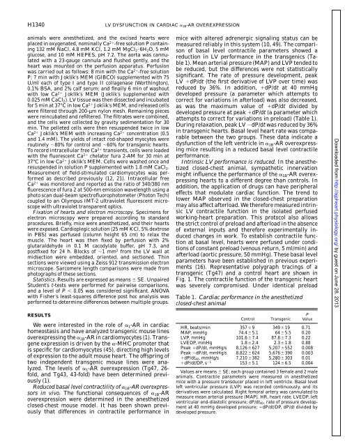
<strong>of</strong> basal level contractile parameters showed a<br />
reduction in LV performance in the transgenics (Table<br />
1). Mean arterial pressure (MAP) and LVP tended to<br />
be reduced, but the differences were not statistically<br />
significant. The rate <strong>of</strong> pressure development, peak<br />
LV dP/dt (the first derivative <strong>of</strong> LVP over time) was<br />
reduced by 36%. In addition, dP/dt at 40 mmHg<br />
developed pressure (a parameter which attempts to<br />
correct for variations in afterload) was also decreased,<br />
as was the maximum value <strong>of</strong> dP/dt divided by<br />
developed pressure at peak dP/dt (a parameter which<br />
attempts to correct for variations in preload) (Table 1).<br />
During relaxation, peak LV dP/dt was reduced by 36%<br />
in transgenic hearts. Basal level heart rate was comparable<br />
between the two groups. These data indicate a<br />
dysfunction <strong>of</strong> the <strong>left</strong> ventricle in <strong>1B</strong>-AR overexpressing<br />
mice resulting in a reduced basal level contractile<br />
performance.<br />
Intrinsic LV performance is reduced. In the anesthetized<br />
closed-chest animal, sympathetic innervation<br />
might influence the performance <strong>of</strong> the <strong>1B</strong>-AR overexpressing<br />
hearts to a different degree than controls. In<br />
addition, the application <strong>of</strong> drugs can have peripheral<br />
effects that modulate cardiac function. The trend to<br />
lower MAP observed in the closed-chest preparation<br />
may also affect afterload. We therefore measured intrinsic<br />
LV contractile function in the isolated perfused<br />
working-heart preparation. This protocol also allows<br />
the strict control <strong>of</strong> preload and afterload in the absence<br />
<strong>of</strong> external inputs and therefore experimentally induced<br />
changes in work. To establish contractile function<br />
at basal level, hearts were perfused under conditions<br />
<strong>of</strong> constant preload (venous return, 5 ml/min) and<br />
afterload (aortic pressure, 50 mmHg). These basal level<br />
parameters have been established in previous experiments<br />
(16). Representative polygraph tracings <strong>of</strong> a<br />
transgenic (Tg47) and a control heart are shown in<br />
Fig. 1. The contractile function <strong>of</strong> the transgenic heart<br />
was severely compromised. Under identical preload<br />
Table 1. Cardiac performance in the anesthetized<br />
closed-chest animal<br />
Control Transgenic<br />
P<br />
Value<br />
HR, beats/min 3579 34919 0.71<br />
MAP, mmHg 74.45.1 645.5 0.20<br />
LVP, mmHg 101.67.4 87.87.3 0.22<br />
LVEDP, mmHg 1.82.4 2.31.8 0.88<br />
Peak dP/dt, mmHg/s 8,126627 5,207552 0.008<br />
Peak dP/dt, mmHg/s 8,822624 5,676390 0.003<br />
dP/dt40, mmHg/s 7,210382 5,280303 0.01<br />
dP/dt/DP, s1 1535.1 1246.5 0.004<br />
Values are means SE; each group contained 3 female and 2 male<br />
animals. Contractile parameters were measured in anesthetized<br />
mice with a pressure transducer placed in <strong>left</strong> ventricle. Basal level<br />
<strong>left</strong> <strong>ventricular</strong> pressure (LVP) was recorded continuously, and its<br />
derivatives were calculated. Right femoral artery was cannulated to<br />
measure mean arterial pressure (MAP). HR, heart rate; LVEDP, <strong>left</strong><br />
<strong>ventricular</strong> end-diastolic pressure; dP/dt40, rate <strong>of</strong> pressure development<br />
at 40 mmHg developed pressure; dP/dt/DP, dP/dt divided by<br />
developed pressure.<br />
Downloaded from<br />
http://ajpheart.physiology.org/<br />
by guest on June 28, 2013