Overexpression of 1B-adrenergic receptor induces left ventricular ...
Overexpression of 1B-adrenergic receptor induces left ventricular ...
Overexpression of 1B-adrenergic receptor induces left ventricular ...

You also want an ePaper? Increase the reach of your titles
YUMPU automatically turns print PDFs into web optimized ePapers that Google loves.
H1342 LV DYSFUNCTION IN CARDIAC <strong>1B</strong>-AR OVEREXPRESSION<br />
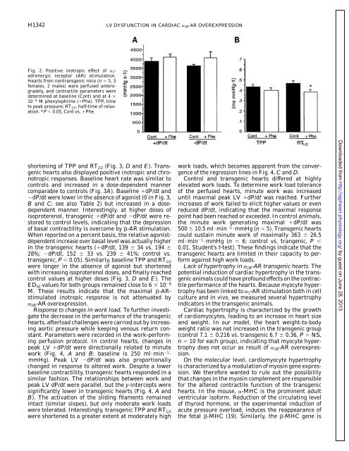
Fig. 2. Positive inotropic effect <strong>of</strong> 1<strong>adrenergic</strong><br />
<strong>receptor</strong> (AR) stimulation.<br />
Hearts from nontransgenic mice (n 5, 3<br />
females, 2 males) were perfused anterogradely,<br />
and contractile parameters were<br />
determined at baseline (Cont) and at 4 <br />
10 6 M phenylephrine (Phe). TPP, time<br />
to peak pressure; RT1/2, half-time <strong>of</strong> relaxation.<br />
*P 0.05, Cont vs. Phe.<br />
shortening <strong>of</strong> TPP and RT 1/2 (Fig. 3, D and E). Transgenic<br />
hearts also displayed positive inotropic and chronotropic<br />
responses. Baseline heart rate was similar to<br />
controls and increased in a dose-dependent manner<br />
comparable to controls (Fig. 3A). Baseline dP/dt and<br />
dP/dt were lower in the absence <strong>of</strong> agonist (0 in Fig. 3,<br />
B and C; see also Table 2) but increased in a dosedependent<br />
manner. Interestingly, at higher doses <strong>of</strong><br />
isoproterenol, transgenic dP/dt and dP/dt were restored<br />
to control levels, indicating that the depression<br />
<strong>of</strong> basal contractility is overcome by -AR stimulation.<br />
When reported on a percent basis, the relative agonistdependent<br />
increase over basal level was actually higher<br />
in the transgenic hearts (dP/dt, 139 34 vs. 194 <br />
28%; dP/dt, 152 33 vs. 239 41%; control vs.<br />
transgenic, P 0.05). Similarly, baseline TPP and RT 1/2<br />
were longer in the absence <strong>of</strong> agonist but shortened<br />
with increasing isoproterenol doses, and finally reached<br />
control values at higher doses (Fig. 3, D and E). The<br />
ED 50 values for both groups remained close to 6 10 9<br />
M. These results indicate that the maximal -ARstimulated<br />
inotropic response is not attenuated by<br />
<strong>1B</strong>-AR overexpression.<br />
Response to changes in work load. To further investigate<br />
the decrease in the performance <strong>of</strong> the transgenic<br />
hearts, afterload challenges were carried out by increasing<br />
aortic pressure while keeping venous return constant.<br />
Parameters were recorded in the work-performing<br />
perfusion protocol. In control hearts, changes in<br />
peak LV dP/dt were directionally related to minute<br />
work (Fig. 4, A and B; baseline is 250 ml·min 1 ·<br />
mmHg). Peak LV dP/dt was also proportionally<br />
changed in response to altered work. Despite a lower<br />
baseline contractility, transgenic hearts responded in a<br />
similar fashion. The relationships between work and<br />
peak LV dP/dt were parallel, but the y-intercepts were<br />
significantly lower in transgenic hearts (Fig. 4, A and<br />
B). The activation <strong>of</strong> the sliding filaments remained<br />
intact (similar slopes), but only moderate work loads<br />
were tolerated. Interestingly, transgenic TPP and RT 1/2<br />
were shortened to a greater extent at moderately high<br />
work loads, which becomes apparent from the convergence<br />
<strong>of</strong> the regression lines in Fig. 4, C and D.<br />
Control and transgenic hearts differed at highly<br />
elevated work loads. To determine work load tolerance<br />
<strong>of</strong> the perfused hearts, minute work was increased<br />
until maximal peak LV dP/dt was reached. Further<br />
increases <strong>of</strong> work failed to elicit higher values or even<br />
reduced dP/dt, indicating that the maximal response<br />
point had been reached or exceeded. In control animals,<br />
the minute work generating maximal dP/dt was<br />
500 10.5 ml·min 1 ·mmHg (n 5). Transgenic hearts<br />
could sustain minute work <strong>of</strong> maximally 363 26.5<br />
ml·min 1 ·mmHg (n 6; control vs. trangenic, P <br />
0.01, Student’s t-test). These findings indicate that the<br />
transgenic hearts are limited in their capacity to perform<br />
against high work loads.<br />
Lack <strong>of</strong> hypertrophy in <strong>1B</strong>-AR transgenic hearts. The<br />
potential induction <strong>of</strong> cardiac hypertrophy in the transgenic<br />
animals could have pr<strong>of</strong>ound effects on the contractile<br />
performance <strong>of</strong> the hearts. Because myocyte hypertrophy<br />
has been linked to 1-AR stimulation both in cell<br />
culture and in vivo, we measured several hypertrophy<br />
indicators in the transgenic animals.<br />
Cardiac hypertrophy is characterized by the growth<br />
<strong>of</strong> cardiomyocytes, leading to an increase in heart size<br />
and weight. In our model, the heart weight-to-body<br />
weight ratio was not increased in the transgenic group<br />
(control 7.1 0.216 vs. transgenic 6.7 0.36, P NS,<br />
n 10 for each group), indicating that myocyte hypertrophy<br />
does not occur as result <strong>of</strong> <strong>1B</strong>-AR overexpression.<br />
On the molecular level, cardiomyocyte hypertrophy<br />
is characterized by a modulation <strong>of</strong> myosin gene expression.<br />
We therefore wanted to rule out the possibility<br />
that changes in the myosin complement are responsible<br />
for the altered contractile function <strong>of</strong> the transgenic<br />
hearts. In the mouse, -MHC is the prominent adult<br />
<strong>ventricular</strong> is<strong>of</strong>orm. Reduction <strong>of</strong> the circulating level<br />
<strong>of</strong> thyroid hormone, or the experimental induction <strong>of</strong><br />
acute pressure overload, <strong>induces</strong> the reappearance <strong>of</strong><br />
the fetal -MHC (19). Similarly, the -MHC gene is<br />
Downloaded from<br />
http://ajpheart.physiology.org/<br />
by guest on June 28, 2013