Introduction: Heather Moss, MD, PhD (Attending) - University of ...
Introduction: Heather Moss, MD, PhD (Attending) - University of ...
Introduction: Heather Moss, MD, PhD (Attending) - University of ...

You also want an ePaper? Increase the reach of your titles
YUMPU automatically turns print PDFs into web optimized ePapers that Google loves.
Compressive Optic Neuropathy: (Continued)<br />
DISCUSSION Thyroid eye disease (TED) is an autoimmune disease which is usually self-limited but can be progressive. It is most commonly<br />
associated with Graves’ disease and hyperthyroidism, but can occur in patients who are euthyroid or hypothyroid. TED incidence is<br />
four times higher in women than men. Diagnosis is usually made clinically but imaging with CT or MRI can be done. Classically this shows<br />
enlargement <strong>of</strong> the EOMs with sparing <strong>of</strong> the tendons. Common clinical characteristics consist <strong>of</strong> inflammation and congestion <strong>of</strong> EOMs,<br />
proptosis, eyelid retraction, lid lag, diplopia, lid edema, chemosis, exposure keratopathy, and possible optic neuropathy. Treatment starts<br />
with establishing a euthyroid state. The literature has shown that corticosteroids are the most effective initial treatment. There is no additional<br />
benefit from early surgical decompression. Intraorbital steroid injections are another possible form <strong>of</strong> treatment which has shown to be effective.<br />
Radiation therapy is another effective treatment option, especially in patients who have failed treatment with steroids.<br />
REFERENCES:<br />
Phillips ME, Marzban MM, Kathuria SS. Treatment <strong>of</strong> thyroid eye disease. Current Treatment Options in Neurology. 2010 Jan;12(1):64-9.<br />
Modjtahedi SP, Modjtahedi BS, Mansury AM, Selva D, Douglas RS, Goldberg RA, Leibovitch I. Pharmacological treatments for thyroid eye<br />
disease. Drugs. 2006;66(13):1685-700.<br />
Alkawas AA, Hussein AM, Shahien EA. Orbital steroid injection versus oral steroid therapy in management <strong>of</strong> thyroid-related ophthalmopathy.<br />
Clinical and Experimental Ophthalmology. 2010 Oct;38(7):692-7.<br />
Abboud M, Arabi A, Salti I, Geara F. Outcome <strong>of</strong> thyroid associated ophthalmopathy treated by radiation therapy. Radiation Oncology. 2011<br />
May;6:46.<br />
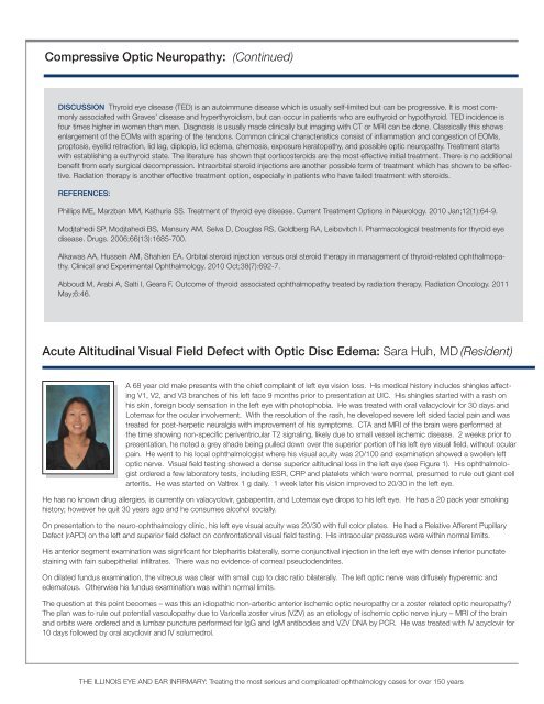
Acute Altitudinal Visual Field Defect with Optic Disc Edema: Sara Huh, <strong>MD</strong> (Resident)<br />
A 68 year old male presents with the chief complaint <strong>of</strong> left eye vision loss. His medical history includes shingles affecting<br />
V1, V2, and V3 branches <strong>of</strong> his left face 9 months prior to presentation at UIC. His shingles started with a rash on<br />
his skin, foreign body sensation in the left eye with photophobia. He was treated with oral valacyclovir for 30 days and<br />
Lotemax for the ocular involvement. With the resolution <strong>of</strong> the rash, he developed severe left sided facial pain and was<br />
treated for post-herpetic neuralgia with improvement <strong>of</strong> his symptoms. CTA and MRI <strong>of</strong> the brain were performed at<br />
the time showing non-specific periventricular T2 signaling, likely due to small vessel ischemic disease. 2 weeks prior to<br />
presentation, he noted a grey shade being pulled down over the superior portion <strong>of</strong> his left eye visual field, without ocular<br />
pain. He went to his local ophthalmologist where his visual acuity was 20/100 and examination showed a swollen left<br />
optic nerve. Visual field testing showed a dense superior altitudinal loss in the left eye (see Figure 1). His ophthalmologist<br />
ordered a few laboratory tests, including ESR, CRP and platelets which were normal, presumed to rule out giant cell<br />
arteritis. He was started on Valtrex 1 g daily. 1 week later his vision improved to 20/30 in the left eye.<br />
He has no known drug allergies, is currently on valacyclovir, gabapentin, and Lotemax eye drops to his left eye. He has a 20 pack year smoking<br />
history; however he quit 30 years ago and he consumes alcohol socially.<br />
On presentation to the neuro-ophthalmology clinic, his left eye visual acuity was 20/30 with full color plates. He had a Relative Afferent Pupillary<br />
Defect (rAPD) on the left and superior field defect on confrontational visual field testing. His intraocular pressures were within normal limits.<br />
His anterior segment examination was significant for blepharitis bilaterally, some conjunctival injection in the left eye with dense inferior punctate<br />
staining with fain subepithelial infiltrates. There was no evidence <strong>of</strong> corneal pseudodendrites.<br />
On dilated fundus examination, the vitreous was clear with small cup to disc ratio bilaterally. The left optic nerve was diffusely hyperemic and<br />
edematous. Otherwise his fundus examination was within normal limits.<br />
The question at this point becomes – was this an idiopathic non-arteritic anterior ischemic optic neuropathy or a zoster related optic neuropathy?<br />
The plan was to rule out potential vasculopathy due to Varicella zoster virus (VZV) as an etiology <strong>of</strong> ischemic optic nerve injury – MRI <strong>of</strong> the brain<br />
and orbits were ordered and a lumbar puncture performed for IgG and IgM antibodies and VZV DNA by PCR. He was treated with IV acyclovir for<br />
10 days followed by oral acyclovir and IV solumedrol.<br />
THE ILLINOIS EYE AND EAR INFIRMARY: Treating the most serious and complicated ophthalmology cases for over 150 years


![CV Joan [W51] - University of Illinois College of Medicine at Chicago ...](https://img.yumpu.com/17336863/1/190x245/cv-joan-w51-university-of-illinois-college-of-medicine-at-chicago-.jpg?quality=85)