INTERNATIONAL JOURNAL OF LEPROSY - Index of
INTERNATIONAL JOURNAL OF LEPROSY - Index of
INTERNATIONAL JOURNAL OF LEPROSY - Index of

You also want an ePaper? Increase the reach of your titles
YUMPU automatically turns print PDFs into web optimized ePapers that Google loves.
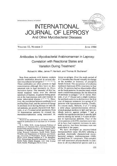
<strong>INTERNATIONAL</strong> <strong>JOURNAL</strong> <strong>OF</strong> <strong>LEPROSY</strong><br />
<strong>INTERNATIONAL</strong><br />
<strong>JOURNAL</strong> <strong>OF</strong> <strong>LEPROSY</strong><br />
And Other Mycobacterial Diseases<br />
VOLUME 52, NUMBER 2 JUNE 1984<br />
Antibodies to Mycobacterial Arabinomannan in Leprosy:<br />
Correlation with Reactional States and<br />
Variation During Treatment'<br />
Richard A. Miller, James P. Harnisch, and Thomas M. Buchanan 2<br />
Sera from patients with leprosy contain<br />
specific antibodies directed at several dis-<br />
9, 15, 17).<br />
tinct mycobacterial antigens ( 1 • 2 . 7 - 8'<br />
These antibodies are <strong>of</strong>ten present in high<br />
concentration although they have no documented<br />
role in host immunity to Mycobacterium<br />
leprae. The intensity <strong>of</strong> this humoral<br />
response varies over the clinical<br />
spectrum <strong>of</strong> leprosy, in general being greatest<br />
at the lepromatous pole and weakest in<br />
polar tuberculoid disease (2 5 . 1 5 ' 21 ). However,<br />
the correlation between antibody level<br />
and bacillary load, and the pattern <strong>of</strong> change<br />
in antibody level during therapy in individual<br />
patients, are poorly characterized. Bjorvatn,<br />
et al. ( 5) tested serial specimens from<br />
16 lepromatous patients by crossed immunoclectrophoresis<br />
using sonicated Al.<br />
' Received for publication on 10 March 1983; accepted<br />
for publication in revised form on 18 October<br />
1983.<br />
2 R. A. Miller, M.D., Acting Assistant Pr<strong>of</strong>essor <strong>of</strong><br />
Medicine; J. P. Harnisch, Chief <strong>of</strong> Dermatology,<br />
Seattle Public Health Hospital; T. M. Buchanan, M.D.,<br />
Pr<strong>of</strong>essor <strong>of</strong> Medicine and Pathology; Chief, Immunology<br />
Research Laboratory, Seattle Public Health<br />
Hospital, and Departments <strong>of</strong> Medicine and Pathology,<br />
University <strong>of</strong> Washington, Seattle, Washington<br />
98195, U.S.A. Reprint requests to: T. M. Buchanan,<br />
M.D., Immunology Research Laboratory, I I th Floor,<br />
Seattle Public Health Hospital, 1131 14th Avenue<br />
South, Seattle, Washington 98114, U.S.A.<br />
Volume 52i NulTther i2 .<br />
Printed<br />
( Ni'10 It LAO I tj 1 Cd<br />
leprae as antigen. Over the study period <strong>of</strong><br />
4-12 months they found virtually no change<br />
in the number or intensity <strong>of</strong> precipitin<br />
bands formed. The occurrence <strong>of</strong> crythema<br />
nodosum leprosum (ENL) reactions in four<br />
<strong>of</strong> the 16 patients had no discernable effect<br />
on the band patterns. In another study which<br />
used a radioimmunoassay for the detection<br />
<strong>of</strong> antibody to antigen 7 <strong>of</strong> M. leprae, Melsom,<br />
et al. ( 12) found only a slight tendency<br />
for antibody activity to decline over the first<br />
year <strong>of</strong> dapsone treatment in a group <strong>of</strong> 15<br />
patients with lepromatous leprosy. Finally,<br />
in a recent study, Mclsom, et al. (") using<br />
a solid phase radioimmunoassay demonstrated<br />
gradual declines in IgG and IgA antibody<br />
activity against antigen 7 and a<br />
smaller but significant decline in specific IgM<br />
activity during the initial 2-4 years <strong>of</strong> therapy<br />
in lepromatous patients. Four <strong>of</strong> the<br />
seven patients with ENL reactions were noted<br />
to have had transient increases in antibody<br />
activity, but these increases were not<br />
quantified in the paper.<br />
We have developed an enzyme-linked<br />
immunosorbent assay (ELISA) utilizing a<br />
purified, chemically characterized mycobacterial<br />
antigen, arabinomannan (AM),<br />
purified from Al. sineginatis. This complex<br />
cell wall associated polysaccharide is common<br />
to all mycobacterial species and is a<br />
133<br />
ID() 0` 5.‘
134^ International Journal <strong>of</strong> Leprosy^ 1984<br />
strong immunogen ' 4). The antibody levels<br />
to this characterized antigen were quantitated<br />
in pretreatment serum specimens obtained<br />
from nine patients with leprosy and<br />
compared with an estimate <strong>of</strong> bacillary load<br />
based on the results <strong>of</strong> six-site scrapings.<br />
Subsequent changes in antibody level over<br />
the initial 12-31 months <strong>of</strong> therapy were<br />
then measured in serial serum specimens<br />
collected from these patients.<br />
MATERIALS AND METIIODS<br />
Enzyme-linked immunosorbent assay.<br />
Arabinomannan (AM) was extracted from<br />
Al. smegmatis, purified, and characterized<br />
biochemically and immunochemically as<br />
previously described ( 13). An ELISA was<br />
performed using a modification <strong>of</strong> the technique<br />
reported by Buchanan ( 6). The AM<br />
was suspended in 0.067 M phosphate buffer,<br />
pH 5.0, at a concentration <strong>of</strong> 20 pg <strong>of</strong><br />
carbohydrate per ml. One hundred pl <strong>of</strong> this<br />
solution was placed in each well <strong>of</strong> a 96well,<br />
flat-bottom polystyrene microtiter<br />
plate (Dynatech, Alexandria, Virginia,<br />
U.S.A.) and incubated at 37°C for 3 hr, then<br />
stored at 4°C. On the day <strong>of</strong> the test the AM<br />
was removed by aspiration, 100 pl <strong>of</strong> phosphate<br />
buffered saline with 5% bovine serum<br />
albumin (BSA) was added to each well, and<br />
the plate was incubated at 37°C for 90 min.<br />
Following a wash with phosphate buffered<br />
saline with 1% BSA (PBS-BSA), the study<br />
sera were added to the wells. Each serum<br />
specimen was diluted 1:100 in PBS-BSA and<br />
assayed in triplicate, except for Patients 8<br />
and 9 whose serum was diluted 1:200. Each<br />
plate also had internal controls consisting<br />
<strong>of</strong>: a) serial dilutions <strong>of</strong> pooled human leprosy<br />
sera, b) standard dilutions <strong>of</strong> pooled<br />
normal human sera, and c) PBS-BSA alone.<br />
The diluted sera were incubated on the plates<br />
for 60 min at room temperature. Following<br />
further washing, peroxidase-conjugated IgG<br />
fraction goat anti-human IgG (Fe fragment,<br />
gamma chain specific) (Cappel Labs, Cochranville,<br />
Pennsylvania, U.S.A.) was added<br />
at a dilution <strong>of</strong> 1:500 in PBS-BSA. Plates<br />
were incubated for 60 min at room temperature.<br />
After washing, H 20,/o-phenylenediamine<br />
was added as a substrate/chromogen,<br />
and the plates were incubated in the<br />
dark at 37°C for approximately 30 min. The<br />
reaction was stopped with 8 N H,S0, and<br />
the 013,,,, was read using a Titertek Multiskan<br />
(Flow Labs, Helsinki, Finland). The<br />
arithmetic mean <strong>of</strong> the three values for each<br />
serum was calculated and used in all subsequent<br />
analyses. Through the linear range<br />
<strong>of</strong> optical densities measured by the ELISA<br />
(approximately 0.4-1.7), a decline <strong>of</strong> 25%<br />
<strong>of</strong> the 01) 4 .), roughly corresponded to a tw<strong>of</strong>old<br />
dilution <strong>of</strong> the scrum.<br />
Patients. All <strong>of</strong> the leprosy patients followed<br />
at the Seattle Public Health Hospital<br />
who met the following criteria were included<br />
in this study: a) banked sera were available<br />
from the date <strong>of</strong> initiation <strong>of</strong> therapy<br />
(except in Patient 9 whose first scrum was<br />
drawn two weeks after the start <strong>of</strong> therapy);<br />
b) stored sera were available over at least<br />
the first full year <strong>of</strong> therapy (range 55-136<br />
weeks, mean 78 weeks); and c) there were<br />
at least four sera available for testing. All<br />
patients were followed by one <strong>of</strong> the investigators<br />
(JH) over the entire period <strong>of</strong><br />
the study. There were seven males and two<br />
females in the study population; age range<br />
was 20-52 years. All clinical diagnoses were<br />
confirmed with skin biopsies from affected<br />
areas for histologic classification. Scrapings<br />
from six separate skin sites (earlobes and<br />
the extensor surfaces <strong>of</strong> the knees and elbows)<br />
were performed at the time <strong>of</strong> diagnosis<br />
using the Carville technique as reported<br />
by Kirchheimer ('"). Based on the<br />
standard Ridley-Jopling criteria ('s), there<br />
were 3 borderline lepromatous (BL), 3 borderline<br />
tuberculoid (BT), and 3 borderline<br />
(BB) cases among the 9 patients. All reactional<br />
episodes (except Patient 8) were confirmed<br />
by biopsy—Patients 4,5, and 6 had<br />
reversal reactions and Patients 8 and 9 had<br />
erythema nodosum leprosum (ENL). The<br />
frequency <strong>of</strong> clinic visits and sera collections<br />
was based on each patient's clinical<br />
course. No patient had a history <strong>of</strong> active<br />
or inactive tuberculosis or atypical mycobacterial<br />
illness.<br />
Serum specimens. Serum was obtained at<br />
most clinic visits. Samples were preserved<br />
with the addition <strong>of</strong> 0.1% sodium azide or<br />
sterilized by passage through a 200 nm filter<br />
(Millipore, Bedford, Massachusetts, U.S.A.),<br />
then aliquoted and stored at —70°C. All sera<br />
were tested on the same day.<br />
Therapeutic regimens. Dapsone and rifampin<br />
therapy was initiated in all patients
Miller, et al.: Anti-arabinomannan Antibodies^135<br />
at the time <strong>of</strong> diagnosis (except Patient 9,<br />
who received dapsone alone for three<br />
months prior to the addition <strong>of</strong> rifampin to<br />
the regimen). In most instances, the dosage<br />
was 100 mg per day <strong>of</strong> dapsone and 600 nig<br />
per day <strong>of</strong> rifampin. One patient (number<br />
5) had rifampin discontinued after six<br />
months because <strong>of</strong> persistently elevated liver<br />
function tests. All reactional episodes were<br />
treated with corticosteroids, usually prednisone<br />
40 mg per day tapered as rapidly as<br />
the clinical course allowed. Patient 9 eventually<br />
required thalidomide for control <strong>of</strong><br />
ENL; this was begun during week 99 <strong>of</strong> therapy.<br />
RESULTS<br />
Correlation <strong>of</strong> ELISA results and six-site<br />
scrapings. Data from the six-site scrapings<br />
and simultaneous antibody levels to AM are<br />
presented in The Table. There is a trend<br />
towards higher levels <strong>of</strong> antibody in BL cases<br />
than in BT, but as noted by other investigators<br />
( 5 - 2 ') there arc both low and high<br />
responders within each clinical and histologic<br />
class. The technique <strong>of</strong> examining<br />
scrapings from six clinically uninvolved skin<br />
sites for quantitation <strong>of</strong> bacillary load may<br />
provide a more accurate assessment <strong>of</strong> antigenic<br />
load than simple classification based<br />
on clinical criteria and the histopathology<br />
<strong>of</strong> leprous lesions ( 10). Correlation <strong>of</strong> antibody<br />
levels to AM measured by ELISA with<br />
the results <strong>of</strong> the six-site scrapings (either<br />
the bacterial index <strong>of</strong> involved sites or the<br />
bacterial index totaled for all six sites) is<br />
better than the comparable correlation with<br />
clinical class <strong>of</strong>disease. The correlation coefficient<br />
<strong>of</strong> the absolute OD 4 .), to the totaled<br />
bacterial index is 0.75 (p = 0.03) by the<br />
Spearman test.<br />
Variation in antibody levels with time in<br />
patients with uncomplicated leprosy. Four<br />
<strong>of</strong> the patients studied (1, 2, 3, and 7) were<br />
free <strong>of</strong> reversal reactions or ENL during the<br />
initial 13-28 months <strong>of</strong> therapy. Their antibody<br />
levels over time are shown in Figure<br />
I. Patient 3 discontinued his dapsone and<br />
rifampin therapy between weeks 18 and 26<br />
with resultant clinical relapse after an initially<br />
excellent clinical response. This may<br />
account for his atypical rise in antibody level<br />
in the week 26 and subsequent sera. The<br />
remaining three patients all had declining<br />
or stable amounts <strong>of</strong> specific antibody after<br />
the initial two months <strong>of</strong> therapy.<br />
Influence <strong>of</strong> ENI, on antibody levels. Two<br />
<strong>of</strong> the patients (8 and 9) experienced ENL<br />
reactions during their initial 1-2 years <strong>of</strong><br />
therapy. Both <strong>of</strong> these patients had extremely<br />
high levels <strong>of</strong> antibody to AM<br />
throughout the study period, requiring additional<br />
dilution <strong>of</strong> their sera prior to assay.<br />
Serial measurement <strong>of</strong> antibody levels<br />
showed gradual declines with no consistent<br />
effect <strong>of</strong> the ENL reactions on the titers (Fig.<br />
1). Patient 9 did have a small increase in<br />
antibody level at the onset <strong>of</strong> ENL but, relative<br />
to high baseline levels, it amounted to<br />
only an 8% increase and is <strong>of</strong> no signifi-<br />
THE TABLE. Pretreatment arabinomannan ELISA and six-site skin smear results train<br />
patients with leprosy.<br />
Patient no.<br />
(age/sex)<br />
Type <strong>of</strong> leprosy<br />
Reactional<br />
state<br />
Pretreatment<br />
OD<br />
Six-site smears<br />
No. sues<br />
positive<br />
Total BIa<br />
1 (41/M) Borderline tuberculoid None 0.116 0 0<br />
2 (45/M) Borderline tuberculoid None 0.182 1<br />
3 (36/M) Borderline None 0.304 0 0<br />
4 (43/M) Borderline lepromatous RR" 0.718 1 1<br />
5 (20/F) Borderline tuberculoid RR 0.857 1 5<br />
6 (26/M) Borderline RR 1.066 0 0<br />
7 (52/M) Borderline None 1.549 3 8<br />
8 (42/F) Borderline lepromatous ENL' 1.704' 5 22<br />
9 (33/M) Borderline lepromatous ENL 1.807' 6 20<br />
Total bacterial index (BI) <strong>of</strong> six sites; each scored 0-6+. Maximum total = 36.<br />
b RR = reversal reaction.<br />
ENL = erythema nodosum leprosum.<br />
d Serum diluted 1:200 because <strong>of</strong> high titer.
1 4<br />
Active<br />
ENL<br />
0^20^40^60^80^100 120 140<br />
Weeks from Initiation <strong>of</strong> Therapy<br />
FIG. 1. Serial arabinomannan antibody levels in<br />
patients with uncomplicated treatment courses (Pa-<br />
tients I, 2, 3 and 7) or with EN I. reactions (Patients 8<br />
and 9). Periods <strong>of</strong> active ENL are delineated by brack-<br />
ets.<br />
cance. There was no evidence to suggest that<br />
antibody variation in patients undergoing<br />
ENL reactions difkred significantly from<br />
that <strong>of</strong> patients with uncomplicated clinical<br />
courses.<br />
Variations in antibody levels during reversal<br />
reactions. Figure 2 presents the pattern<br />
<strong>of</strong> variation in antibody levels during<br />
reversal reactions as measured in the AM<br />
ELISA. The graphs are standardized for the<br />
three patients (4, 5 and 6) by defining the<br />
date <strong>of</strong> clinical onset <strong>of</strong> the reversal reaction<br />
as "day 0". Corticosteroids were begun on<br />
all patients within one week <strong>of</strong> this date and,<br />
in most cases, there was rapid and steady<br />
improvement in symptoms. During the 4-6<br />
weeks preceding the onset <strong>of</strong> the reversal<br />
reactions, the antibody levels in all had declined<br />
to a value below the pretreatment<br />
level. Coincident with or shortly after the<br />
development <strong>of</strong> the clinical symptoms <strong>of</strong><br />
reversal reaction, all patients had abrupt increases<br />
in the quantity <strong>of</strong> specific antibody<br />
to AM which persisted for several weeks,<br />
eventually peaking between six and 23 weeks<br />
International Journal <strong>of</strong> Leprosy<br />
1.4 -<br />
0.8 -<br />
1.4 -<br />
1.4 -<br />
0.8 -<br />
0 5 -<br />
- 20 0^20^40^60<br />
Weeks prior or subsequent<br />
to onset <strong>of</strong> reversal reaction.<br />
1984<br />
Fie. 2. Serial arabinomannan antibody levels in<br />
the three patients with reversal reactions. Arrows mark<br />
the onset <strong>of</strong> the reactional state. Note: The OD scale<br />
(ordinate axis) is discontinuous, but covers the same<br />
range <strong>of</strong> values for each patient.<br />
after the reactional state began. In all cases<br />
the reversal reaction was clinically resolving<br />
at the time <strong>of</strong> peak antibody levels. Thereafter,<br />
the antibody levels fell steadily in all<br />
three patients, and reached levels below the<br />
pretreatment value in two <strong>of</strong> them.<br />
DISCUSSION<br />
The correlation <strong>of</strong> the antibody level with<br />
the bacterial index <strong>of</strong> the six-site smears<br />
implies that the level <strong>of</strong> antibody may serve<br />
as a rough measure <strong>of</strong> the amount <strong>of</strong> AM<br />
accessible to the host immune system. This<br />
relationship appears to hold true across the<br />
clinical spectrum <strong>of</strong> leprosy in the population<br />
studied. The documented increase in<br />
seropositivity as one moves along the spectrum<br />
towards polar lepromatous disease has<br />
led to speculation that antibody levels reflect<br />
antigen load, but none <strong>of</strong> the previous<br />
studies attempted to relate antibody titer to<br />
an objective measure <strong>of</strong> bacillary load.
52, 2^Miller, et al.: Anti-arabinoinannan Antibodies^137<br />
The finding that antibody titers to AM<br />
from Al. smegmatis remain constant or decline<br />
at a slow rate is in agreement with the<br />
two studies in the literature that tested serial<br />
sera on the same patients (5. 12 ) and the<br />
three studies that reported only mean antibody<br />
levels in patient groups during treatment<br />
( 3' 16 ' 21 ). This persistence <strong>of</strong> antibody<br />
despite adequate therapy and excellent clinical<br />
response is likely due to the continued<br />
presence in the host <strong>of</strong> mycobacterial antigens.<br />
Mouse foot pad inoculation studies<br />
( 1 ") have confirmed the validity <strong>of</strong> the bacterial<br />
index (BI) and morphological index<br />
(MI), and it is now firmly established that<br />
nonviable mycobacterial "skeletons" can<br />
persist well into therapy and present a<br />
chronic antigenic stimulus to the host.<br />
Using the AM assay, the pattern <strong>of</strong> change<br />
in antibody titer during ENL reactions was<br />
not distinguishable from that seen in uncomplicated<br />
cases. The other studies which<br />
examined serum specimens during ENL reactions<br />
also found no consistent variation<br />
in antibody levels when compared with those<br />
<strong>of</strong> uncomplicated cases ( 3 ' 5 ' ' 2). Circulating<br />
and fixed immune complexes are involved<br />
in the pathogenesis <strong>of</strong> ENL ( 2()) and it is<br />
noteworthy that only those patients with the<br />
highest antibody levels, and presumably<br />
greater bacterial loads, experienced this<br />
complication.<br />
The pattern observed in the three patients<br />
with reversal reaction was quite distinct from<br />
that seen in the other six patients. Each<br />
manifested a triphasic pattern with an initial<br />
fall in antibody preceding the clinical<br />
development <strong>of</strong> the reversal reaction, followed<br />
by a sharp rise coincident with the<br />
appearance <strong>of</strong> symptoms, and finally a second<br />
decline, occurring several weeks to<br />
months after the onset <strong>of</strong> the reaction. These<br />
variations in antibody level did not correlate<br />
with changes in steroid dosage, and the<br />
peak levels occurred at a time when the clinical<br />
symptoms <strong>of</strong> the reactional state were<br />
well controlled. This triphasic pattern may<br />
be correlated with the rapid improvement<br />
in effective cell-mediated immunity against<br />
M. leprae ("), with resultant release <strong>of</strong> antigens,<br />
which is the hallmark <strong>of</strong> the reversal<br />
reaction.<br />
Little data arc available from prior studies<br />
on the variation <strong>of</strong> humoral response to<br />
other antigens during reversal reactions.<br />
Melsom, et al. (". 12) reported that one <strong>of</strong><br />
the patients involved in their study developed<br />
increasing antibody titers to Al. leprae<br />
antigen 7 during a reversal reaction, but the<br />
patient was subsequently shown to have active<br />
tuberculosis. Abe, et al., using a fluorescent<br />
leprosy antibody absorption test,<br />
studied two patients with reversal reactions<br />
( 3), one <strong>of</strong> whom manifested a sharp increase<br />
in IgG antibody titer at the onset <strong>of</strong><br />
the reversal reaction, followed by a decline<br />
in titer three and six months later. This was<br />
distinct from the pattern they observed in<br />
ENL or in uncomplicated cases. Further<br />
cases will have to be studied to confirm these<br />
preliminary findings, but the marked contrast<br />
between the behavior <strong>of</strong> the antibody<br />
response to AM in our three patients with<br />
reversal reactions when compared to that<br />
<strong>of</strong> the six patients with other clinical courses<br />
appears to be significant.<br />
Further studies with more patients will<br />
be <strong>of</strong> interest to determine whether the level<br />
<strong>of</strong> antibody to AM in a pretreatment serum<br />
allows prediction <strong>of</strong> the relative risk <strong>of</strong> developing<br />
a reactional state. In this study,<br />
two <strong>of</strong> three patients with pretreatment<br />
ELISA values greater than 1.5 developed<br />
ENL, and all three <strong>of</strong> the patients with values<br />
<strong>of</strong> 0.7-1.1 developed reversal reactions<br />
(The Table, Figs. 1 and 2). In contrast, the<br />
three patients with pretreatment serum<br />
ELISA values less than 0.35 experienced no<br />
reactional states during more than one year<br />
<strong>of</strong> follow up. Since the antibody level to AM<br />
correlates directly with the estimated bacillary<br />
load, this implies that patients with the<br />
fewest organisms are the least likely to develop<br />
reactional states. Patients with moderate<br />
numbers <strong>of</strong> organisms may be the most<br />
likely to develop reversal reactions, and<br />
those with the greatest numbers may be the<br />
most susceptible to developing ENL. If ongoing<br />
research confirms the utility <strong>of</strong> this<br />
assay in defining subgroups at high risk for<br />
the development <strong>of</strong> reactional states, it may<br />
facilitate the design <strong>of</strong> studies aimed at decreasing<br />
the incidence <strong>of</strong> these unpleasant<br />
and dangerous complications.<br />
SUMMARY<br />
An enzyme-linked immunosorbent assay<br />
was used to measure antibody to mycobac-
138^ international Journal <strong>of</strong> Leprosy^ 1984<br />
terial arabinomannan in serial scrum specimens<br />
obtained over the initial 12-31<br />
months <strong>of</strong> therapy from nine patients with<br />
leprosy. The antibody level in pretreatment<br />
sera was directly proportional to the quantity<br />
<strong>of</strong> Mycobacterium leprac present in each<br />
patient as assessed by six-site scrapings<br />
(r = 0.75). The three patients with the lowest<br />
antibody levels (OD 0.1-0.3) had uncomplicated<br />
courses and their levels declined<br />
slowly with treatment. Three patients<br />
with intermediate antibody levels (OD 0.7-<br />
1.1) each experienced a reversal reaction<br />
during therapy; serial antibody titers in all<br />
three followed a triphasic pattern over the<br />
course <strong>of</strong> the reaction. The two patients who<br />
developed erythema nodosum leprosum<br />
during therapy had extremely high levels <strong>of</strong><br />
antibody initially (OD > 1.5), which fell<br />
slowly with time and which were unaffected<br />
by the reactional state. The pretreatment<br />
antibody level to arabinomannan reflects the<br />
amount <strong>of</strong> Al. leprac present and may have<br />
predictive value for the development <strong>of</strong> reactional<br />
states.<br />
RESUMEN<br />
Se use un inmunoensayo enzimatico para mcdir la<br />
concentracion de anticuerpo contra una arabionoma-<br />
nana micobacteriana en especimenes seriados de suero<br />
obtenidos durante los 12 a 31 meses iniciales do terapia<br />
en 9 pacientes con lepra. El nivel de anticuerpo en los<br />
sueros de pretratamiento fue directamente proporcio-<br />
nal a Ia cantidad de Mycobacterium leprac presente en<br />
cada paciente segan se determin6 por el analisis bac-<br />
teriolOgico en 6 sitios de abrasiOn (r = 0.75). Los 3<br />
pacientes con los niveles mas bajos de anticuerpos (DO<br />
0.1-0.3) evolucionaron sin mostrar complicaciones y<br />
sus niveles decayeron lentamente con el tratamiento.<br />
Tres pacientes que tuvieron niveles intermedios de<br />
anticuerpos (DO 0.7-1.1) prescntaron algan tipo de<br />
reacciOn reversa durante Ia terapia; en todos los casos,<br />
los titulos seriados de anticuerpos siguieron un patron<br />
trifäsico durante el curso de Ia reaction. Los dos pa-<br />
cientes que dcsarrollaron critema nodoso leproso du-<br />
rante la terapia tuvieron niveles de anticuerpo extre-<br />
madamente elevados al inicio (DO > 1.5), mismos que<br />
decayeron lentarnente con el tiempo sin ser afectados<br />
por el estado reaccional. El nivel de anticuerpos contra<br />
arabinomanana antes de iniciarse el tratamiento relleja<br />
Ia cantidad de M. leprae presente y pucde toner un<br />
valor predictivo del desarrollo de estados reaccionales.<br />
RESUME<br />
Une evaluation par unc methode d'immunosorbant,<br />
associee a unc enzyme, a etc: utilisee pour mesurer un<br />
anticorps arabinomannan mycobacterien dans des se-<br />
ries d'echantillons de serum obtenus au cours de la<br />
periode initiate de la therapeutique chez ncuf malades<br />
atteints de l&pre, periode qui s'etendait sur 12 a 13<br />
mois. Le taus d'antilnotiques dans le serum preleve<br />
avant traitement etait directemcnt proportionnel 6 la<br />
quantite de A/Ivo/were/lam hp/we qui etait presente<br />
chez chaque malade, ainsi qu'on pouvait l'estimer par<br />
des prelevements cutanes effectues en six endroits (r<br />
0,75). Les trois malades presentant les taus d'anticorps<br />
les plus faibles (01) 0,1-0,3) presentaient one evolution<br />
non compliquee et cur taux montrait une diminution<br />
lento avec lc traitement. "Frois malades presentant des<br />
taus d'anticorps intermediaires (01) 0,7-1,1) avaicnt<br />
chacun soullert (rune "reaction reverse" au cours du<br />
traitement. Chez chacun de ces trois malades les titres<br />
d'anticorps en series avaient present& on pr<strong>of</strong>it en trois<br />
phases au cours de la reaction. Les deux malades qui<br />
ont developpe un erytheme noueux lepreux au cours<br />
de la therapcutique prescntaient des taus extremement<br />
clews d'anticorps au debut (OD < 1,5), taus qui soot<br />
retombes lentement avec le temps, et qui n'ont pas et&<br />
affectes par Film reactionnel. Lc taux d'anticorps ara-<br />
binomannan avant traitement relletait Ia quantite de<br />
11. leprae qui emit presente, et pourrait avoir une va-<br />
lour de prediction quint au developpement d'etats<br />
reactionnels.<br />
Acknowledgments. The authors thank Charlotte<br />
Leitch and Susan Dinning for valuable technical as-<br />
sistance.<br />
This study was supported in part by Grant Al-16290<br />
and Contract Al-92624 from the National Institute <strong>of</strong><br />
Allergy and Infectious Diseases, National Institutes <strong>of</strong><br />
Health; by Federal Health Services Project SEA-78-<br />
17; by the Immunology <strong>of</strong> Leprosy component <strong>of</strong> the<br />
UNDP/World Bank/WHO Special Programme for Re-<br />
search in Tropical Diseases; and by the Rockefeller<br />
Foundation for Research on Great Neglected Diseases.<br />
REFERENCES<br />
1. ABE, M. Anti-.I/. /mat' antibodies in leprosy pa-<br />
tients as demonstrated by indirect immun<strong>of</strong>luo-<br />
rescence. Abstract in Int. J. Lepr. 41 (1973) 549.<br />
2. Aim, M., !ZUNI!, S., SMno. T. and MATHUR, S. K.<br />
Early serodiagnosis <strong>of</strong> leprosy by indirect immu-<br />
notluorescence. Lepr. India 48 (1976) 272-276.<br />
3. Am, M., MINAGAWA, F. and Yosnimi, Y. Ther-<br />
apeutic effect <strong>of</strong> rifampicin (REP) on leprosy. IV.<br />
Serological examination. Repura 43 (1974) 45-51.<br />
4. AZUN1A, I., KINIURA, H., NIINAKA, T., AOKI, T. and<br />
YANIAMURA, Y. Chemical and immunological<br />
studies on mycobactcrial polysaccharides. I. Pu-<br />
rification and properties <strong>of</strong> polysaccharides from<br />
human tubercle bacilli. J. Bacteriol. 95 (1968) 263-<br />
271.<br />
5. 13.1012VATN, B., NAM'S, B. and KRONVALL., G. Sta-<br />
bility <strong>of</strong> individual antimycobacterial precipita-<br />
tion patterns during treatment for lepromatous<br />
leprosy. Int. J. Lepr. 46 (1978) 144-148.<br />
6. BUCHANAN, T. M. Antigen-specific serotyping <strong>of</strong>
57 , 7 ^ Miller, et al.: Anti-arabinomannan Antibodies^139<br />
A'eisseria gonorrhocac. I. Use <strong>of</strong> an enzyme-linked<br />
immunosorbent assay to quantitate pilus antigens<br />
on gonococci. J. Infect. Dis. 138 (1978) 319-325.<br />
7. CALDwEl.i., H. D., KIRCIIIITIMER, W. F. and<br />
BUCHANAN, T. M. Identification <strong>of</strong> a Mycobacterium<br />
leprae specific protein antigen(s) and its<br />
possible application for the serodiagnosis <strong>of</strong> leprosy.<br />
Int. J. Lepr. 47 (1979) 477-483.<br />
8. HARBou, M., CLOSS, O., Bitim., G., KRONVALL, G.<br />
and Axii.sEN, N. H. Mycobacterium leprae specific<br />
antibodies detected by radioimmunoassay.<br />
Scand. J. Immunol. 7 (1978) 111-120.<br />
9. KAPLAN, M. a and CliAsE, M. W. Antibodies to<br />
mycobacteria in human tuberculosis. II. Response<br />
to nine defined mycobacterial antigens with evidence<br />
for an antibody common to tuberculosis and<br />
lepromatous leprosy. J. Infect. Dis. 142 (1980) 835-<br />
843.<br />
10. KIRCHHEIMER, W. F. Leprosy. Am. J. Med. Technol.<br />
40 (1974) 474-478.<br />
1 I. MELSOM, R., 'JARBOE, NI. and NA.AES, B. Class<br />
specific anti-Mycobacterium leprae antibody assay<br />
in lepromatous leprosy (I3L-LL) patients during<br />
the first two to four years <strong>of</strong> DDS treatment. Int.<br />
J. Lepr. 50 (1982) 271-281.<br />
12. MELSOM, R., NAM'S, B., HARBOE, M. and (loss,<br />
O. Antibody activity against Mycobacterium leprae<br />
antigen 7 during the lirst year <strong>of</strong> DDS treatment<br />
in lepromatous (BL-LL) leprosy. Lepr. Rev.<br />
49 (1978) 17-29.<br />
13. MILLER, R. A., DISSANAYAKE, S. and BUCHANAN,<br />
T. M. Development <strong>of</strong> an enzyme-linked immunosorbent<br />
assay using arabinomannan from<br />
Mycobacterium smet,onatis: A potentially useful<br />
screening test for the diagnosis <strong>of</strong> incubating leprosy.<br />
Am. J. Trop. Med. 32 (1983) 555-564.<br />
14. MisAki, A., AZUNIA, 1. and YAM:WI:RA, Y. Structural<br />
and immunochemical studies on D-arabino-<br />
D-mannans and D-mannans <strong>of</strong> Mycobacterium<br />
tuberculosis and other Mycobacterium species. J.<br />
I3iochem. (Tokyo) 82 (1977) 1759-1770.<br />
15. MYRVANG, B., FEEK, C. M. and GoDAL, T. Antimycobacterial<br />
antibodies in sera from patients<br />
throughout the clinico-pathologic disease spectrum<br />
<strong>of</strong> leprosy. Acta Pathol. Microbiol. Scand.<br />
[B] 82 (1974) 701-706.<br />
16. REES, R. J. W., CHATITREIT, K. R., PEPYS, J. and<br />
TEE, R. D. Sonic immunologic aspects <strong>of</strong> leprosy.<br />
Am. Rev. Respir. Dis. 92 Suppl (1965) 139-149.<br />
17. REIcii, C. V. Rapid effective measure <strong>of</strong> a humoral<br />
substance(s) reacting specifically with Mycobacterium<br />
leprae antigens. Infect. Immun. 10<br />
(1974) 963-965.<br />
18. RIDLEY, D. S. and JOPLIN:0, W. H. Classification<br />
<strong>of</strong> leprosy according to immunity. A five-group<br />
system. Int. J. Lepr. 34 (1966) 255-273.<br />
19. SANSONETTI, P. and LAGRANGE, P. H. The immunology<br />
<strong>of</strong> leprosy: Speculations on the leprosy<br />
spectrum. Rev. Infect. Dis. 3 (1981) 422-469.<br />
20. WATERS, M. F. R., TURK, J. L. and WEMANIBU,<br />
S. N. C. Mechanisms <strong>of</strong> reaction in leprosy. Int.<br />
J. Lepr. 39 (1971) 417-428.<br />
21. YODER, L., NAAFS, B., HARI30E, M. and BJUNE, G.<br />
Antibody activity against Mycobacterium leprae<br />
antigen 7 in leprosy: Studies on variation in antibody<br />
content throughout the spectrum and on<br />
the effect <strong>of</strong> DDS treatment <strong>of</strong> relapse in 13T leprosy.<br />
Lepr. Rev. 50 (1979) 113-121.