Publications_files/Roberts and Azizi 2011.pdf
Publications_files/Roberts and Azizi 2011.pdf
Publications_files/Roberts and Azizi 2011.pdf

Create successful ePaper yourself
Turn your PDF publications into a flip-book with our unique Google optimized e-Paper software.
The Journal of Experimental Biology 214, 353-361<br />
© 2011. Published by The Company of Biologists Ltd<br />
doi:10.1242/jeb.038588<br />
Introduction<br />
Most biologists are familiar with the idea that a kangaroo’s hop is<br />
powered by spring-like tendons that allow it to literally bounce<br />
along the ground (Alex<strong>and</strong>er <strong>and</strong> Vernon, 1975). This mechanism<br />
is obvious to any observer, so much so that the kangaroo’s hop has<br />
become the iconic image of elastic mechanisms in locomotion. Yet<br />
in many ways this image sells springs short. It is becoming<br />
increasingly clear that the role of elastic mechanisms in movement<br />
extends well beyond obviously springy gaits such as hopping,<br />
influencing the mechanics, energetics <strong>and</strong> control of a wide range<br />
of activities. The springs are diverse as well, including not just<br />
tendons but connective tissue elements that bind muscles together<br />
<strong>and</strong> molecular springs within the muscles themselves. Although<br />
they may not always be as obvious as a kangaroo’s bouncing gait,<br />
elastic mechanisms are likely to play a significant role in all<br />
vertebrate locomotor systems.<br />
A spring element, by definition, follows a very simple behavior.<br />
Springs deform when a force is applied <strong>and</strong> recoil to their resting<br />
shape when the force is released. Materials can act like springs<br />
when loaded in tension, like a rubber b<strong>and</strong>, or in compression, like<br />
a rubber ball. Both kinds of loading are important in nature. When<br />
springy materials deform, they store energy in the form of elastic<br />
strain energy, <strong>and</strong> when they recoil this energy is released. The<br />
amount of energy stored depends on the material stiffness <strong>and</strong> the<br />
deformation. Compared with many biological tissues, springs are<br />
‘dumb’; their behavior is determined by a single force-length<br />
relationship, <strong>and</strong> cannot be modified or controlled directly. Springs<br />
also cannot produce mechanical energy; they can only return<br />
energy loaded previously by an external source. By way of<br />
comparison, any given muscle can produce mechanical energy, <strong>and</strong><br />
COMMENTARY<br />
Flexible mechanisms: the diverse roles of biological springs in vertebrate movement<br />
Thomas J. <strong>Roberts</strong>1, * <strong>and</strong> Emanuel <strong>Azizi</strong>2 1 2 Department of Ecology <strong>and</strong> Evolutionary Biology, Brown University, Providence, RI 02912, USA <strong>and</strong> Department of Biology, The<br />
College of New Jersey, Ewing, NJ 08628, USA<br />
*Author for correspondence (roberts@brown.edu)<br />
Summary<br />
The muscles that power vertebrate locomotion are associated with springy tissues, both within muscle <strong>and</strong> in connective tissue<br />
elements such as tendons. These springs share in common the same simple action: they stretch <strong>and</strong> store elastic strain energy<br />
when force is applied to them <strong>and</strong> recoil to release energy when force decays. Although this elastic action is simple, it serves a<br />
diverse set of functions, including metabolic energy conservation, amplification of muscle power output, attenuation of muscle<br />
power input, <strong>and</strong> rapid mechanical feedback that may aid in stability. In recent years, our underst<strong>and</strong>ing of the mechanisms <strong>and</strong><br />
importance of biological springs in locomotion has advanced significantly, <strong>and</strong> it has been demonstrated that elastic mechanisms<br />
are essential for the effective function of the muscle motors that power movement. Here, we review some recent advances in our<br />
underst<strong>and</strong>ing of elastic mechanisms, with an emphasis on two proposed organizing principles. First, we review the evidence that<br />
the various functions of biological springs allow the locomotor system to operate beyond the bounds of intrinsic muscle<br />
properties, including metabolic <strong>and</strong> mechanical characteristics, as well as motor control processes. Second, we propose that an<br />
energy-based framework is useful for interpreting the diverse functions of series-elastic springs. In this framework, the direction<br />
<strong>and</strong> timing of the flow of energy between the body, the elastic element <strong>and</strong> the contracting muscle determine the function served<br />
by the elastic mechanism (e.g. energy conservation vs power amplification). We also review recent work demonstrating that<br />
structures such as tendons remodel more actively <strong>and</strong> behave more dynamically than previously assumed.<br />
Key words: elastic, locomotion, tendon.<br />
THE JOURNAL OF EXPERIMENTAL BIOLOGY<br />
353<br />
can operate over a wide range of combinations of forces <strong>and</strong><br />
lengths, all controlled <strong>and</strong> fine-tuned by intricate feed-forward <strong>and</strong><br />
feedback signals from the nervous system. What is so fascinating<br />
about elastic mechanisms is that, from such a simple <strong>and</strong> arguably<br />
limited mechanism, nature has produced a remarkable variety of<br />
applications.<br />
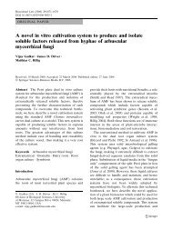
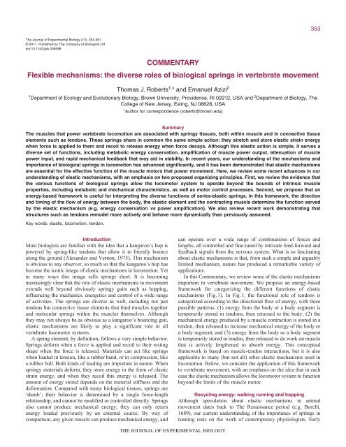
In this Commentary, we review some of the elastic mechanisms<br />
important in vertebrate movement. We propose an energy-based<br />
framework for categorizing the different functions of elastic<br />
mechanisms (Fig.1). In Fig.1, the functional role of tendons is<br />
categorized according to the directional flow of energy, with three<br />
possible patterns: (1) energy from the body or a body segment is<br />
temporarily stored in tendons, then returned to the body; (2) the<br />
mechanical energy produced by a muscle contraction is stored in a<br />
tendon, then released to increase mechanical energy of the body or<br />
a body segment; <strong>and</strong> (3) energy from the body or a body segment<br />
is temporarily stored in tendon, then released to do work on muscle<br />
that is actively lengthened to absorb energy. This conceptual<br />
framework is based on muscle-tendon interactions, but it is also<br />
applicable to many (but not all) other elastic mechanisms used in<br />
locomotion. Below, we consider the application of this framework<br />
to vertebrate movement, with an emphasis on the idea that in each<br />
case the elastic mechanism allows the locomotor system to function<br />
beyond the limits of the muscle motor.<br />
Recycling energy: walking running <strong>and</strong> hopping<br />
Although speculation about elastic mechanisms in animal<br />
movement dates back to The Renaissance period (e.g. Borelli,<br />
1689), our current underst<strong>and</strong>ing of the importance of springs in<br />
running rests on the work of contemporary physiologists. Early
354<br />
T. J. <strong>Roberts</strong> <strong>and</strong> E. <strong>Azizi</strong><br />
Task<br />
Energy flow<br />
Function<br />
Activities<br />
studies of spring mechanisms in terrestrial locomotion proceeded<br />
on two fronts. First, studies combining careful anatomical (e.g.<br />
tendon dimensions), mechanical (e.g. force plate recordings) <strong>and</strong><br />
mathematical approaches showed that a significant fraction of the<br />
work done in a step could be provided by the spring-like action of<br />
tendons, rather than by muscle work (Alex<strong>and</strong>er <strong>and</strong> Vernon,<br />
1975). In other studies, combined measurements of the mechanical<br />
work of running <strong>and</strong> the energy input necessary to sustain this<br />
work, as measured by oxygen consumption, resulted in very high<br />
efficiency values that could only be explained if elastic mechanisms<br />
contributed some of this work ‘for free’. These energetic studies<br />
began with human subjects (Cavagna et al., 1964) <strong>and</strong> later<br />
included broad <strong>and</strong> comprehensive comparative analyses (Cavagna<br />
et al., 1977; Heglund et al., 1982). The conclusion from both<br />
mechanical <strong>and</strong> energetic studies was that much of the cyclical<br />
work done during running could be attributed to the energy stored<br />
<strong>and</strong> released by elastic structures with each step (Fig.1A).<br />
Since then, the idea that running is a ‘bouncing gait’ powered<br />
by spring-like limbs has become a cornerstone for models of<br />
running mechanics <strong>and</strong> energetics (Dickinson et al., 2000). Key<br />
features of the dynamics of running, including ground reaction<br />
force patterns, the trajectory of the center of mass, <strong>and</strong> the change<br />
in foot contact time with speed, are consistent with a simple<br />
spring–mass model of running for humans (McMahon <strong>and</strong> Cheng,<br />
1990; Blickhan, 1989), <strong>and</strong> for other runners <strong>and</strong> hoppers (Farley<br />
et al., 1993). Direct measurements of muscle function have<br />
demonstrated the importance of the stretch <strong>and</strong> recoil of tendons on<br />
the length changes <strong>and</strong> power output of active muscles during<br />
running (<strong>Roberts</strong> et al., 1997) <strong>and</strong> hopping (Biewener et al., 1998).<br />
A<br />
Energy conservation<br />
Body Tendon Body<br />
Metabolic economy<br />
Running<br />
Hopping<br />
Walking<br />
B<br />
Power amplification<br />
Muscle Tendon Body<br />
Power production<br />
Jumping<br />
Acceleration<br />
Incline running<br />
Ballistic feeding<br />
Power attenuation<br />
Body Tendon Muscle<br />
Energy absorption<br />
Decline running<br />
Deceleration<br />
More recently, empirical <strong>and</strong> theoretical approaches have<br />
demonstrated that the use of elastic mechanisms is not limited to<br />
running but also occurs during walking. Historically, conceptual<br />
models of energy-saving mechanisms in locomotion have<br />
categorized walking as a ‘pendulum gait’, in which energy is<br />
conserved through the pendular exchange of kinetic <strong>and</strong> potential<br />
energy as the walker vaults over a strut-like leg with each step<br />
(Cavagna et al., 1977; Dickinson et al., 2000). The dichotomy<br />
between the spring mechanism of running <strong>and</strong> the pendular<br />
mechanism of walking was challenged by Lee <strong>and</strong> Farley (Lee <strong>and</strong><br />
Farley, 1998), who used kinematic data to show that the<br />
compression of the leg (as measured by the distance from the hip<br />
to the toe) during walking in humans was only 26% less than that<br />
during running. Ultrasound data has provided direct evidence of the<br />
importance of tendon action during walking; triceps surae muscles<br />
in humans are able to operate nearly isometrically (i.e. at constant<br />
length) during walking owing to the stretch <strong>and</strong> recoil of the<br />
Achilles tendon (Fukunaga et al., 2001; Lichtwark et al., 2007).<br />
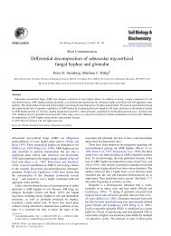
Recent models have also demonstrated that some key dynamic<br />
features of walking, such as the pattern of ground reaction force,<br />
are not consistent with a simple inverted pendulum model, but can<br />
be explained by a spring-loaded inverted pendulum (Geyer et al.,<br />
2006) (Fig.2). Together, these studies suggest that elastic<br />
mechanisms are an essential part of both walking <strong>and</strong> running gaits.<br />
Do all runners use an elastic mechanism to conserve energy? The<br />
literature is mixed on this question. In particular, the importance of<br />
elastic mechanisms in small animals has been questioned. A<br />
comparison of the elastic strain energy stored <strong>and</strong> recovered in the<br />
Achilles tendons of hopping kangaroo rats showed that the fraction<br />
C<br />
L<strong>and</strong>ing<br />
Fig.1. A schematic illustrating how the directional flow of energy in muscle–tendon systems determines mechanical function. (A)Mechanical energy is<br />
conserved (i.e. muscle work is reduced) when elastic structures store <strong>and</strong> recover cyclic changes in the mechanical energy of the body or an appendage.<br />
(B)Tendons loaded directly by the work of muscle contraction can release that energy rapidly to the body. If the energy is released more rapidly than it is<br />
stored, muscle power can be amplified. (C)A rapid decline in the mechanical energy of the body or an appendage can be temporarily stored as elastic strain<br />
energy, followed by the release of this strain energy to do work on active muscles. This mechanism has the potential to reduce peak power input to<br />
muscles, thereby functioning as a power attenuator. In the figure, red indicates the flow of energy between active muscle contraction, tendon strain energy<br />
<strong>and</strong> body kinetic/potential energies.<br />
THE JOURNAL OF EXPERIMENTAL BIOLOGY
A<br />
Walking<br />
B<br />
Walking<br />
C<br />
Running<br />
Model Stance dynamics<br />
Inverted pendulum<br />
Spring–mass<br />
system<br />
Spring–mass<br />
system<br />
GRF (bw)<br />
GRF (bw)<br />
GRF (bw)<br />
of the mechanical energy oscillations of the body that could be<br />
provided by tendon springs was relatively small in kangaroo rats<br />
(14%–24%) when compared with the same values for kangaroos<br />
(21%–36%) (Biewener et al., 1981). A comprehensive comparative<br />
study also provided evidence that the tendons of small animals are<br />
relatively stiff <strong>and</strong> therefore have a lower capacity for energy<br />
storage when compared with large animals’ tendons (Pollock <strong>and</strong><br />
Shadwick, 1994a; Pollock <strong>and</strong> Shadwick, 1994b). However, a<br />
recent scaling analysis demonstrated that although small mammals’<br />
tendons can be relatively stiff, the scaling of muscle mechanical<br />
advantage indicates that they also experience higher forces<br />
(Bullimore <strong>and</strong> Burn, 2005). Scaling relationships also indicate that<br />
small animals do less work per stride, thus the absolute amount of<br />
energy that must be stored <strong>and</strong> recovered to contribute to the work<br />
of each step is reduced in smaller animals. Taken together, these<br />
scaling relationships predict that the fraction of locomotor work<br />
1<br />
0<br />
Fy<br />
Fx<br />
0 50 100<br />
Stance time (%)<br />
Fig.2. Simple spring–mass models can reproduce key features of the<br />
dynamics of running <strong>and</strong> walking. Walking has historically been<br />
characterized as an inverted pendulum gait, in which the exchange of<br />
elastic <strong>and</strong> potential energy helps to power the motion of the body center<br />
of mass as it vaults over a rigid leg. (A)A simple inverted pendulum<br />
produces a ground reaction force (GRF) pattern (red) that is a poor<br />
approximation of the GRF observed in walking humans (black). bw, body<br />
weights. (B)Ground reaction forces developed during walking are<br />
consistent with an inverted pendulum model, if the model includes a leg<br />
spring. (C)The parabolic pattern of vertical ground reaction forces (Fy) <strong>and</strong><br />
the sine-wave pattern of horizontal forces (Fx) in running are produced by a<br />
simple spring–mass model, <strong>and</strong> absolute values of forces can be matched<br />
with appropriate values for leg spring stiffness <strong>and</strong> angle of attack. Figure<br />
modified from figs1 <strong>and</strong> 3 of Geyer et al. (Geyer et al., 2006).<br />
1<br />
0<br />
1<br />
0<br />
Fy<br />
Fy<br />
Fx<br />
Fx<br />
THE JOURNAL OF EXPERIMENTAL BIOLOGY<br />
Elastic mechanisms in locomotion<br />
355<br />
that can be provided by tendon energy storage <strong>and</strong> recovery is<br />
greater in small runners than in large (Bullimore <strong>and</strong> Burn, 2005).<br />
Good evidence exists for the action of elastic mechanisms in<br />
running arthropods, suggesting that size per se is not a barrier to<br />
the effective use of springs (Dudek <strong>and</strong> Full, 2006).<br />
What is the benefit of a springy gait? The most obvious answer<br />
is that work done by tendons does not have to be done by muscles,<br />
<strong>and</strong> muscle work is metabolically expensive. However, the<br />
utilization of tendon elasticity does not come completely without<br />
cost. Tendons operate in series with muscles, <strong>and</strong> can only act as<br />
useful springs when muscles generate force. Force generation by<br />
muscles requires metabolic energy, thus there is a cost to operating<br />
tendon springs. It has been proposed that the net metabolic benefit<br />
of tendon elasticity in walking <strong>and</strong> running is best understood in<br />
the context of two properties of skeletal muscle (<strong>Roberts</strong> et al.,<br />
1997; <strong>Roberts</strong>, 2002). The first is the ‘Fenn effect’, which states<br />
that active muscles use more energy when performing work than<br />
when only generating force (Fenn, 1924). Thus, to the extent that<br />
tendons allow muscles to generate force without doing work (or<br />
while doing less work), they reduce the rate of energy consumption<br />
of each active muscle fiber. Second <strong>and</strong> less well recognized is the<br />
influence that tendon mechanisms can have on the recruited muscle<br />
volume during walking <strong>and</strong> running. Regardless of the work<br />
performed, walkers <strong>and</strong> runners must produce enough force to<br />
support body weight. Owing to the force–velocity properties of<br />
muscles, force can be produced with fewer active muscle fibers if<br />
the muscle operates at low or zero (i.e. isometric) shortening<br />
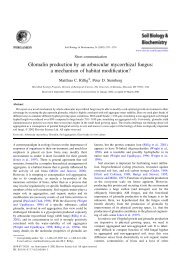
velocity (<strong>Roberts</strong> et al., 1997). This effect of muscle shortening<br />
velocity on recruitment has been demonstrated in studies of turkey<br />
ankle extensors, in which the volume of muscle recruited to<br />
generate a unit force, as estimated from electromyography, is<br />
directly proportional to shortening velocity (Gabaldon et al., 2008)<br />
(Fig.3). Human hopping experiments have also demonstrated a<br />
dramatic increase in muscle activity for a given force output when<br />
humans hop on extremely soft surfaces that undermine effective<br />
elastic-energy storage (Moritz <strong>and</strong> Farley, 2005).<br />
Recently, interest in elastic mechanisms in walkers <strong>and</strong> runners<br />
has turned to the potential importance of springs in motor control<br />
<strong>and</strong> stability. Some of this interest has been stimulated by<br />
roboticists, who must make a robot that can walk without falling<br />
down before they can worry about its energy efficiency.<br />
Mathematical models support the idea that the elastic behavior of<br />
the leg can stabilize movement, meaning that an elastic system has<br />
some ability to recover from a perturbation with limited or no<br />
change in control strategies (Blickhan et al., 2007; Ghigliazza et<br />
al., 2005). Experimental work has demonstrated responses of<br />
human runners to perturbations, such as a sudden change in running<br />
or hopping surface stiffness, that appear to be due, at least in part,<br />
to the spring-like behavior of the leg. Moritz <strong>and</strong> Farley (Moritz<br />
<strong>and</strong> Farley, 2004) demonstrated that human runners faced with a<br />
surprise change in surface stiffness show changes in leg stiffness<br />
that are faster than typical reflex responses <strong>and</strong> that precede<br />
changes in electromyograph (EMG) activity. They concluded that<br />
this response was due to passive mechanical reactions of the springlike<br />
limb (Moritz <strong>and</strong> Farley, 2004). These rapid feedback<br />
mechanisms might involve the elastic action of tendons, but the<br />
spring-like behavior of limbs might reflect the action of muscles as<br />
well as elastic structures. Muscles undergoing a stretch–shorten<br />
cycle could produce some of the spring-like function of the leg, <strong>and</strong><br />
this action might provide some of the rapid mechanical feedback<br />
observed experimentally (Ferris et al., 1998). The response of<br />
guinea fowl hindlimbs to sudden perturbations support the idea that
356<br />
T. J. <strong>Roberts</strong> <strong>and</strong> E. <strong>Azizi</strong><br />
some ‘elastic’ responses of the support limb are actually explained<br />
by active muscle function (Daley <strong>and</strong> Biewener, 2006), <strong>and</strong> a<br />
detailed forward-dynamics model of human hopping suggests that<br />
the elastic elements in series with muscles provide only one of<br />
several possible explanations for rapid stabilizing responses (van<br />
der Krogt et al., 2009). More work is needed to determine the extent<br />
to which elastic structures promote stability in terrestrial gaits.<br />
Recycling energy: swimming <strong>and</strong> flying<br />
Compared with terrestrial locomotion, examples of elastic<br />
mechanisms are relatively scarce in studies of swimming <strong>and</strong> flying<br />
vertebrates. This discrepancy may be explained in part by the<br />
relative number of studies of these different locomotor modes, but<br />
it is also possible that the nature of swimming <strong>and</strong> flying mechanics<br />
limits the usefulness of elastic mechanisms. Compared with<br />
running or walking, swimming <strong>and</strong> flying require a substantial loss<br />
of energy to the environment to provide propulsion <strong>and</strong> overcome<br />
drag. Work lost to the environment to overcome drag cannot be<br />
stored <strong>and</strong> recovered to help power cyclical movements.<br />
Nevertheless, at least two potential benefits of elastic mechanisms<br />
in flight <strong>and</strong> swimming have been identified.<br />
In flying animals, it has been proposed that elastic mechanisms<br />
might aid in providing the inertial work required to accelerate <strong>and</strong><br />
decelerate the wing with each wing beat. In birds, the down stroke<br />
to upstroke transition might be aided by the storage <strong>and</strong> recovery<br />
of elastic energy in the tendon of supracoracoideus, a wing elevator<br />
(Hedrick et al., 2004). Direct evidence for this mechanism is<br />
currently lacking, <strong>and</strong> it is also possible that the kinetic energy lost<br />
as the wing is decelerated is imparted to the environment to provide<br />
useful aerodynamic forces (Hedrick et al., 2004).<br />
Swimmers <strong>and</strong> flyers may also potentially benefit from the<br />
temporal redistribution of muscle work via elastic elements. Such<br />
a mechanism might allow for relatively constant <strong>and</strong> efficient<br />
muscle power production, even at points in the locomotor cycle<br />
when there is a mismatch between available muscle power <strong>and</strong> the<br />
opportunity for generating useful propulsive forces (Gosline <strong>and</strong><br />
Shadwick, 1983; Pabst, 1996). Alternatively, the release of energy<br />
stored elastically might allow for power development at points in<br />
the locomotor cycle when muscle operating lengths or velocities<br />
are unfavorable for producing power. It has been proposed, for<br />
example, that the release of stored energy in the skin of sharks<br />
occurs at the point at which muscles are stretched to long lengths<br />
<strong>and</strong> reduced in their capacity to produce force owing to the<br />
muscle’s length–tension properties (Wainwright et al., 1978).<br />
These kinds of mechanisms would fall broadly under the category<br />
of energy flow depicted in Fig.1B, because the timing of muscular<br />
work is decoupled from the timing of locomotor work by the<br />
temporary storage of muscle work as elastic strain energy (although<br />
power is not necessarily amplified).<br />
Playing with power: amplification <strong>and</strong> attenuation<br />
Elastic mechanisms can act as power amplifiers, by storing muscle<br />
work slowly <strong>and</strong> releasing it rapidly. This mechanism is distinct<br />
from the role of series elastic elements in walking <strong>and</strong> running<br />
because the source of the elastic strain energy is muscle contraction,<br />
rather than transient changes in the body’s kinetic or potential<br />
energy (Fig.1). Power amplification mechanisms work because<br />
springs <strong>and</strong> muscles have different intrinsic power limits. Skeletal<br />
muscles are limited in their maximum power output, ultimately by<br />
rate limits to enzymatic processes associated with actomyosin<br />
cross-bridge cycling. The mechanical function of tendons <strong>and</strong> other<br />
spring elements has a structural basis, <strong>and</strong> therefore is not subject<br />
Force (N)<br />
iEMG / force impulse<br />
1.5<br />
1.0<br />
0.5<br />
THE JOURNAL OF EXPERIMENTAL BIOLOGY<br />
1<br />
0<br />
A<br />
10 N<br />
10 N<br />
0 0.3 0.6 0.9<br />
B<br />
0<br />
–0.05 0 0.05 0.1<br />
Velocity (V/Vmax)<br />
Fig.3. Tendons can reduce the metabolic cost of muscle activity during<br />
running by reducing the volume of muscle that must be active to produce<br />
force. (A)A schematic to show how the shortening velocity of a muscle<br />
affects the muscle volume required to generate a given force. A<br />
hypothetical force–velocity curve is given for a single motor unit in a limb<br />
muscle composed of approximately 30 such units. Each motor unit is<br />
capable of producing 1N of force, when isometric. If the muscle must<br />
develop 10N of force (during stance phase support, for example), this force<br />
can be produced with 10 active motor units if the muscle is isometric. If the<br />
muscle contracts at an intermediate shortening velocity, at which muscle<br />
power is maximized, the force output of each active motor unit is reduced<br />
to ~0.30N, <strong>and</strong> the entire muscle must be active to develop the same 10N<br />
of muscle force. (B)Data from measurements of average muscle<br />
shortening velocity <strong>and</strong> recruited muscle volume required to generate force<br />
in running turkeys support the hypothesis presented in A. The rectified<br />
integrated EMG signal (iEMG) per unit force developed is used as a proxy<br />
for recruited muscle volume. The average iEMG for level (circles) <strong>and</strong><br />
incline (triangles) running at various speeds is directly proportional to the<br />
average relative shortening velocity during stance (modified from Gabaldon<br />
et al., 2008).<br />
to such limitations on power output. Muscle work applied to elastic<br />
elements over the course of a relatively slow muscular contraction<br />
can be released rapidly to produce transient power outputs that<br />
exceed the capacity of the muscle (Fig.1B). The energy released<br />
by the tendon is equal to (or slightly less, given some tendon<br />
energy loss) the work done by the muscle, but it is released in<br />
a shorter amount of time to produce higher power outputs<br />
(powerwork/time). This mechanism makes it possible to uncouple<br />
the work of the muscle from the work done on the body or limbs,<br />
<strong>and</strong> allows for power outputs that exceed the maximum power<br />
capacity of the muscle. It is important to note that the term<br />
‘amplification’ is potentially misleading, in that familiar electronic<br />
power amplifiers work by adding energy to a power source. Elastic<br />
mechanisms in animals do not add energy to the system, but rather<br />
amplify power only in the sense that they release energy more<br />
rapidly than it is stored.
Muscle-mass specific power (W kg –1 )<br />
600<br />
400<br />
200<br />
0<br />
–100<br />
–50<br />
100 Toe-off<br />
Jumping is an activity for which power amplification is<br />
potentially beneficial because the time available to produce power<br />
once the acceleration of the body begins is limited, <strong>and</strong> the time<br />
available to generate power before the jump begins is not. Poweramplifying<br />
mechanisms are typically detected in jumpers by<br />
comparing the power developed during a jump, as measured by<br />
inverse dynamics or force-plate ergometry, to the total power<br />
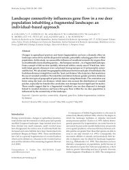
available from the recruited muscles. Evidence for an amplifying<br />
mechanism exists when the peak power output of a jump exceeds<br />
the power-producing capacity of the muscles involved (Fig.4).<br />
Systems using elastic-based power amplification also typically<br />
display an asymmetric power profile, with most of the power<br />
applied to the body late in the jump (Aerts, 1997; <strong>Roberts</strong> <strong>and</strong><br />
Marsh, 2003; Henry et al., 2005) (Fig.4). An elastic mechanism for<br />
muscle power amplification also appears to be essential to jumping<br />
success in bushbabies (Aerts, 1997) <strong>and</strong> frogs (Peplowski <strong>and</strong><br />
Marsh, 1997; <strong>Roberts</strong> <strong>and</strong> Marsh, 2003), <strong>and</strong> even in animals not<br />
specialized for jumping, such as birds (Henry et al., 2005) (Fig.4)<br />
<strong>and</strong> humans (Bobbert, 2001).<br />
The shuttling of energy through tendon springs may provide a<br />
mechanism to decouple the timing of muscle work production <strong>and</strong><br />
body movement that is useful for a wide range of activities. Hof<br />
proposed that the brief burst of power at the ankle late in the stance<br />
phase of walking in humans involves an elastic ‘catapult’<br />
mechanism powered by elastic energy storage in the Achilles<br />
tendon (Hof et al., 1983). This idea is supported by a recent<br />
successful design for an ankle prosthesis, which incorporates<br />
elasticity both in series <strong>and</strong> in parallel with the ankle motor, in part<br />
to provide the necessary power density late in the stance phase of<br />
0<br />
50<br />
Time (ms)<br />
Fig.4. Pro<strong>files</strong> of amplified power. Muscle-mass specific power output in a<br />
jumping guinea fowl. The power developed during a jump shows an<br />
asymmetric profile with peak power occurring late in the jump. In addition,<br />
the power achieved during the jump exceeds the estimated peak power<br />
output of the hindlimb muscles (dotted line) by more than a factor of two.<br />
Taken together, the patterns observed in this jump are consistent with the<br />
use of elastic mechanisms to amplify power output (modified from Henry et<br />
al., 2005).<br />
EMG (mV)<br />
THE JOURNAL OF EXPERIMENTAL BIOLOGY<br />
2<br />
1<br />
A<br />
B<br />
Elastic mechanisms in locomotion<br />
Ballistic<br />
mouth<br />
opening<br />
}<br />
0 0<br />
0 0.1 0.2 0.3 0.4 0.5<br />
Time (s)<br />
357<br />
Fig.5. Power amplification in ballistic tongue projection. The jaw depressor<br />
of toads is active for 250ms prior to mouth opening. Ballistic mouth<br />
opening occurs in about 20ms with no EMG activity. This observed pattern<br />
is consistent with the use of elastic mechanisms that are loaded slowly<br />
during the pre-activation phase <strong>and</strong> released quickly during the ballistic<br />
phase. Gape angle is indicated in black <strong>and</strong> EMG signal in red (modified<br />
from Lappin et al., 2006).<br />
walking (Au et al., 2008). A particularly dramatic catapult-like<br />
mechanism also occurs during limb swing in horses. Power output<br />
of the biceps brachii is amplified by more than 50 times during a<br />
rapid burst of power to initiate limb protraction (Wilson et al.,<br />
2003). Accelerating animals may also benefit from the temporal<br />
redistribution of muscle work via elastic elements. For example,<br />
high power outputs in accelerating turkeys provide evidence of the<br />
use of elastic mechanisms (<strong>Roberts</strong> <strong>and</strong> Scales, 2002). These<br />
examples come from systems that are amenable to study or for<br />
which the amplification of power is particularly dramatic, but it is<br />
likely that elastic mechanisms resulting in modest power<br />
amplification are widespread.<br />
Some feeding mechanisms also make effective use of elastic<br />
power amplification, <strong>and</strong> to our knowledge are the only vertebrate<br />
motions for which the kind of anatomical latch/trigger mechanism<br />
common to invertebrate catapult mechanisms (e.g. Bennet-Clark,<br />
1975) has been identified. Some species of toads, salam<strong>and</strong>ers <strong>and</strong><br />
chameleons use catapult-like mechanisms to rapidly protrude their<br />
tongues during feeding. Ballistic tongue projection mechanisms in<br />
salam<strong>and</strong>ers have evolved independently at least three times, <strong>and</strong><br />
these systems are capable of power outputs that are an order of<br />
magnitude greater than that of the muscles associated with tongue<br />
projection (Deban et al., 2007). Toads also use an impressive<br />
amplification system associated with rapid jaw opening. The<br />
catapult-like action of the depressor m<strong>and</strong>ibulae in toads is<br />
reflected in the decoupling of the timing of muscle activity, which<br />
begins in the 250ms period before jaw movement, <strong>and</strong> the rapid<br />
jaw depression event that lasts only 20ms (Lappin et al., 2006)<br />
(Fig.5). Some very rapid feeding mechanisms in fish also appear<br />
75<br />
50<br />
25<br />
Gape angle (deg)
358<br />
Force (N)<br />
Power (W kg –1 )<br />
Length change (mm)<br />
T. J. <strong>Roberts</strong> <strong>and</strong> E. <strong>Azizi</strong><br />
20<br />
15<br />
10<br />
5<br />
0<br />
150<br />
100<br />
50<br />
0<br />
–50<br />
–100<br />
–150<br />
0<br />
0.5<br />
1.0<br />
1.5<br />
2.0<br />
2.5<br />
3.0<br />
A<br />
B<br />
C<br />
Stimulus<br />
+<br />
0 0.1 0.2 0.3<br />
Time (s)<br />
0.4 0.5 0.6<br />
Fig.6. Internal cycling of energy in a muscle–tendon unit. (A)Muscle force,<br />
(B) fascicle power <strong>and</strong> (C) fascicle length in a fixed-end contraction (i.e. the<br />
muscle–tendon unit is isometric) illustrate the possible negative<br />
consequences of internal energy cycling. The mechanical work produced<br />
by the muscle is loaded into the tendon (positive power phase) <strong>and</strong> then<br />
released back into the muscle fascicles (negative power phase). No<br />
external work is performed, but fascicles produce <strong>and</strong> absorb work. This<br />
type of futile energy cycling could occur in vivo if the timing <strong>and</strong> magnitude<br />
of muscle activity is poorly matched to the kinetics <strong>and</strong> kinematics of<br />
movement or relative to the timing of release of a catch mechanism.<br />
Measurements were taken on frog plantaris muscle, using sonomicrometry<br />
to measure fascicle length <strong>and</strong> a servomotor to measure muscle force<br />
(<strong>Azizi</strong> <strong>and</strong> <strong>Roberts</strong>, 2010).<br />
to use an elastic power-amplifying mechanism. Pipefish generate<br />
muscle mass specific power outputs of more than 5000Wkg –1 to<br />
rapidly elevate their head towards prey (van Wassenbergh et al.,<br />
2008), <strong>and</strong> modeling evidence indicates that rapid jaw depression<br />
in a suction-feeding cichlid involves a catapult-like latch <strong>and</strong><br />
trigger mechanism (Aerts et al., 1987). This kind of decoupling in<br />
the timing of muscle work <strong>and</strong> movement would seem to require a<br />
true catapult mechanism, in which the storage <strong>and</strong> release of energy<br />
is controlled by a latch/trigger mechanism.<br />
What is the benefit of temporarily storing muscle work in elastic<br />
structures before releasing it to effect movement? The most obvious<br />
benefit, already described, is that energy storage <strong>and</strong> release in<br />
springs allows power outputs that exceed the power available from<br />
the muscle motors. However, there are several other less obvious<br />
potential benefits of the uncoupling of muscle work from work on<br />
the body. During locomotor movements, the storage of muscle<br />
work in elastic elements may allow muscles to perform work during<br />
a period (of a stride cycle, for example) when the application of<br />
power to the body or limbs is constrained by kinematics or kinetics.<br />
Alternatively, elastic mechanisms may allow muscles to produce<br />
power at lengths <strong>and</strong>/or velocities that are most favorable for<br />
performance given muscle force–velocity or length–tension<br />
properties (e.g. Wainwright et al., 1978). Finally, muscle power<br />
–<br />
Longitudinal stiffness (N mm –1 )<br />
Rest<br />
THE JOURNAL OF EXPERIMENTAL BIOLOGY<br />
A<br />
B<br />
Passive Active<br />
450<br />
400 Passive<br />
350<br />
300<br />
250<br />
200<br />
150<br />
100<br />
Aactive<br />
50<br />
0<br />
Shorten Lengthen<br />
–4 –2 0 2 4 6 8 10<br />
Transverse strain (%L0)<br />
Fig.7. Variation in aponeurosis stiffness with biaxial loading. (A)A<br />
schematic of aponeurosis behavior during active <strong>and</strong> passive force<br />
production. (B)The longitudinal stiffness of aponeuroses (along the muscle<br />
line of action) as a function of transverse strain. L0 is the resting length of<br />
the aponeurosis. When the tissue is loaded biaxially during active force<br />
production the effective longitudinal stiffness of the elastic structure<br />
increases. This figure highlights the capacity of aponeuroses to function<br />
across range of stiffnesses dynamically (modified from <strong>Azizi</strong> <strong>and</strong> <strong>Roberts</strong>,<br />
2009).<br />
output is strongly dependent on temperature, with typical Q10<br />
values of 2 or more (Bennett, 1985). For ectotherms, the temporal<br />
separation of muscle work from work done in movement may free<br />
performance from potential temperature constraints. This was<br />
demonstrated recently for the elastic tongue projection mechanism<br />
in chameleons (Anderson <strong>and</strong> Deban, 2010). The mechanical<br />
power developed to project the tongue decreased by only about<br />
50% over a 20°C decrease in temperature, whereas the power<br />
developed by the tongue retractor mechanism decreased more than<br />
600% across the same temperature range (Anderson <strong>and</strong> Deban,<br />
2010). The use of thermally insensitive elastic mechanisms may<br />
provide organisms an exp<strong>and</strong>ed thermal niche for select behaviors<br />
(Anderson <strong>and</strong> Deban, 2010).<br />
Muscles not only produce mechanical power, they also absorb it.<br />
The role of elastic mechanisms in energy absorption has received less<br />
attention than for power generation, but tendons may play an<br />
important role in activities that require the dissipation of mechanical<br />
energy. Studies on isolated muscle–tendon preparations (Griffiths,<br />
1991) <strong>and</strong> in vivo studies have demonstrated that rapid stretches<br />
applied to muscle–tendon units can be taken up by the stretch of<br />
tendons, sometimes so effectively that the muscle remains isometric<br />
(Reeves <strong>and</strong> Narici, 2003) or shortens (Griffiths, 1991) during the<br />
‘eccentric’ (muscle–tendon unit lengthening) event. Griffiths<br />
(Griffiths, 1991) proposed that this action provides a mechanical<br />
buffer against damage to muscles, which are particularly susceptible<br />
to damage when muscle fibers are actively lengthened (Proske <strong>and</strong><br />
Morgan, 2001; Lieber <strong>and</strong> Friden, 2002). Of course, tendons cannot<br />
completely insulate muscles from active lengthening. The net
absorption of energy can only be achieved by active lengthening of<br />
muscle fibers, as the energy temporarily stored in stretched elastic<br />
elements must be released. Just as muscle fibers must be the ultimate<br />
source of energy in power-producing events, muscle fibers must be<br />
the sink for energy when muscle–tendon units act to reduce the<br />
energy of the body or a body segment. Thus, the initial stretch of<br />
tendon in an activity like a jump l<strong>and</strong>ing is followed by a period in<br />
which the tendon releases its potential energy by recoiling to stretch<br />
active muscle fibers (Fig.1C). Is there an advantage to having the<br />
work done on the muscle by the tendon rather than direct absorption<br />
from the environment? This mechanism may be important in that it<br />
provides for attenuation of power input to muscle contractile<br />
elements (<strong>Roberts</strong> <strong>and</strong> <strong>Azizi</strong>, 2010). Just as power amplification by<br />
tendons allows for power outputs beyond a muscle’s capacity, power<br />
attenuation via tendons may allow muscle–tendon systems to absorb<br />
energy at a rate beyond the muscle’s maximum capacity for energy<br />
absorption. This elastic mechanism can result in reductions in the<br />
peak power input, lengthening velocity, <strong>and</strong> force experienced by<br />
lengthening muscles. These factors have been associated with muscle<br />
damage (Proske <strong>and</strong> Morgan, 2001; Lieber <strong>and</strong> Friden, 2002), thus<br />
the shuttling of energy through tendons before it is absorbed by<br />
muscles may provide a protective mechanism.<br />
Challenges associated with biological springs<br />
This Commentary, like most of the literature, focuses on the<br />
beneficial effects of elastic mechanisms. Yet the placement of<br />
passive springs in series with active actuators presents some<br />
potential challenges to locomotor control, energetics <strong>and</strong><br />
mechanics. Human-engineered powertrains are typically designed<br />
to maximize stiffness in coupling the motor to the load in order to<br />
provide precision of position control, rapid force development, <strong>and</strong><br />
to reduce the chance of unstable oscillations (Pratt <strong>and</strong> Williamson,<br />
1995). In nature, some features of elastic systems must in part be<br />
designed to avoid some of these potential tradeoffs, <strong>and</strong> to<br />
minimize their consequences. To give one illustration of the<br />
potentially negative consequences of series elastic springs, Fig.6<br />
presents muscle power, force <strong>and</strong> length for a single fixed-end<br />
contraction. In this contraction the origin <strong>and</strong> insertion of the<br />
muscle are fixed, <strong>and</strong> therefore the entire muscle–tendon unit<br />
remains isometric. Upon stimulation, muscle fascicles shorten as<br />
the tendon is stretched, <strong>and</strong> subsequently lengthen as the tendon<br />
recoils. The pattern in Fig.6 illustrates three potentially significant<br />
consequences of tendon action in this ‘isometric’ contraction. First,<br />
the rate of force rise is slowed by the fact that in the process of<br />
stretching the tendon, the muscle shortens <strong>and</strong> its force output is<br />
reduced by muscle force–velocity properties. Second, the muscle<br />
actually develops significant power <strong>and</strong> does work to develop force<br />
(Fig.6B), even though the system does no external work. In the<br />
context of the schematic of Fig.1, this example provides a fourth<br />
pattern of energy flow, from muscle to tendon <strong>and</strong> back to muscle<br />
again. Muscles consume more energy when they generate force<br />
while performing work, thus an ‘isometric’ contraction in a system<br />
with a tendon is presumably more costly than if the system operated<br />
without a tendon. A final effect is that the energy loaded into the<br />
tendon spring is released to stretch the muscle fascicles <strong>and</strong> do work<br />
on them. This illustrates a control challenge inherent in systems<br />
powered by muscle–tendon units. Energy loaded into tendons has<br />
the potential to backfire, recoiling against the muscle itself rather<br />
than unloading its energy to power movement. The pattern of<br />
muscle activation – for example, in power-amplifying mechanisms<br />
– is presumably tuned to ensure that energy flows in the desired<br />
direction in the muscle–tendon system.<br />
THE JOURNAL OF EXPERIMENTAL BIOLOGY<br />
Elastic mechanisms in locomotion<br />
359<br />
The nature of the springs<br />
As our underst<strong>and</strong>ing of the uses of springs in animal movement<br />
has advanced, so has our underst<strong>and</strong>ing of the nature of the springs.<br />
Familiar tendon springs, connective tissue elements (e.g.<br />
epimysium, perimysium) that hold muscles together, <strong>and</strong> the<br />
molecular constituents of muscles themselves all provide springlike<br />
actions that may significantly influence muscle <strong>and</strong> locomotor<br />
function. Many of these springs are also quite dynamic, <strong>and</strong> key<br />
parameters, such as stiffness, can be modulated via both rapid <strong>and</strong><br />
long-term mechanisms.<br />
For tendon springs to operate effectively, their mechanical<br />
properties must be matched to their function. A key parameter for<br />
elastic mechanisms is tendon stiffness, <strong>and</strong> there is increasing<br />
evidence that the stiffness of a tendon is ‘tuned’ by remodeling to<br />
allow for the effective operation of the muscle–tendon-load system.<br />
Several studies have now documented significant increases in<br />
tendon stiffness in response to long-term exercise (Buchanan <strong>and</strong><br />
Marsh, 2001; Arampatzis et al., 2007). There is not yet a consensus<br />
on whether such exposure to a loading regime results in changes in<br />
tendon dimensions (Kasashima et al., 2002), material properties<br />
(Buchanan <strong>and</strong> Marsh, 2001), or both (Seynnes et al., 2009). What<br />
is clear from these studies, however, is that tendon properties are<br />
more plastic than previously thought. The dynamic response of a<br />
tendon to loading has been demonstrated even in very short<br />
timescales. Recent studies have documented increased blood flow<br />
<strong>and</strong> metabolic energy consumption in tendons during periods of<br />
heavy exercise (Kjaer et al., 2005; Hannukainen et al., 2005),<br />
illustrating the robust metabolic infrastructure available to promote<br />
changes in tendon mechanical properties.<br />
Variation in tendon stiffness over very short time scales, such as<br />
during a single muscle contraction, also appears to occur in some<br />
muscle–tendon systems. For many muscles, a significant portion of<br />
the tendon is an aponeurosis. Sheet-like aponeuroses are continuous<br />
with the free portion of the tendon <strong>and</strong> extend over the muscle belly<br />
to provide an attachment surface for muscle fascicles. Several studies<br />
have noted what might be considered unusual behavior for a<br />
structural spring: the stiffness of the aponeurosis (as measured by<br />
muscle force <strong>and</strong> aponeurosis longitudinal strain) appears to be<br />
different during active contractions when compared with passive<br />
loading (Zuurbier et al., 1994; Lieber et al., 2000; <strong>Azizi</strong> <strong>and</strong> <strong>Roberts</strong>,<br />
2009) (Fig.7). The explanation for this behavior appears to be that<br />
transverse (e.g. orthogonal to the line of action) expansion of the<br />
muscle during active contraction loads aponeuroses biaxially, which<br />
tends to alter their effective stiffness measured in the longitudinal<br />
direction (<strong>Azizi</strong> <strong>and</strong> <strong>Roberts</strong>, 2009) (Fig.7). This mechanism<br />
provides for the possibility of rapid <strong>and</strong> dynamic changes in effective<br />
tendon stiffness during muscle contractions. The implications of this<br />
dynamic modulation of stiffness for the energetics <strong>and</strong> mechanics of<br />
locomotion have yet to be fully explored.<br />
Relatively poorly understood is the extent to which the elasticity<br />
of connective tissue structures within the muscle has significance<br />
for muscle or locomotor function. The hierarchical organization of<br />
muscle tissue relies on organized connective tissue wrappings –<br />
epimysium, perimysium <strong>and</strong> endomysium – that together comprise<br />
an extensive connective tissue skeleton within muscles. The<br />
elasticity of intramuscular connective tissues is part of the ‘parallel’<br />
elasticity of muscles that acts to resist passive stretch. Intramuscular<br />
connective tissues are considered crucial for the lateral transmission<br />
of force between adjacent fibers (Huijing, 1999), different heads of<br />
the same muscle (Huijing et al., 1998) or two synergistic muscles<br />
(Huijing et al., 2007). In addition, these structures can encapsulate<br />
muscle groups in tightly packed compartments <strong>and</strong> may serve an
360<br />
T. J. <strong>Roberts</strong> <strong>and</strong> E. <strong>Azizi</strong><br />
important function in maintaining intramuscular pressure (Purslow,<br />
2002) <strong>and</strong> controlling muscle shape changes (<strong>Azizi</strong> et al., 2008)<br />
during contraction. What is currently unclear is the degree to which<br />
intramuscular connective tissues utilize elastic mechanisms as they<br />
transmit <strong>and</strong> bear the loads generated by muscle.<br />
Elastic function is also present in the molecular springs of the<br />
sarcomere. Recent biophysical studies have established the presence<br />
of elasticity in several regions of the actomyosin cross-bridge (Veigel<br />
et al., 1998). Myofilament stiffness has been shown to be non-linear<br />
(Edman, 2009), <strong>and</strong> to vary dynamically depending on the crossbridge<br />
state (Brenner et al., 1982) or the pattern of length changes<br />
(Mantovani, 1999). Similarly, the mechanical behavior of the giant<br />
cytoskeletal protein titin has been characterized as spring-like<br />
(Granzier <strong>and</strong> Labeit, 2006). Early observations had suggested a<br />
largely passive role for titin’s elasticity (Maruyama et al., 1977).<br />
However, titin stiffness has been shown to increase in the presence<br />
of calcium <strong>and</strong> may therefore contribute more dynamically to the<br />
force capacity of the sarcomere upon activation (Labeit et al., 2003).<br />
Changes in titin isoforms, which vary in their stiffness, may also<br />
occur in response to long-term exposure to loading in order to better<br />
tune this molecular spring to a muscle’s mechanical function<br />
(Lindstedt et al., 2002). Our rapidly advancing knowledge of the<br />
molecular springs of muscles will provide exciting opportunities for<br />
future integrated studies that aim to reveal the function of these elastic<br />
structures during movement.<br />
Acknowledgements<br />
We thank the Brown Morphology Group for helpful comments on a version of this<br />
manuscript, <strong>and</strong> Greg Sawicki, Nicolai Konow, <strong>and</strong> Henry Astley for useful<br />
discussions <strong>and</strong> insights. The authors gratefully acknowledge support from the<br />
NIH (RO1AR46499 to T.J.R. <strong>and</strong> F32AR054246 to E.A.) <strong>and</strong> NSF (IOS0642428<br />
to T.J.R.). Deposited in PMC for release after 12 months.<br />
References<br />
Aerts, P. (1997). Vertical jumping in Galago senegalensis: the quest for an obligate<br />
mechanical power amplifier. Philos. Trans. R. Soc. Lond. B Biol. Sci. 353, 1607-<br />
1620.<br />
Aerts, P. A., Osse, J. W. M. <strong>and</strong> Verraes, W. (1987). Model of jaw depression during<br />
feeding in Astatotilapia elegans (Teleostei: Cichlidae): mechanisms for energy<br />
storage <strong>and</strong> triggering. J. Morphol. 194, 85-109.<br />
Alex<strong>and</strong>er, R. M. <strong>and</strong> Vernon, A. (1975). The mechanics of hopping by kangaroos<br />
(Macropodidae). J. Zool. 177, 265-303.<br />
Anderson, C. V. <strong>and</strong> Deban, S. M. (2010). Ballistic tongue projection in chameleons<br />
maintains high performance at low temperature. Proc. Natl. Acad. Sci. USA 107,<br />
5495-5499.<br />
Arampatzis, A., Karamanidis, K., Morey-Klapsing, G., De Monte, G. <strong>and</strong> Stafilidis,<br />
S. (2007). Mechanical properties of the triceps surae tendon <strong>and</strong> aponeurosis in<br />
relation to intensity of sport activity. J. Biomech. 40, 1946-1952.<br />
Au, S., Berniker, M. <strong>and</strong> Herr, H. (2008). Powered ankle-foot prosthesis to assist<br />
level-ground <strong>and</strong> stair-descent gaits. Neural. Netw. 21, 654-666.<br />
<strong>Azizi</strong>, E. <strong>and</strong> <strong>Roberts</strong>, T. J. (2009). Biaxial strain <strong>and</strong> variable stiffness in aponeuroses.<br />
J. Physiol. 587, 4309-4318.<br />
<strong>Azizi</strong>, E. <strong>and</strong> <strong>Roberts</strong>, T. J. (2010). Muscle performance during frog jumping: influence<br />
of elasticity on muscle operating lengths. Proc. Biol. Sci. 277, 1523-1530.<br />
<strong>Azizi</strong>, E., Brainerd, E. L. <strong>and</strong> <strong>Roberts</strong>, T. J. (2008). Variable gearing in pennate<br />
muscles. Proc. Natl. Acad. Sci. USA 105, 1745-1750.<br />
Bennet-Clark, H. C. (1975). The energetics of the jump of the locust Schistorcerca<br />
gregaria. J. Exp. Biol. 63, 53-83.<br />
Bennett, A. F. (1985). Temperature <strong>and</strong> muscle. J. Exp. Biol. 115, 333-344.<br />
Biewener, A. A., Alex<strong>and</strong>er, R. M. <strong>and</strong> Heglund, N. C. (1981). Elastic energy storage<br />
in the hopping of kangaroo rats (Dipodomys spectabilis). J. Zool. 195, 369-383.<br />
Biewener, A. A., Konieczynski, D. D. <strong>and</strong> Baudinette, R. V. (1998). In vivo muscle<br />
force–length behavior during steady-speed hopping in tammar wallabies. J. Exp. Biol.<br />
201, 1681-1694.<br />
Blickhan, R. (1989). The spring-mass model for running <strong>and</strong> hopping. J. Biomech. 2,<br />
1217-1227.<br />
Blickhan, R., Seyfarth, A., Geyer, H., Grimmer, S., Wagner, H. <strong>and</strong> Günther, M.<br />
(2007). Intelligence by mechanics. Philos. Transact. A Math. Phys. Eng. Sci. 365, 199.<br />
Bobbert, M. F. (2001). Dependence of human squat jump performance on the series<br />
elastic compliance of the triceps surae: a simulation study. J. Exp. Biol. 204, 533-542.<br />
Borelli, G. A. (1689). De Motu Animalium. Translated by P. Maquet, 1989. New York:<br />
Springer-Verlag.<br />
Brenner, B., Schoenberg, M., Chalovich, J. M., Greene, L. E. <strong>and</strong> Eisenberg, E.<br />
(1982). Evidence for cross-bridge attachment in relaxed muscle at low ionic strength.<br />
Proc. Natl. Acad. Sci. USA 79, 7288-7291.<br />
THE JOURNAL OF EXPERIMENTAL BIOLOGY<br />
Buchanan, C. I. <strong>and</strong> Marsh, R. L. (2001). Effects of long-term exercise on the<br />
biomechanical properties of the Achilles tendon of guinea fowl. J. Appl. Physiol. 90,<br />
164-171.<br />
Bullimore, S. R. <strong>and</strong> Burn, J. F. (2005). Scaling of elastic energy storage in mammalian<br />
limb tendons: do small mammals really lose out? Biol. Lett. 1, 57-59.<br />
Cavagna, G. A., Saibene, F. P. <strong>and</strong> Margaria, R. (1964). Mechanical work in running. J.<br />
Appl. Phys. 19, 249-256.<br />
Cavagna, G. A., Heglund, N. C. <strong>and</strong> Taylor, C. R. (1977). Mechanical work in terrestrial<br />
locomotion; two basic mechanisms for minimizing energy expenditure. Am. J. Physiol.<br />
233, R243-R261.<br />
Daley, M. A. <strong>and</strong> Biewener, A. A. (2006). Running over rough terrain reveals limb<br />
control for intrinsic stability. Proc. Natl. Acad. Sci. USA 103, 15681-15686.<br />
Deban, S. M., O’Reilly, J. C., Dicke, U. <strong>and</strong> van Leeuwen, J. L. (2007). Extremely<br />
high-power tongue projection in plethodontid salam<strong>and</strong>ers. J. Exp. Biol. 210, 655-667.<br />
Dickinson, M. H., Farley, C. T., Full, R. J., Koehl, M. A. R., Kram, R. <strong>and</strong> Lehman, S.<br />
(2000). How animals move: an integrative view. Science 288, 100-106.<br />
Dudek, D. M. <strong>and</strong> Full, R. J. (2006). Passive mechanical properties of legs from running<br />
insects. J. Exp. Biol. 209, 1502-1515.<br />
Edman, K. A. (2009). Non-linear myofilament elasticity in frog intact muscle fibres. J.<br />
Exp. Biol. 212, 1115-1119.<br />
Farley, C. T., Glasheen, J. <strong>and</strong> McMahon, T. A. (1993). Running springs: speed <strong>and</strong><br />
animal size. J. Exp. Biol. 185, 71-86.<br />
Fenn, W. O. (1924). The relation between the work performed <strong>and</strong> the energy liberated<br />
in muscular contraction. J. Physiol. 58, 373-395.<br />
Ferris, D. P., Louie, M. <strong>and</strong> Farley, C. T. (1998). Running in the real world: adjusting<br />
leg stiffness for different surfaces. Proc. Biol. Sci. 265, 989-994.<br />
Fukunaga, T., Kubo, K., Kawakami, Y., Fukashiro, S., Kanehisa, H. <strong>and</strong> Maganaris,<br />
C. N. (2001). In vivo behaviour of human muscle tendon during walking. Proc. R. Soc.<br />
Lond. B Biol. Sci. 268, 229-233.<br />
Gabaldon, A. M., Nelson, F. E. <strong>and</strong> <strong>Roberts</strong>, T. J. (2008). Relative shortening velocity<br />
in locomotor muscles: turkey ankle extensors operate at low V/Vmax. Am. J. Physiol.<br />
Regul. Integr. Comp. Physiol. 294, R200-R210.<br />
Geyer, H., Seyfarth, A. <strong>and</strong> Blickhan, R. (2006). Compliant leg behaviour explains<br />
basic dynamics of walking <strong>and</strong> running. Proc. Biol. Sci. 273, 2861-2867.<br />
Ghigliazza, R. M., Altendorfer, R., Holmes, P. <strong>and</strong> Koditschek, D. (2005). A simply<br />
stabilized running model. SIAM Rev. Soc. Ind. Appl. Math. 47, 519.<br />
Gosline, J. M. <strong>and</strong> Shadwick, R. E. (1983). The role of elastic energy storage<br />
mechanisms in swimming: an analysis of mantle elasticity in escape jetting in the<br />
squid, Loligo opalescens. Can. J. Zool. 61, 1421-1431.<br />
Granzier, H. L. <strong>and</strong> Labeit, S. (2006). The giant muscle protein titin is an adjustable<br />
molecular spring. Exerc. Sport. Sci. Rev. 34, 50-53.<br />
Griffiths, R. I. (1991). Shortening of muscle fibers during stretch of the active cat medial<br />
gastrocnemius muscle: the role of tendon compliance. J. Physiol. (Lond.) 436, 219-<br />
236.<br />
Hannukainen, J., Kalliokoski, K. K., Nuutila, P., Fujimoto, T., Kemppainen, J.,<br />
Viljanen, T., Laaksonen, M. S., Parkkola, R., Knuuti, J. <strong>and</strong> Kjaer, M. (2005). In<br />
vivo measurements of glucose uptake in human Achilles tendon during different<br />
exercise intensities. Int. J. Sports. Med. 26, 727-731.<br />
Hedrick, T. L., Usherwood, J. R. <strong>and</strong> Biewener, A. A. (2004). Wing inertia <strong>and</strong> wholebody<br />
acceleration: an analysis of instantaneous aerodynamic force production in<br />
cockatiels (Nymphicus holl<strong>and</strong>icus) flying across a range of speeds. J. Exp. Biol. 207,<br />
1689-1702.<br />
Heglund, N. C., Fedak, M. A., Taylor, C. R. <strong>and</strong> Cavagna, G. A. (1982). Energetics<br />
<strong>and</strong> mechanics of terrestrial locomotion IV. Total mechanical energy changes as a<br />
function of speed <strong>and</strong> body size in birds <strong>and</strong> mammals. J. Exp. Biol. 97, 57-66.<br />
Henry, H. T., Ellerby, D. J. <strong>and</strong> Marsh, R. L. (2005). Performance of guinea fowl<br />
Numida meleagris during jumping requires storage <strong>and</strong> release of elastic energy. J.<br />
Exp. Biol. 208, 3293.<br />
Hof, A. L., Geelen, B. A. <strong>and</strong> Van den Berg, J. (1983). Calf muscle moment, work <strong>and</strong><br />
efficiency in level walking; role of series elasticity. J. Biomech. 16, 523-537.<br />
Huijing, P. A. (1999). Muscle as a collagen fiber reinforced composite: a review of force<br />
transmission in muscle <strong>and</strong> whole limb. J. Biomech. 32, 329-345.<br />
Huijing, P. A., Baan, G. C. <strong>and</strong> Rebel, G. T. (1998). Non-myotendinous force<br />
transmission in rat extensor digitorum longus muscle. J. Exp. Biol. 201, 682-691.<br />
Huijing, P. A., van de Langenberg, R. W., Meesters, J. J. <strong>and</strong> Baan, G. C. (2007).<br />
Extramuscular myofascial force transmission also occurs between synergistic muscles<br />
<strong>and</strong> antagonistic muscles. J. Electromyogr. Kinesiol. 17, 680-689.<br />
Kasashima, Y., Smith, R. K., Birch, H. L., Takahashi, T., Kusano, K. <strong>and</strong> Goodship,<br />
A. E. (2002). Exercise-induced tendon hypertrophy: cross-sectional area changes<br />
during growth are influenced by exercise. Equine. Vet. J. Suppl., 264-268.<br />
Kjaer, M., Langberg, H., Miller, B. F., Boushel, R., Crameri, R., Koskinen, S.,<br />
Heinemeier, K., Olesen, J. L., Døssing, S., Hansen, M. et al. (2005). Metabolic<br />
activity <strong>and</strong> collagen turnover in human tendon in response to physical activity. J.<br />
Musculoskelet. Neuronal. Interact. 5, 41-52.<br />
Labeit, D., Watanabe, K., Witt, C., Fujita, H., Wu, Y., Lahmers, S., Funck, T., Labeit,<br />
S. <strong>and</strong> Granzier, H. (2003). Calcium-dependent molecular spring elements in the giant<br />
protein titin. Proc. Natl. Acad. Sci. USA 100, 13716-13721.<br />
Lappin, A. K., Monroy, J. A., Pilarski, J. Q., Zepnewski, E. D., Pierotti, D. J. <strong>and</strong><br />
Nishikawa, K. C. (2006). Storage <strong>and</strong> recovery of elastic potential energy powers<br />
ballistic prey capture in toads. J. Exp. Biol. 209, 2535-2553.<br />
Lee, C. R. <strong>and</strong> Farley, C. T. (1998). Determinants of the center of mass trajectory in<br />
human walking <strong>and</strong> running. J. Exp. Biol. 201, 2935-2944.<br />
Lichtwark, G. A., Bougoulias, K. <strong>and</strong> Wilson, A. M. (2007). Muscle fascicle <strong>and</strong> series<br />
elastic element length changes along the length of the human gastrocnemius during<br />
walking <strong>and</strong> running. J. Biomech. 40, 157-164.<br />
Lieber, R. L. <strong>and</strong> Friden, J. (2002). Mechanisms of muscle injury gleaned from animal<br />
models. Am. J. Phys. Med. Rehabil. 81, S70-S79.<br />
Lieber, R. L., Leonard, M. E. <strong>and</strong> Brown-Maupin, C. G. (2000). Effects of muscle<br />
contraction on the load-strain properties of frog aponeurosis <strong>and</strong> tendon. Cells Tissues<br />
Organs 166, 48-54.
Lindstedt, S. L., Reich, T. E., Keim, P. <strong>and</strong> LaStayo, P. C. (2002). Do muscles<br />
function as adaptable locomotor springs? J. Exp. Biol. 205, 2211-2216.<br />
Mantovani, M., Cavagna, G. A. <strong>and</strong> Heglund, N. C. (1999). Effect of stretching on<br />
undamped elasticity in muscle fibres from Rana temporaria. J. Muscle. Res. Cell Motil.<br />
20, 33-43.<br />
Maruyama, K., Matsubara, S., Natori, R., Nonomura, Y., Kimura, S., Ohashi, K.,<br />
Murakami, F., H<strong>and</strong>a, S. <strong>and</strong> Eguchi, G. (1977). Connectin, an elastic protein of<br />
muscle: characterization <strong>and</strong> function. J. Biochem. 82, 317.<br />
McMahon, T. A. <strong>and</strong> Cheng, G. C. (1990). The mechanics of running: how does<br />
stiffness couple with speed? J. Biomech. 23, 65-78.<br />
Moritz, C. T. <strong>and</strong> Farley, C. T. (2004). Passive dynamics change leg mechanics for an<br />
unexpected surface during human hopping. J. Appl. Physiol. 97, 1313-1322.<br />
Moritz, C. T. <strong>and</strong> Farley, C. T. (2005). Human hopping on very soft elastic surfaces:<br />
implications for muscle pre-stretch <strong>and</strong> elastic energy storage in locomotion. J. Exp.<br />
Biol. 208, 939.<br />
Pabst, D. (1996). Springs in swimming animals. Integr. Comp. Biol. 36, 723.<br />
Peplowski, M. M. <strong>and</strong> Marsh, R. L. (1997). Work <strong>and</strong> power output in the hindlimb<br />
muscles of cuban tree frogs Osteopilus septentrionalis during jumping. J. Exp. Biol.<br />
200, 2861-2870.<br />
Pollock, C. M. <strong>and</strong> Shadwick, R. E. (1994a). Allometry of muscle, tendon, <strong>and</strong> elastic<br />
energy storage capacity in mammals. Am. J. Physiol. 266, R1022-R1031.<br />
Pollock, C. M. <strong>and</strong> Shadwick, R. E. (1994b). Relationship between body mass <strong>and</strong><br />
biomechanical properties of limb tendons in adult mammals. Am. J. Physiol. 266,<br />
R1016-R1021.<br />
Pratt, G. A. <strong>and</strong> Williamson, M. M. (1995). Series elastic actuators. Proc of IEEE/RSJ.<br />
International Conference on Intelligent Robots <strong>and</strong> Systems 95, ‘Human Robot<br />
Interaction <strong>and</strong> Cooperative Robots’, 1, 399-406.<br />
Proske, U. <strong>and</strong> Morgan, D. L. (2001). Muscle damage from eccentric exercise:<br />
mechanism, mechanical signs, adaptation <strong>and</strong> clinical applications. J. Physiol. 537,<br />
333-345.<br />
Purslow, P. P. (2002). The structure <strong>and</strong> functional significance of variations in the<br />
connective tissue within muscle. Comp. Biochem. Physiol. A Mol. Integr. Physiol. 133,<br />
947-966.<br />
THE JOURNAL OF EXPERIMENTAL BIOLOGY<br />
Elastic mechanisms in locomotion<br />
361<br />
Reeves, N. D. <strong>and</strong> Narici, M. V. (2003). Behavior of human muscle fascicles during<br />
shortening <strong>and</strong> lengthening contractions in vivo. J. Appl. Physiol. 95, 1090-1096.<br />
<strong>Roberts</strong>, T. J. (2002). The integrated function of muscles <strong>and</strong> tendons during<br />
locomotion. Comp. Biochem. Physiol. A Mol. Integr. Physiol. 133, 1087-1099.<br />
<strong>Roberts</strong>, T. J. <strong>and</strong> <strong>Azizi</strong>, E. (2010). The series-elastic shock absorber: tendons<br />
attenuate muscle power during eccentric actions. J. Appl. Physiol. 109, 396-404.<br />
<strong>Roberts</strong>, T. J. <strong>and</strong> Marsh, R. L. (2003). Probing the limits to muscle-powered<br />
accelerations: lessons from jumping bullfrogs. J. Exp. Biol. 206, 2567-2580.<br />
<strong>Roberts</strong>, T. J. <strong>and</strong> Scales, J. A. (2002). Mechanical power output during running<br />
accelerations in wild turkeys. J. Exp. Biol. 205, 1485-1494.<br />
<strong>Roberts</strong>, T. J., Marsh, R. L., Wey<strong>and</strong>, P. G. <strong>and</strong> Taylor, C. R. (1997). Muscular force<br />
in running turkeys: the economy of minimizing work. Science 275, 1113-1115.<br />
Seynnes, O. R., Erskine, R. M., Maganaris, C. N., Longo, S., Simoneau, E. M.,<br />
Grosset, J. F. <strong>and</strong> Narici, M. V. (2009). Training-induced changes in structural <strong>and</strong><br />
mechanical properties of the patellar tendon are related to muscle hypertrophy but not<br />
to strength gains. J. Appl. Physiol. 107, 523-530.<br />
van der Krogt, M. M., de Graaf, W. W., Farley, C. T., Moritz, C. T., Richard Casius, L.<br />
J. <strong>and</strong> Bobbert, M. F. (2009). Robust passive dynamics of the musculoskeletal<br />
system compensate for unexpected surface changes during human hopping. J. Appl.<br />
Physiol. 107, 801-808.<br />
Van Wassenbergh, S., Strother, J. A., Flammang, B. E., Ferry-Graham, L. A. <strong>and</strong><br />
Aerts, P. (2008). Extremely fast prey capture in pipefish is powered by elastic recoil. J.<br />
R. Soc. Interface 5, 285-296.<br />
Veigel, C., Bartoo, M. L., White, D. C., Sparrow, J. C. <strong>and</strong> Molloy, J. E. (1998). The<br />
stiffness of rabbit skeletal actomyosin cross-bridges determined with an optical<br />
tweezers transducer. Biophys. J. 75, 1424-1438.<br />
Wainwright, S. A., Vosburgh, F. <strong>and</strong> Hebrank, J. H. (1978). Shark Skin: Function in<br />
Locomotion. Science (New York, NY). 202, 747.<br />
Wilson, A. M., Watson, J. C. <strong>and</strong> Lichtwark, G. A. (2003). Biomechanics: A catapult<br />
action for rapid limb protraction. Nature 421, 35-36.<br />
Zuurbier, C. J., Everard, A. J., van der Wees, P. <strong>and</strong> Huijing, P. A. (1994). Lengthforce<br />
characteristics of the aponeurosis in the passive <strong>and</strong> active muscle condition <strong>and</strong><br />
in the isolated condition. J. Biomech. 27, 445-453.