Ophthalmology Update - Cleveland Clinic
Ophthalmology Update - Cleveland Clinic
Ophthalmology Update - Cleveland Clinic
Create successful ePaper yourself
Turn your PDF publications into a flip-book with our unique Google optimized e-Paper software.
8<br />
OPHTHALMOLOGY UPDATE 2009 SPECIAL EDITION<br />
Case Study: DSAEK to Treat Amantadine-associated Corneal Edema<br />
Christopher T. Hood, MD<br />
Roger H.S. Langston, MD<br />
William J. Dupps, Jr.,<br />
MD, PhD<br />
Presentation:<br />
A 45-year-old Caucasian woman presented to the<br />
Cole Eye Institute for management of corneal<br />
edema. She described experiencing six months of<br />
blurry vision in both eyes that was worse in the<br />
morning and improved slightly throughout the day.<br />
She denied redness, pain or photophobia. She was<br />
being treated with Muro 128 ointment four times<br />
daily in the right eye upon referral.<br />
She denied any history of ocular trauma, surgery<br />
or inflammatory disease. Her medical history was<br />
significant for a longstanding diagnosis of multiple<br />
sclerosis, for which she was taking baclofen, methyl-<br />
phenidate, glatiramer acetate injection, neurontin,<br />
amantadine, escitalopram oxalate and bupropion.<br />
She denied any family history of eye disease.<br />
Examination:<br />
On examination, visual acuity was 20/800 in the right<br />
eye and 20/400 in the left eye. Pupils were equal<br />
in size and reactive, without an afferent pupillary<br />
defect. Extraocular movements were full. Intraocular<br />
pressures were 12 mm Hg in the right eye and 10<br />
mm Hg in the left eye. Anterior segment examination<br />
demonstrated normal eyelids, sclera and conjunc-<br />
tiva. Bilateral diffuse stromal and epithelial edema<br />
was observed with marked Descemet membrane<br />
folds and pre-Descemet membrane opacification<br />
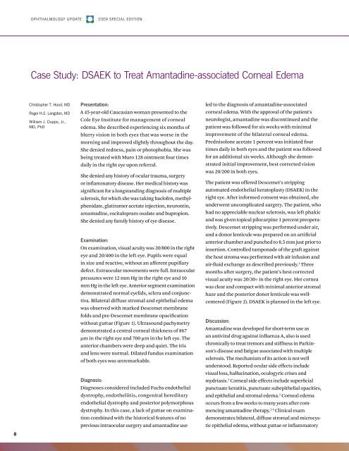
without guttae (Figure 1). Ultrasound pachymetry<br />
demonstrated a central corneal thickness of 867<br />
µm in the right eye and 700 µm in the left eye. The<br />
anterior chambers were deep and quiet. The iris<br />
and lens were normal. Dilated fundus examination<br />
of both eyes was unremarkable.<br />
Diagnosis:<br />
Diagnoses considered included Fuchs endothelial<br />
dystrophy, endotheliitis, congenital hereditary<br />
endothelial dystrophy and posterior polymorphous<br />
dystrophy. In this case, a lack of guttae on examina-<br />
tion combined with the historical features of no<br />
previous intraocular surgery and amantadine use<br />
led to the diagnosis of amantadine-associated<br />
corneal edema. With the approval of the patient’s<br />
neurologist, amantadine was discontinued and the<br />
patient was followed for six weeks with minimal<br />
improvement of the bilateral corneal edema.<br />
Prednisolone acetate 1 percent was initiated four<br />
times daily in both eyes and the patient was followed<br />
for an additional six weeks. Although she demonstrated<br />
initial improvement, best corrected vision<br />
was 20/200 in both eyes.<br />
The patient was offered Descemet’s stripping<br />
automated endothelial keratoplasty (DSAEK) in the<br />
right eye. After informed consent was obtained, she<br />
underwent uncomplicated surgery. The patient, who<br />
had no appreciable nuclear sclerosis, was left phakic<br />
and was given topical pilocarpine 1 percent preoperatively.<br />
Descemet stripping was performed under air,<br />
and a donor lenticule was prepared on an artificial<br />
anterior chamber and punched to 8.5 mm just prior to<br />
insertion. Controlled tamponade of the graft against<br />
the host stroma was performed with air infusion and<br />
air-fluid exchange as described previously. 1 Three<br />
months after surgery, the patient’s best corrected<br />
visual acuity was 20/30+ in the right eye. Her cornea<br />
was clear and compact with minimal anterior stromal<br />
haze and the posterior donor lenticule was wellcentered<br />
(Figure 2). DSAEK is planned in the left eye.<br />
Discussion:<br />
Amantadine was developed for short-term use as<br />
an antiviral drug against influenza A, also is used<br />
chronically to treat tremors and stiffness in Parkinson’s<br />
disease and fatigue associated with multiple<br />
sclerosis. The mechanism of its action is not well<br />
understood. Reported ocular side effects include<br />
visual loss, hallucination, oculogyric crises and<br />
mydriasis. 2 Corneal side effects include superficial<br />
punctuate keratitis, punctuate subepithelial opacities,<br />
and epithelial and stromal edema. 2 Corneal edema<br />
occurs from a few weeks to many years after commencing<br />
amantadine therapy. 2-6 <strong>Clinic</strong>al exam<br />
demonstrates bilateral, diffuse stromal and microcystic<br />
epithelial edema, without guttae or inflammatory


