

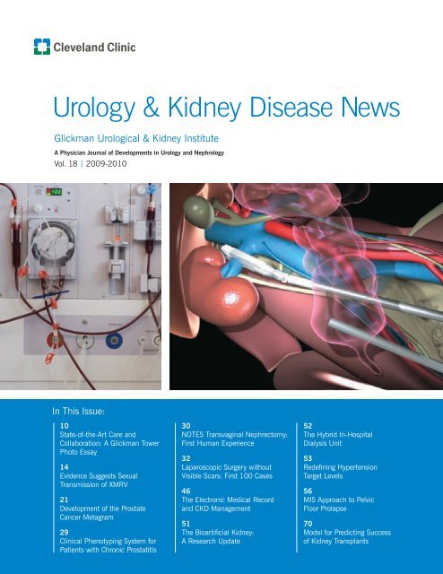
Urology & Kidney Disease News Fall 2009 - Cleveland Clinic
Urology & Kidney Disease News Fall 2009 - Cleveland Clinic
Urology & Kidney Disease News Fall 2009 - Cleveland Clinic

Create successful ePaper yourself
Turn your PDF publications into a flip-book with our unique Google optimized e-Paper software.
<strong>Urology</strong> & <strong>Kidney</strong> <strong>Disease</strong> <strong>News</strong><br />
Glickman Urological & <strong>Kidney</strong> Institute<br />
A Physician Journal of Developments in <strong>Urology</strong> and Nephrology<br />
Vol. 18 | <strong>2009</strong>-2010<br />
In This Issue:<br />
10<br />
State-of-the-Art Care and<br />
Collaboration: A Glickman Tower<br />
Photo Essay<br />
14<br />
Evidence Suggests Sexual<br />
Transmission of XMRV<br />
21<br />
Development of the Prostate<br />
Cancer Metagram<br />
29<br />
<strong>Clinic</strong>al Phenotyping System for<br />
Patients with Chronic Prostatitis<br />
30<br />
NOTES Transvaginal Nephrectomy:<br />
First Human Experience<br />
32<br />
Laparoscopic Surgery without<br />
Visible Scars: First 100 Cases<br />
46<br />
The Electronic Medical Record<br />
and CKD Management<br />
51<br />
The Bioartificial <strong>Kidney</strong>:<br />
A Research Update<br />
52<br />
The Hybrid In-Hospital<br />
Dialysis Unit<br />
53<br />
Redefining Hypertension<br />
Target Levels<br />
56<br />
MIS Approach to Pelvic<br />
Floor Prolapse<br />
70<br />
Model for Predicting Success<br />
of <strong>Kidney</strong> Transplants
On the Cover<br />
From the development of the<br />
kidney dialysis machine to<br />
pioneering the bioartificial<br />
kidney, <strong>Cleveland</strong> <strong>Clinic</strong> has a<br />
long history of innovation in<br />
the treatment of end-stage<br />
renal disease. To read about<br />
our latest activities, see pages<br />
49-53. And for a look at our<br />
new 21-bed state-of-the-art<br />
dialysis unit, see the<br />
Glickman Tower photo essay<br />
on page 10.<br />
Natural orifice translumenal<br />
endoscopic surgery (NOTES)<br />
brings forth a change in the<br />
intellectual and physical<br />
approach to urologic surgery.<br />
<strong>Cleveland</strong> <strong>Clinic</strong> was the first<br />
to perform a NOTES transvaginal<br />
nephrectomy in a<br />
human. For details on the<br />
procedure and its outcomes,<br />
see page 30.<br />
<strong>Urology</strong> & <strong>Kidney</strong> <strong>Disease</strong> <strong>News</strong><br />
Chairman’s Report .....................................................................4<br />
<strong>News</strong> from the Glickman Urological and <strong>Kidney</strong> Institute<br />
New Staff .................................................................................6<br />
Upcoming Conferences ..............................................................7<br />
Staff Awards and Appointments ..................................................8<br />
From the Medical Editor: Care in the 21st Century .......................9<br />
Glickman Tower Photo Essay ....................................................10<br />
Prostate Cancer<br />
XMRV is Found in Prostatic Secretions:<br />
Evidence Suggests Sexual Transmission .....................................14<br />
A Novel Stereotactic Prostate Biopsy<br />
and Treatment Guidance System ...............................................15<br />
Magnetic Resonance Imaging of the Prostate .............................16<br />
Analysis of T1c Prostate Cancers Treated<br />
at Very Low Prostate-Specific Antigen Levels ..............................17<br />
A Nomogram for Predicting Upgrading in Patients<br />
with Low and Intermediate Grade Prostate Cancer<br />
in the Era of Extending Prostate Sampling ..................................18<br />
The Influence of Body Mass Index on<br />
Prostate Cancer Screening in a Large Cohort ..............................20<br />
Metagram for Tailoring Prostate Cancer Management ..................21<br />
Active Surveillance with Selective<br />
Delayed Intervention for Prostate Cancer ....................................22<br />
Active Surveillance: Are the Epstein Criteria Suitable<br />
for Defining Biologically Insignificant Prostate Cancer? ................23<br />
Brachytherapy and External Beam Radiotherapy<br />
for Prostate Cancer: A Comparison ............................................24<br />
Establishing Treatment Success Criteria for Cryosurgical<br />
Ablation of Prostate Cancer: An Analysis of the COLD Registry.....26<br />
Benign Prostatic <strong>Disease</strong><br />
980: The New Hot Number in BPH Therapy .............................28<br />
<strong>Clinic</strong>al Phenotyping of Patients With<br />
Chronic Prostatitis/Chronic Pelvic Pain Syndrome<br />
and Correlation With Symptom Severity .....................................29<br />
Robotics and Laparoscopy<br />
NOTES (Natural Orifice Translumenal Endoscopic Surgery)<br />
Transvaginal Nephrectomy: First Human Experience ...................30<br />
NOTES (Natural Orifice Translumenal Endoscopic Surgery)<br />
Renal Cryoablation in the Porcine Model....................................31<br />
clevelandclinic.org/urology
3 <strong>Urology</strong> & <strong>Kidney</strong> <strong>Disease</strong> <strong>News</strong><br />
Laparoscopic Surgery Without Visible Scars:<br />
Experience with the First 100 Cases .........................................32<br />
Application of 2-mm Needlescopic Instruments<br />
in Urological Laparoscopic Surgery ............................................34<br />
Stereotactic Ablation of Renal Tumors........................................36<br />
Robotic Laparoendoscopic Single Site Surgery<br />
Using GelPort® as the Access Platform .....................................37<br />
Laparoendoscopic Single Site Pyeloplasty:<br />
A Comparison to Standard Laparoscopic Technique ....................38<br />
Single-Port Single-Surgeon Robotic-Assisted<br />
Laparoscopic Reconstructive <strong>Urology</strong>.........................................40<br />
Pediatric <strong>Urology</strong><br />
Technical Modifications of Robotic-Assisted Pyeloplasty in Children<br />
Resulting in Minimal Morbidity and Improved Efficacy ................41<br />
<strong>Kidney</strong> Cancer<br />
Guidelines for Management of the <strong>Clinic</strong>al Stage 1 Renal Mass ...42<br />
Managing Patients with Renal Tumors and Venous Thrombus ......43<br />
Partial Nephrectomy for Renal Masses 7 cm or Less:<br />
The New Standard in Nephron-Sparing Surgery .........................44<br />
Neoadjuvant VEGF-targeted Therapy in Renal Cell Carcinoma .....45<br />
Chronic <strong>Kidney</strong> <strong>Disease</strong><br />
Optimal Use of the Electronic Medical Record<br />
in Chronic <strong>Kidney</strong> <strong>Disease</strong> ........................................................46<br />
The Development of a Glomerulonephritis Board<br />
Can Help in the Treatment of Individual Cases as Well<br />
as in Developing Effective Treatment Regimens ..........................47<br />
Developing Evidence-Based Treatment Options<br />
for <strong>Kidney</strong> <strong>Disease</strong>s .................................................................48<br />
Understanding the Molecular Mechanism in Anemia of CKD .......48<br />
Renal Failure<br />
The New Centers for Medicare and Medicaid Services<br />
Conditions for Coverage Regulations ..........................................49<br />
ICU Nephrology: Change and Optimism ....................................50<br />
Bioartificial <strong>Kidney</strong> Making Milestones .......................................51<br />
The Hybrid In-Hospital Dialysis Unit:<br />
Current and Future Challenges ..................................................52<br />
Hypertension<br />
Redefining Hypertension for Tomorrow: Target Levels ..................53<br />
Resistant Endocrine Hypertension .............................................55<br />
Female <strong>Urology</strong><br />
Minimally Invasive Approaches to Female Pelvic Organ Prolapse..56<br />
Electrostimulation after Delivery Injury to<br />
Treat Urinary Incontinence........................................................57<br />
Bladder Neck Coaptation as a Predictor of<br />
Successful Injection for Female Stress Incontinence ....................58<br />
Expression of Urinary Chemokines<br />
in Patients with Overactive Bladder ...........................................60<br />
Bladder Cancer<br />
Lack of Pathologic Down-Staging with Neoadjuvant<br />
Chemotherapy for Muscle-Invasive Urothelial Carcinoma<br />
of the Bladder: A Contemporary Series ......................................61<br />
Studying the Effects of Smoke on Bladder Cancer Treatment .......62<br />
Urethral Reconstruction<br />
Urethral Reconstruction in Older Men Results in<br />
High Patient Satisfaction and Excellent Urinary Function .............64<br />
Functional and Quality of Life Outcomes in Patients<br />
Undergoing Transperineal Repair with Gracilis Muscle<br />
Interposition for Complex Rectourethral Fistula ...........................66<br />
Sexual Health<br />
Peyronie’s <strong>Disease</strong>: An Update on the Role of Diagnostics ...........68<br />
Increasing Penile Length at Time of<br />
Penile Prosthesis Implantation ..................................................69<br />
Transplantation<br />
Prediction in Transplantation .....................................................70<br />
<strong>Kidney</strong> Function in Living <strong>Kidney</strong> Donors ...................................72<br />
Special Programs<br />
Lifestyle Modification Program Shows<br />
Promise for Prostate Cancer Patients .........................................73<br />
The Novick Center for <strong>Clinic</strong>al and<br />
Translational Research Manages High<br />
Volume of Research within the Institute .....................................73<br />
<strong>Urology</strong> Residency Program Expansion in Progress .....................74<br />
Services for Physicians .............................................................75<br />
Services for Patients.................................................................75<br />
Author Index ...........................................................................76
4 <strong>Urology</strong> & <strong>Kidney</strong> <strong>Disease</strong> <strong>News</strong><br />
Chairman’s Report<br />
Eric A. Klein, MD<br />
Chairman, <strong>Cleveland</strong> <strong>Clinic</strong> Glickman<br />
Urological & <strong>Kidney</strong> Institute<br />
The last 12 months have been eventful ones for The Glickman<br />
Urological & <strong>Kidney</strong> Institute. On September 12, 2008,<br />
we dedicated the new, high-tech, state-of-the-art Glickman<br />
Tower, a 12-story facility on the main <strong>Cleveland</strong> <strong>Clinic</strong><br />
campus. The institute occupies the top four floors of the<br />
tower, comprising more than 200,000 square feet of space<br />
for clinical and academic activities. The Glickman Tower<br />
has been carefully designed to provide patients with care for<br />
all urological and kidney diseases, to optimize every aspect<br />
of the patient experience, and to create an environment in<br />
which physicians and scientists from different disciplines<br />
can work together synergistically in patient care, education<br />
and research.<br />
Highlights of the new Glickman Tower include space for<br />
physician academic offices and staff support, work space for<br />
residents and fellows, an integrated patient care and clinical<br />
research area for the Center for <strong>Kidney</strong>/Pancreas Transplantation,<br />
a new 21-bay outpatient dialysis unit, and dedicated<br />
space for the support staff of the Center for Quality Patient<br />
Care, Center for Outcomes Research, and the newly named<br />
Novick Center for <strong>Clinic</strong>al and Translational Research. The<br />
7th and 8th floors comprise 80 patient exam rooms, including<br />
seven dedicated for pediatric urology. Patient, nursing<br />
and physician movements are monitored by a built-in Versus<br />
Information System, an RFID-based real-time locating system<br />
that displays patient wait times and location on easily<br />
accessible screens to optimize flow through the outpatient<br />
clinic. The 9th floor includes a new urodynamics lab, 12 procedure<br />
rooms, and four operating rooms equipped for general<br />
anesthesia. A state-of-the-art 88-seat amphitheater resides<br />
on the first floor, equipped with Internet access, video links<br />
to the operating theatre, and satellite-link capabilities for<br />
world-wide real-time transmission of lectures and surgery.<br />
(See the photos of the Glickman Tower, page 10-13.)<br />
Sadly, just a few weeks after dedicating the Glickman Tower,<br />
Andrew C. Novick, MD, the inaugural chair of the Glickman<br />
Urological & <strong>Kidney</strong> Institute, died suddenly and unexpectedly<br />
of complications related to lymphoma. Andy was a true<br />
surgical pioneer in the areas of renal transplantation, surgical<br />
revascularization of diseased kidneys, bench surgery and<br />
the surgical management of kidney cancer. Under his tutelage,<br />
the Department of <strong>Urology</strong> went from six to 61 faculty<br />
between 1985 and 2008, and he laid the foundation for the<br />
integration of urology and nephrology to become the multidisciplinary<br />
institute, integrating patient care, resident and<br />
fellow education, and basic and clinical research. John Barry,<br />
MD, summed up Andy’s career best, writing on learning of<br />
Andy’s death, “Well done, Andy, well done.” (See more in the<br />
tribute to Dr. Novick, included with this mailing.)<br />
In April <strong>2009</strong>, Inderbir Gill, MD, a pioneer in laparoscopic<br />
urologic procedures, departed <strong>Cleveland</strong> to assume the<br />
Chair of the Institute of <strong>Urology</strong> at the University of Southern<br />
California. D. Karl Montague, MD, currently serves as Interim<br />
Chair of the Department of <strong>Urology</strong>, and a new permanent<br />
chair of the department is expected to be named by year’s<br />
end. J. Stephen Jones, MD, remains as Chair of the Department<br />
of Regional <strong>Urology</strong>; Martin Schreiber, MD, chairs the<br />
Department of Nephrology, and Lawrence Hakim, MD, is the<br />
Chair of the Department of <strong>Urology</strong>, <strong>Cleveland</strong> <strong>Clinic</strong> Florida.<br />
New Center Directors in the last year include Howard Goldman,<br />
MD, Director of the Center for Quality Patient Care;<br />
Jihad Kaouk, MD, Director of the Center for Laparoscopic<br />
and Robotic Surgery; Jeffrey Palmer, MD, Director of the<br />
Center for Pediatric and Adolescent <strong>Urology</strong>; Daniel Shoskes,<br />
MD, Director of the Novick Center for <strong>Clinic</strong>al and Translational<br />
Research; and Andrew Stephenson, MD, Acting<br />
Director of the Center for Urologic Oncology.<br />
Several individuals and programs were awarded important<br />
recognition in the last year. William Fissell, MD, of the Department<br />
of Nephrology, received a Career Development<br />
Award from the American Society of Nephrology to foster his<br />
work on development of a bioartificial kidney, which could<br />
someday allow for continuous outpatient dialysis using a<br />
device the size of a 12-ounce aluminum can. Karl Montague,<br />
MD, received the ACGME’s Courage to Teach Award in recognition<br />
of his dedication during a long career as an educator<br />
and residency director; Andrew Stephenson, MD, received a<br />
grant from the Robert Wood Johnson Foundation to study<br />
decision analysis techniques for helping patients decide on<br />
the best treatment for their prostate cancer; and we were<br />
named a Center of Excellence for Prostate Cancer by Medical<br />
Economics. At the annual meeting of the American Urological<br />
Association held in Chicago in April <strong>2009</strong>, our residents,<br />
fellows, and faculty were awarded 5 “Best Paper” honors and<br />
the “Best Video” award. Both the Department of Nephrology<br />
and the collective departments of <strong>Urology</strong> ranked highly (5th<br />
and 2nd, respectively) in U.S.<strong>News</strong> & World Report rankings.
A four-story grand hall in the new Glickman Tower features artwork, a café and an educational health resource center for patients. For more<br />
photos, see the Glickman Tower photo essay, page 10.<br />
Our educational programs remain a significant focus. In<br />
the milieu of the largest post-graduate medical education<br />
program in the world at <strong>Cleveland</strong> <strong>Clinic</strong>, at any given time<br />
we have six nephrology fellows, 24 urology residents and 20<br />
urology fellows (distributed among eight individual fellowship<br />
programs). Many of these individuals go on to become<br />
academic leaders in their fields. Based on an application approved<br />
by the Residency Review Committee last year, in July<br />
<strong>2009</strong> the urology residency was able to expand to five per year<br />
(see story, page 74). This expansion will help to cover continued<br />
growth in clinical and research activities, and will enable<br />
the development of a community-based in- and outpatient<br />
rotation for upper level residents. In the six-year residency,<br />
we continue to offer a dedicated year of protected time devoted<br />
to basic, translational or outcomes-related investigation,<br />
one of the few remaining programs in the country to do so.<br />
The demand for clinical services offered by our urology and<br />
nephrology programs remains robust, reflecting the strong<br />
regional and national reputation of our physician staff.<br />
During 2008, we performed 20,975 urological operations.<br />
Our departments of urology collectively recorded a total of<br />
91,582 outpatient visits, while our Department of Nephrology<br />
recorded 26,424 outpatient visits and 12,188 dialysis treatments.<br />
Despite the changes we have experienced in the last year, we<br />
remain excited by the opportunity for the institute to continue<br />
a legacy of significant innovation and contributions<br />
to our core mission of patient care, education and research.<br />
We are pleased to share current activities with our colleagues<br />
and friends in this second annual issue of <strong>Urology</strong> & <strong>Kidney</strong><br />
<strong>Disease</strong> <strong>News</strong>.<br />
Eric A. Klein, MD<br />
clevelandclinic.org/urology<br />
5
6 <strong>Urology</strong> & <strong>Kidney</strong> <strong>Disease</strong> <strong>News</strong><br />
<strong>News</strong> from the Glickman Urological and <strong>Kidney</strong> Institute<br />
New Staff The Glickman Urological & <strong>Kidney</strong> Institute<br />
welcomes the following new staff members:<br />
<strong>Urology</strong><br />
Anthony N. Avallone, MD, FACS,<br />
received his medical degree and underwent<br />
postgraduate training at the<br />
University of California, San Francisco.<br />
He completed fellowship training at<br />
the University of Texas M.D. Anderson<br />
Cancer Center and received additional<br />
training at L’Institut Mutualiste<br />
Montsouris in Paris.<br />
Dr. Avallone’s specialty interests include urologic oncology,<br />
general urology, laparoscopic and robotic surgery.<br />
Prior to joining <strong>Cleveland</strong> <strong>Clinic</strong>, Dr. Avallone was Director<br />
of Urologic Oncology at the Center for Cancer Care within<br />
the Goshen Health System in Goshen, Ind.<br />
Ryan K. Berglund, MD, received his<br />
medical degree from the Columbia<br />
University College of Physicians and<br />
Surgeons in New York. He underwent<br />
postgraduate training at <strong>Cleveland</strong><br />
<strong>Clinic</strong> and Memorial Sloan-Kettering<br />
Cancer Center in New York.<br />
Dr. Berglund’s specialty interests include adrenal cancer,<br />
bladder cancer, elevated PSA, general urology, kidney cancer,<br />
penile cancer, prostate cancer, prostate/BPH, prostatitis,<br />
testis cancer, and ureteropelvic junction obstruction.<br />
Shih-Chieh Jeff Chueh, MD, PhD,<br />
earned his medical degree at the<br />
Taipei Medical University in Taiwan.<br />
He received his doctorate degree from<br />
the Postgraduate Institute of <strong>Clinic</strong>al<br />
Medicine, College of Medicine, National<br />
Taiwan University in Taipei.<br />
Prior to joining <strong>Cleveland</strong> <strong>Clinic</strong>, Dr. Chueh practiced at<br />
National Taiwan University Hospital and was a professor<br />
in the Department of <strong>Urology</strong> at the National Taiwan<br />
University College of Medicine. Dr. Chueh’s specialty interests<br />
include laparoscopic surgery and renal transplantation.<br />
He is helping to lead the Glickman Urological & <strong>Kidney</strong><br />
Institute’s renal transplant program at the Charleston<br />
Area Medical Center in West Virginia.<br />
David Levy, MD, received his medical<br />
degree from Chicago Medical School<br />
and underwent postgraduate training at<br />
the University of Texas M.D. Anderson<br />
Cancer Center and University Hospitals<br />
of <strong>Cleveland</strong>.<br />
Dr. Levy’s specialty interests include<br />
adrenal cancer, bladder cancer, kidney cancer, elevated<br />
PSA, general urology, kidney stone disease, prostate cancer,<br />
penile cancer, priapism/hydrocele, prostate/BPH, prostate<br />
cancer, testis cancer, ureteropelvic junction obstruction,<br />
and vasectomy.<br />
Hadley Wood, MD, received her<br />
medical degree from Johns Hopkins<br />
University School of Medicine and went<br />
through postgraduate training at<br />
<strong>Cleveland</strong> <strong>Clinic</strong>.<br />
Dr. Wood’s specialties include genitourinary<br />
prosthetics, impotence (surgical<br />
only), male urinary incontinence, prostate /BPH, peyronies,<br />
priapism/hydrocele, lower urinary tract reconstructive<br />
surgery, penile cancer, and urethral stricture.<br />
Nephrology<br />
Jesse Schold, PhD, earned his doctorate<br />
in Health Services Research, Management<br />
and Policy from the College of<br />
Public Health and Health Professions at<br />
the University of Florida in Gainesville.<br />
He received a master’s degree in statistics<br />
and mathematics education from<br />
North Carolina State University. He has<br />
a bachelor’s degree in psychology from Emory University in<br />
Atlanta.<br />
Prior to joining <strong>Cleveland</strong> <strong>Clinic</strong>, Dr. Schold was an assistant<br />
professor at the University of Florida. His specialty<br />
interests are outcomes research and medical informatics.<br />
Dr. Schold will be Director of Outcomes Research and Medical<br />
Informatics for <strong>Kidney</strong>/<strong>Kidney</strong> Pancreas Transplantation.<br />
He will have joint appointments in the Department of<br />
Quantitative Health Sciences in the Lerner Research Institute,<br />
Division of Research, the Department of Nephrology<br />
and Hypertension, and the Transplant Center.
Upcoming Conferences<br />
October 29-30, <strong>2009</strong><br />
1st International Symposium on<br />
Robotic <strong>Kidney</strong> and Adrenal Surgery<br />
Course Director: Jihad Kaouk, MD<br />
A comprehensive 2-day course, live-cases and hands-on lab<br />
for practicing and academic urologists seeking to acquire<br />
or enhance their abilities to perform robotic renal and<br />
adrenal surgery.<br />
Space is limited to the first 80 registrations.<br />
November 11, <strong>2009</strong><br />
Defining the Role of<br />
XMRV in Human Cancer<br />
Directors: Robert A. Silverman, PhD,<br />
and Eric A. Klein, MD<br />
This symposium will bring together<br />
the world’s leading researchers to<br />
share the latest discoveries on XMRV,<br />
a novel virus discovered in men<br />
genetically predisposed to prostate cancer.<br />
November 12-15, <strong>2009</strong><br />
World Congress on Hypospadias<br />
and Disorders of Sex Development<br />
Co-Chairs: Jeffrey S. Palmer, MD, FACS, FAAP, and Joao<br />
Luiz Pippi Salle, MD<br />
This multi-professional conference (urology, gynecology,<br />
endocrinology, social work, medical genetics, genetic<br />
counseling, psychology and psychiatry) will have a portion<br />
of time dedicated to each specialty and a plenary session<br />
for all specialists.<br />
Sponsored by <strong>Cleveland</strong> <strong>Clinic</strong>, this congress will take<br />
place both at the The Hosptial for Sick Children (SickKids)<br />
and at the Marriott Hotel Toronto Eaton Centre.<br />
April 23-24, 2010<br />
Please check our websites for more details on these programs as they become available.<br />
clevelandclinic.org/urology, or clevelandclinic.org/nephrology<br />
5th Annual Ambulatory<br />
<strong>Urology</strong> Symposium<br />
Directors: J. Stephen Jones, MD, FACS, and Edmund<br />
Sabanegh, MD<br />
This course is intended to update urologists on ambulatory<br />
urology from both a practice and medical standpoint.<br />
<strong>Cleveland</strong> <strong>Clinic</strong> Excels in<br />
Latest U.S. <strong>News</strong> Rankings<br />
<strong>Urology</strong> ranked No. 2 in the nation;<br />
<strong>Kidney</strong> disorders ranked No. 5<br />
The <strong>Cleveland</strong> <strong>Clinic</strong> Glickman Urological<br />
& <strong>Kidney</strong> Institute’s urology program was<br />
ranked among the top 2 programs in the United<br />
States for the 10th consecutive year by U.S.<strong>News</strong> & World<br />
Report. The institute’s kidney disorders program ranked<br />
5th in the nation.<br />
The <strong>2009</strong> “America’s Best Hospitals” survey recognized<br />
<strong>Cleveland</strong> <strong>Clinic</strong> as one of the nation’s best hospitals<br />
overall, ranking the hospital as No. 4 in the country.<br />
<strong>Cleveland</strong> <strong>Clinic</strong> ranked in 15 of the 16 specialties<br />
surveyed by the magazine.<br />
Twelve of its specialties were listed among the top 10 in<br />
the United States. For details, visit clevelandclinic.org.<br />
clevelandclinic.org/urology<br />
7<br />
<strong>News</strong> from the Glickman Urological and <strong>Kidney</strong> Institute
8 <strong>Urology</strong> & <strong>Kidney</strong> <strong>Disease</strong> <strong>News</strong><br />
<strong>News</strong> from the Glickman Urological and <strong>Kidney</strong> Institute<br />
Staff Awards and Appointments<br />
Kenneth W. Angermeier, MD, was named president of the<br />
Society of Genitourinary Reconstructive Surgeons. He will<br />
hold the post through May 2010.<br />
William Fissell, MD, received the Carl W. Gottschalk Research<br />
Scholar Grant from the American Society of Nephrology.<br />
The grant provides $100,000 per year for two years.<br />
J. Stephen Jones, MD, FACS, received the <strong>2009</strong>-2010 Silver<br />
Platter Award for Contribution to Excellence in the Field of<br />
Cryosurgery from the International Society of Cryosurgery<br />
in St. Petersburg, Russia.<br />
Eric Klein, MD, Chairman of the Glickman Urological &<br />
<strong>Kidney</strong> Institute, was named president of the Society of<br />
Urologic Oncology. Dr. Klein will hold a two-year term.<br />
Drogo K. Montague, MD, Director of the Center for Genitourinary<br />
Reconstruction, and Interim Chairman of <strong>Urology</strong>,<br />
received the Parker J. Palmer Courage to Teach award in<br />
March. Dr. Montague was one of only 11 residency program<br />
directors in the nation to receive the award, which is<br />
granted annually by the Accreditation Council for Graduate<br />
Medical Education. The award honors outstanding teachers<br />
and leaders in graduate medical education. Dr. Montague<br />
was program director for the urology residency program<br />
from 1985 until 2006. He is currently associate program<br />
director.<br />
Joseph Nally, MD, was named the National <strong>Kidney</strong> Foundation’s<br />
new Vice Chair for Public Policy for the <strong>Kidney</strong> <strong>Disease</strong><br />
Outcomes Quality Initiative.<br />
Andrew Stephenson, MD, was one of 20 physicians selected<br />
for the Physician Faculty Scholars Program Class of 2012 by<br />
The Robert Wood Johnson Foundation. Dr. Stephenson will<br />
develop and test a Markov-based decision analysis model<br />
for patients with localized prostate cancer.<br />
Institute Leadership <strong>News</strong><br />
Eric Klein, MD, Chairman of the Glickman Urological<br />
& <strong>Kidney</strong> Institute, now holds the Andrew C. Novick,<br />
MD, Distinguished Chair in <strong>Urology</strong>. The chair, established<br />
in 2005 by <strong>Cleveland</strong> <strong>Clinic</strong> benefactors Babs<br />
and Carl Glickman, Gloria and Irving Fine, Eugenia<br />
and Dr. William Kiser, and Ronald Weinberg, was<br />
rededicated to Dr. Klein in June.<br />
J. Stephen Jones, MD, FACS, Drogo K. Montague, MD,<br />
and Martin S. Schreiber, Jr., MD, have been named<br />
Vice Chairmen of the Glickman Urological & <strong>Kidney</strong><br />
Institute. They will maintain their Departmental<br />
Chair positions as well.<br />
Staff, fellows and residents of the Glickman<br />
Urological & <strong>Kidney</strong> Institute submitted 939<br />
abstracts for the <strong>2009</strong> meeting of the American<br />
Urological Association. Of those, 372 were accepted<br />
and published. Sixteen of our physicians served as<br />
faculty on post-graduate courses.<br />
Christina Ching, MD, received the Most Bizarre and Interesting<br />
Case award at the North Central Section of the AUA<br />
meeting, September 2008.<br />
Hai-Hong Jiang, MD, PhD, was named an AUA Foundation<br />
Research Scholar for <strong>2009</strong> for his research: Electrostimulation<br />
After Delivery Injury to Treat Urinary Incontinence<br />
John Kefer, MD, received an Outstanding Paper award from<br />
the Engineering & <strong>Urology</strong> Society annual meeting in April.<br />
John Kefer, MD, received an Outstanding Paper award at<br />
the annual meeting of the AUA, <strong>2009</strong>.<br />
Wesley Kong, MD, received a Best Poster Award at the annual<br />
meeting of the AUA, <strong>2009</strong>.<br />
Brian Lane, MD, received a Best Poster Award at the annual<br />
meeting of the AUA, <strong>2009</strong>.<br />
Una Lee, MD, was awarded the Bruce Hubbard Stewart<br />
Memorial Award, <strong>2009</strong>.<br />
Pravin Rao, MD, received a Best Poster Award at the annual<br />
meeting of the AUA, <strong>2009</strong>.<br />
Matthew Simmons, MD, was awarded the George and Grace<br />
Crile Traveling Fellowship award, <strong>2009</strong>.<br />
Vairavan Subramanian, MD, won the Resident Bowl Competition<br />
of the North Central Section of the AUA meeting in<br />
September 2008.<br />
Vairavan Subramanian, MD, received a Best Poster Award at<br />
the annual meeting of the AUA, <strong>2009</strong>.<br />
Anil Thomas, MD, won best podium for the adrenal/kidney/<br />
ureter benign and malignant podium session at the North<br />
Central Section of the AUA meeting in September 2008.<br />
Christopher Weight, MD, won 2nd prize at the annual meeting<br />
of the Ohio <strong>Urology</strong> Society, <strong>2009</strong>.<br />
Christopher Weight, MD, won best poster from the <strong>Kidney</strong>/<br />
Ureter Malignant poster session at the North Central Section<br />
of the AUA meeting, September 2008.<br />
Christopher Weight, MD, received the <strong>2009</strong> SLS Outstanding<br />
Laparoendoscopic Resident Surgeon award.
Care in the 21st Century:<br />
One Size Does Not—and Should Not—Fit All<br />
J. Stephen Jones, MD, FACS<br />
Medical Editor, <strong>Urology</strong> & <strong>Kidney</strong><br />
<strong>Disease</strong> <strong>News</strong><br />
<strong>Urology</strong> & <strong>Kidney</strong> <strong>Disease</strong> <strong>News</strong> has arisen from its predecessors,<br />
Nephrology <strong>News</strong> and <strong>Urology</strong> <strong>News</strong>, as a tangible example<br />
of the specialties’ integration into Glickman Urological<br />
and <strong>Kidney</strong> Institute. The role of medical editor has become<br />
even more exciting by the breadth of these two specialties<br />
at <strong>Cleveland</strong> <strong>Clinic</strong>, and I am thrilled to share the second<br />
integrated edition with you.<br />
The work depicted in this issue demonstrates several clear<br />
trends within the institute. Innovation to improve patient<br />
care has only grown during these economically challenging<br />
times, especially notable in the areas of robotic and<br />
laparoscopic surgery, bioartificial kidney, and female urology.<br />
Patient care initiatives continue to receive ever sharper<br />
focus on aligning the power of technology with the brainpower<br />
of collaborative care. Examples in this issue involve<br />
prediction modeling, outcomes research, neoadjuvant<br />
therapy initiatives, clinical guidelines development, and<br />
lifestyle modification programs. This alignment becomes<br />
particularly evident when considering the innovative models<br />
of care for patients with benign renal conditions and<br />
hypertension. Meanwhile, traditional major surgical care<br />
remains at the center of everyday life in urology at <strong>Cleveland</strong><br />
<strong>Clinic</strong>, as shown by reports on reconstructive urology and<br />
sexual health.<br />
This brings us to the most significant trend found across<br />
most of these pages, but most evident in the prostate cancer<br />
section—the inexorable migration away from universal<br />
application of major intervention, and toward individualized<br />
intervention. The goal is to balance disease-specific<br />
risk with the ability to manage cancer with the least cost,<br />
effort, time and justifiable morbidity possible. As a result,<br />
patients in our practice currently are offered a spectrum of<br />
treatments from open radical prostatectomy to minimally<br />
invasive options, many of which are explored herein.<br />
Whereas almost all patients early in the PSA era underwent<br />
radical prostatectomy or several weeks of radiation therapy,<br />
the reports in this issue describe experience with mini-<br />
mally invasive options ranging from robotic assisted radical<br />
prostatectomy to focal cryotherapy. As it becomes ever<br />
more evident that most patients with prostate cancer will<br />
not die as a result of their disease, potentially regardless of<br />
treatment approach, we continue to enthusiastically pursue<br />
these options that minimize morbidity.<br />
It would be impossible to count the number of raised<br />
eyebrows when we began performing focal cryotherapy in<br />
2005 on a highly selective basis for patients whose repeat,<br />
or “staging,” saturation biopsy confirmed the presence of<br />
small, unilateral, low-grade tumors that appeared to pose<br />
low risk of disease progression. Nevertheless, four years<br />
later we routinely host surgeons from a number of the nation’s<br />
premier institutions to observe the program, which<br />
ultimately has led several to model their own focal therapy<br />
initiatives based on its success.<br />
Regardless of how minimally invasive focal therapy is, it remains<br />
invasive. Thus, the fastest growing program at <strong>Cleveland</strong><br />
<strong>Clinic</strong> is now active surveillance with selective deferred<br />
intervention. We have almost 400 patients who would<br />
traditionally be treated with radical therapy but instead<br />
are under active surveillance—intermittent PSA, biopsy<br />
and MRI—for up to six years. To date, not a single patient<br />
has developed incurable disease, and far fewer than half of<br />
the men who started on the program have gone on to active<br />
treatment. This experience is markedly different from<br />
reports that suggest that even if supported by the urologist,<br />
few men will be willing to simply observe their prostate<br />
cancer. Quite contrary, we believe that the single most<br />
important factor in whether men remain on surveillance is<br />
how accurately the information is conveyed to them, and we<br />
have not found this to be difficult to achieve at all.<br />
Nevertheless, we find that many physicians believe that<br />
active surveillance presents unacceptable medicolegal risk.<br />
I had an epiphany on this when challenged by a large group<br />
of highly qualified urologists at a recent meeting. I asked<br />
whether anyone had ever heard of a urologist being sued<br />
for active surveillance. All quiet. I then asked if any of them<br />
had been sued—or knew someone who had—for treating a<br />
patient with prostate cancer. Everyone in the room immediately<br />
recognized the reality—there is a clear medicolegal<br />
risk with treatment; by contrast, properly selected men who<br />
undergo surveillance for prostate cancer are at low risk of<br />
untoward effects, and I am not aware of a single patient<br />
who has sued the urologist for offering him all reasonable<br />
management options and helping him understand which<br />
ones will best meet his individual goals.<br />
I hope you enjoy this publication, which highlights all the<br />
work from our institute, and I encourage you to contact us<br />
with your thoughts and suggestions.<br />
clevelandclinic.org/urology<br />
9<br />
From the Medical Editor
10 <strong>Urology</strong> & <strong>Kidney</strong> <strong>Disease</strong> <strong>News</strong><br />
THE GLICKMAN TOWER:<br />
A PHOTO ESSAY<br />
TRANSFORMING CARE, ENHANCING COLLABORATION<br />
Last fall, the Glickman Urological & <strong>Kidney</strong> Institute moved into the newly built Glickman Tower on<br />
<strong>Cleveland</strong> <strong>Clinic</strong>’s main campus. The building provides a tangible environment for the hospital’s innovative,<br />
recently established model of care that brings together clinical areas organized by organ and disease<br />
systems. One of the many institutes formed under this new model of care, the Glickman Urological &<br />
<strong>Kidney</strong> Institute unites our nephrologists and urologists. Together, under the roof of the new tower, these<br />
specialists deliver top-quality, innovative patient care, while collaborating on education and research.
disorders.<br />
<br />
Technology, Inc., real-time location information system<br />
<br />
<br />
ments,<br />
tests and procedures.<br />
clevelandclinic.org/urology<br />
<br />
<br />
<br />
for appointments.<br />
<br />
<br />
<br />
new technology (see article, page 15), housed<br />
in a state-of-the-art dedicated room.<br />
11
12 <strong>Urology</strong> & <strong>Kidney</strong> <strong>Disease</strong> <strong>News</strong><br />
At 12 stories, the Glickman Tower is the tallest<br />
building on <strong>Cleveland</strong> <strong>Clinic</strong>’s main campus.<br />
Its 200,000 square feet contain 77 physician<br />
<br />
rooms, 12 outpatient procedure rooms and 74<br />
exam rooms, all organized in clusters to protect<br />
patient privacy. An auditorium is fully equipped<br />
with technology for telemedicine and video<br />
conferencing. The tower features a chapel and<br />
meditation room where patients and families<br />
can relax and spend quiet time.<br />
A state-of-the-art conference room on the 10th<br />
<br />
<br />
surrounding area.<br />
<br />
surround the “core” – an area that allows patients<br />
to enter the rooms from an outer corridor, keeping<br />
<br />
-<br />
<br />
agement<br />
of hypertension.
clevelandclinic.org/urology<br />
The 21-bed dialysis unit is<br />
<br />
windows offering scenic views<br />
of the outdoors.<br />
<br />
dialysate made on the premises.<br />
13
14 <strong>Urology</strong> & <strong>Kidney</strong> <strong>Disease</strong> <strong>News</strong><br />
Prostate Cancer<br />
XMRV is Found in Prostatic Secretions:<br />
Evidence Suggests Sexual Transmission<br />
Seunghee Hong, Jaydip Das Gupta, PhD, Christopher J.<br />
Weight, MD, Carvell Nguyen, MD, Robert H. Silverman,<br />
PhD, and Eric A. Klein, MD<br />
Viruses are etiologic agents of various human cancers, including<br />
cervical carcinoma (HPV), Kaposi sarcoma (HHV-8),<br />
hepatocellular carcinoma (HBV and HCV) and adult T-cell<br />
leukemia (HTLV-1). Genetic and epidemiologic evidence<br />
suggest that prostate cancer may also have an infectious<br />
etiology, although a causative agent has not been identified.<br />
Several years ago we described the discovery of a novel gammaretrovirus,<br />
XMRV, which has become a candidate human<br />
tumor virus based on its association in human prostate<br />
tumors with a reduced activity variant of the antiviral gene,<br />
RNASEL (also known as hereditary prostate cancer 1 or<br />
HPC1) and because it is a member of a viral family known to<br />
cause leukemias and lymphomas in certain mammals.<br />
Little is known about how XMRV establishes infections in<br />
the prostate. In this study we obtained expressed prostatic<br />
secretions (EPS) by manually milking prostates after radical<br />
prostatectomy of prostate cancer patients. XMRV Env RNA<br />
was detected in EPS by qRT-PCR and verified by nested RT-<br />
PCR in EPS from 4 of 32 (12.5%) unselected prostate cancer<br />
cases (Figure 1). Additional experiments showed that 1) a<br />
fragment of prostatic acid phosphatase, the predominant<br />
protein in human semen, promotes the infectivity of XMRV<br />
in both prostatic stromal and epithelial cells by 44 – 90 fold<br />
(Figure 2); 2) intact human semen has a similar effect; and<br />
3) XMRV infects both HelLa cells (derived from cervical<br />
carcinoma) and human foreskin fibroblasts. Together the<br />
results suggest that XMRV is sexually transmitted, and that<br />
intrinsic properties of the prostatic milieu promote efficient<br />
infection of the prostate. Efforts are ongoing to determine<br />
XMRV is carried in the lower genital tract of women<br />
and if it is oncogenic in the prostate.<br />
This study was published online in the Journal of Virology,<br />
April 29, <strong>2009</strong>.<br />
For references, please email the editor.<br />
Key Point:<br />
XMRV, a novel gammaretrovirus associated with prostate<br />
cancer, is found in expressed prostatic secretions and its<br />
infectivity is enhanced by prostatic acid phosphatase and<br />
semen. These observations suggest that XMRV may be<br />
sexually transmitted.<br />
<br />
with prostate cancer.<br />
<br />
of prostatic stromal and epithelial cells.
A Novel Stereotactic Prostate Biopsy and Treatment Guidance System<br />
Georges-Pascal Haber, MD, and Jihad Kaouk, MD<br />
In an effort to decrease false-negative rates of transrectal<br />
ultrasound (TRUS) guided prostate biopsy, the Glickman<br />
Urological & <strong>Kidney</strong> Institute is investigating the use of a<br />
novel stereotactic prostate biopsy and treatment guidance<br />
system.<br />
TargetScan Touch TM system (Envisioneering Medical Technologies,<br />
St. Louis, Mo.) uses a stationary 3-D TRUS probe<br />
with an attached biopsy needle holder and guide.<br />
First, a 3-D map of the prostate is created; then a computer<br />
algorithm calculates an optimum biopsy scheme using the<br />
measured dimensions of the prostate. The system then<br />
uses a fixed template that allows the physician to biopsy the<br />
prostate at specific locations.<br />
The instrument can be used for 12-core template biopsy<br />
and saturation biopsy if indicated. TargetScan Touch TM<br />
system is designed to facilitate precise targeting, mapping<br />
and sampling of multiple biopsy sites, and to store mapped<br />
images for accurate repeat biopsies, which could be used<br />
to target the same region of the prostate in the future if<br />
needed. This would be particularly useful in patients with<br />
suspicious histology, such as atypia, on initial biopsy. The<br />
3-D model created may be used for future focal therapy,<br />
such as cryotherapy and photodynamic therapy.<br />
Our primary objective in investigating TargetScan Touch TM<br />
will be to compare rates of detection of prostate cancer by<br />
handheld TRUS-guided vs. TargetScan Touch TM -guided<br />
prostate biopsy, in terms of: a) total 12 core detection rate<br />
and b) rate of detection of cancer in suspicious lesions. Our<br />
secondary objectives will include measuring: a) the rates<br />
of detection of hypo- and hyperechoic lesions by handheld<br />
vs. TargetScan TRUS), b) among patients who undergo<br />
prostatectomy, the rates of pathologically significant vs.<br />
insignificant prostate cancer detected by handheld vs.<br />
TargetScan TRUS biopsies, and c) patients’ perceptions<br />
of pain during the two techniques and overall satisfaction<br />
of the procedures, by means of responses to numerical pain<br />
and satisfaction scales.<br />
There is an urgent need for better performance of prostate<br />
biopsies on several levels. First, even the latest handheld<br />
TRUS-guided biopsy protocols miss a significant number of<br />
potentially aggressive tumors, compelling a second biopsy<br />
in the face of a high PSA level or other risk factors, or even<br />
delaying diagnosis until the cancer is at an advanced, less<br />
curable, stage. A better targeted initial biopsy may reduce<br />
the false-negative rate and lessen the need for a repeat<br />
biopsy.<br />
Key Point:<br />
We are investigating TargetScan Touch, a novel stereotactic<br />
prostate biopsy and treatment guidance system to<br />
compare its rate of prostate cancer detection to a handheld<br />
TRUS-guided prostate biopsy.<br />
This new technology appears promising. If preliminary data<br />
<br />
may have implications on improving cancer mapping, clinical<br />
staging and planning for focal therapy.<br />
Second, when an atypical histological phenotype, such as<br />
high-grade PIN, is found in a given biopsy core, knowledge<br />
of the precise location of that core should allow better<br />
targeted re-biopsy to determine whether a coexistent cancer<br />
is in that region.<br />
Third, emerging data suggest that systematic prostate biopsies<br />
enable better predictions of tumor grade and volume<br />
than random biopsies; increasing the precision of systematic<br />
biopsies will likely improve predictions more and may<br />
lead to better treatment decisions by physicians.<br />
Finally, more precise localization of prostate tumors may be<br />
necessary for the development of targeted focal therapy for<br />
men with low risk, unifocal prostate cancer lesions, many<br />
of whom may be currently overtreated. Following focal<br />
ablation of a prostate tumor, the necessary post-operative<br />
biopsy to assess treatment efficacy would be facilitated by<br />
knowledge of the precise coordinates of the pre-operative<br />
biopsy core that located the tumor. The TargetScan Touch TM<br />
platform has the potential to improve prostate biopsies<br />
toward all of those ends.<br />
clevelandclinic.org/urology<br />
15<br />
Prostate Cancer
16 <strong>Urology</strong> & <strong>Kidney</strong> <strong>Disease</strong> <strong>News</strong><br />
Prostate Cancer<br />
Magnetic Resonance Imaging of the Prostate<br />
Brian R. Herts, MD, and Joseph Veniero, MD, PhD<br />
Prostate magnetic resonance (MR) provides high-resolution<br />
images of the prostate, seminal vesicles and nearby pelvic<br />
structures and organs including the bladder and rectum.<br />
High-resolution T2-weighted imaging (T2WI), diffusionweighted<br />
imaging (DWI), MR spectroscopy (MRS), and<br />
dynamic contrast-enhanced (DCE) T1-weighted imaging all<br />
have been shown to have success in detecting prostate cancer.<br />
A combination of all four of these techniques is used to<br />
maximize the sensitivity and specificity of the examination.<br />
We perform the examination utilizing only the highest field<br />
strength magnets available in routine clinical use (3-Tesla),<br />
and images are acquired both prior to and after IV gadolinium<br />
contrast. We use an endorectal coil whenever possible<br />
to improve the image quality.<br />
Prostate MR may be indicated for evaluation of patients<br />
with biopsy-proven or suspected prostate cancer; primary<br />
uses and contraindications for prostate MR are described<br />
below.<br />
Prostate MR is suitable for<br />
srectal<br />
ultrasound (TRUS) guided biopsy in patients with<br />
concerning rises in PSA but without documented prostate<br />
cancer<br />
<br />
possibility of multifocal disease or disease spread before<br />
Figure 1 <br />
considering focal therapy<br />
<br />
extension outside the prostatic capsule and involvement of<br />
either the seminal vesicles or neurovascular bundle<br />
<br />
for lymphadenopathy, rectal or bladder invasion<br />
<br />
prostate cancer therapy (cryotherapy or prostatectomy)<br />
Contraindications to Prostate MR<br />
<br />
mask prostate cancer on MR. If possible, patients should<br />
not undergo prostate MR after a recent biopsy.<br />
<br />
patients with pacemakers, defibrillations and implanted<br />
ferromagnetic metallic foreign bodies. The presence of<br />
other implanted medical devices is evaluated on a case-bycase<br />
basis. Most modern urological prostheses create no<br />
concern for MRI.<br />
<br />
(GFR less than 30 ml/min/1.73m2) are not recommended<br />
to have contrast-enhanced MRI exams. However, tailored<br />
non-contrast enhanced studies can be performed on these<br />
patients.
Analysis of T1c Prostate Cancers Treated<br />
<br />
Andrew J. Stephenson, MD, J. Stephen Jones, MD, FACS,<br />
Adrian V. Hernandez, MD, PhD, Jay P. Ciezki, MD, Michael<br />
C. Gong, MD, and Eric A. Klein, MD<br />
Historically, patients with a normal digital rectal examination<br />
(DRE) and a serum prostate-specific antigen (PSA) level<br />
>4.0 ng/ml were recommended to undergo prostate biopsy<br />
given the 20–30% risk of prostate cancer (pCA).Compared to<br />
cancers detected at PSA >4.0, cancers detected in the 2.6–4.0<br />
PSA range are more likely to be organ-confined without a<br />
substantial difference in the rate of indolent cancer. As such,<br />
a lowering of the PSA threshold for biopsy to 2.5 has been<br />
advocated to increase the detection of clinically significant<br />
cancers at a more curable stage, and this had been adopted<br />
in the guidelines of some professional societies.The Prostate<br />
Cancer Prevention Trial (PCPT) has challenged the validity<br />
of any PSA threshold for biopsy, since no value had sufficient<br />
sensitivity and specificity for the detection of pCA, and a continuum<br />
of risk at all PSA values was observed among patients<br />
who were biopsied without accepted indications.<br />
We analyzed the outcome of patients with T1c pCA treated<br />
at very low PSA levels (
18 <strong>Urology</strong> & <strong>Kidney</strong> <strong>Disease</strong> <strong>News</strong><br />
Prostate Cancer<br />
A Nomogram for Predicting Upgrading in Patients with Low and Intermediate Grade<br />
Prostate Cancer in the Era of Extending Prostate Sampling<br />
Ayman S. Moussa, MD, Michael W.<br />
Kattan, PhD, Changhong Yu, and J.<br />
Stephen Jones, MD, FACS<br />
The Gleason score (GS) is considered<br />
the most important factor in characterizing<br />
the biological potential of<br />
a prostate cancer and plays a major<br />
role in treatment decision making.<br />
However, the GS assigned at biopsy<br />
exhibits a limited correlation with the<br />
GS assigned following radical prostatectomy<br />
(RP). In previous studies, we<br />
found that 50% of patients with GS<br />
6 and 26.2 % of patients with GS > 7<br />
experienced postoperative upgrading.<br />
For men with biopsy GS 6 and/or 7<br />
(3+4), it is important not to underestimate<br />
the disease risk and to assess<br />
the accuracy of biopsy GS in predict-<br />
Figure 1: GS upgrade nomogram<br />
ing the actual pathological GS. Otherwise,<br />
patients with unrecognized<br />
high-grade cancer may choose less<br />
aggressive treatment options and be<br />
at increased risk of adverse oncologic<br />
outcomes.<br />
To address this issue, we examined<br />
the rate of Gleason score upgrading<br />
between biopsy and final pathology<br />
in a large cohort of patients and constructed<br />
a nomogram to predict the<br />
probability of biopsy Gleason sum<br />
upgrading. Our objective was to provide<br />
clinicians with a tool that could<br />
estimate the probability of a more<br />
aggressive prostate cancer variant,<br />
which could suggest a change in the<br />
selected treatment.<br />
Key Points:<br />
A study of 1,017 prostate cancer<br />
patients with theoretically low<br />
<br />
at diagnostic biopsy found that<br />
nearly one-third will be upgraded<br />
<br />
pathology.<br />
Our nomogram for predicting the<br />
probability of Gleason sum upgrading<br />
based on multiple variables had<br />
a predictive accuracy of 68%. It<br />
can provide important additional<br />
information to aid the urologist and<br />
ing,<br />
especially for patients considering<br />
non-surgical options.<br />
<br />
patients seen at our institution. Each scale<br />
position has corresponding prognostic<br />
-<br />
<br />
<br />
<br />
<br />
Gleason score. For age, use age at time<br />
<br />
-<br />
<br />
<br />
<br />
<br />
American, (C) for Caucasian and (O) for<br />
<br />
<br />
<br />
-<br />
<br />
<br />
percent cancer in any core: use the current<br />
<br />
<br />
the highest secondary grade present in the
nomogram performance with larger distance indicating worse<br />
<br />
<br />
<br />
The study included 1,017 patients who underwent transrectal<br />
ultrasound biopsy and radical prostatectomy from<br />
2000-2007. Their prostate biopsies showed GS 6 and GS 7<br />
(3+4). The majority of patients received extended biopsies<br />
of 10 or more cores.<br />
The final pathological GS was upgraded in 336 patients<br />
(33%), downgraded in 58 (5.7%), and unchanged in 623<br />
(61.3%). Notably 51.1% of GS 6 patients were upgraded to<br />
GS 7 or higher and 26.1% of GS 7 (3+4) patients were upgraded<br />
to GS 7 (4+3) or higher, both potentially placing the<br />
patient in a higher risk category.<br />
In constructing our nomogram, we incorporated multiple<br />
predictor variables that were selected based on an intensive<br />
medical literature review. The variables evaluated included<br />
age, race, preoperative prostatic specific antigen (PSA), abnormal<br />
digital rectal examination (DRE), number of cores<br />
taken, number of positive cores, maximum percent cancer<br />
in any core, number of previous biopsies, prostate cancer<br />
volume, clinical stage, atypical small acinar proliferation<br />
(ASAP), inflammation, high-grade prostatic intraepithelial<br />
neoplasia (HGPIN), and perineural invasion.<br />
PSA, increasing age, presence of perineural invasion, absence<br />
of inflammation, clinical stage and total prostate<br />
volume were found to be significant predictors of GS upgrading.<br />
The amount of cancer at biopsy did not predict for<br />
tumor upgrading when the extent of biopsy was considered,<br />
<br />
of predictors for upgrading Gleason score<br />
<br />
Age 0.006*<br />
DRE <br />
Race <br />
<br />
<br />
<br />
20 <strong>Urology</strong> & <strong>Kidney</strong> <strong>Disease</strong> <strong>News</strong><br />
Prostate Cancer<br />
<br />
<br />
(Biopsy GS)<br />
<br />
<br />
<br />
<br />
<br />
<br />
<br />
<br />
<br />
<br />
G 5* G 6 G 8 <br />
<br />
D<br />
<br />
<br />
D<br />
<br />
<br />
<br />
<br />
S<br />
<br />
<br />
D<br />
<br />
<br />
U<br />
<br />
<br />
S<br />
<br />
(66.5)<br />
D: downgrade, S: same, U: upgrade; Data in parentheses are percentage<br />
<br />
<br />
U<br />
11<br />
<br />
U<br />
<br />
<br />
* GS 5 is no longer a viable GS with the modern approach, but these were early in the series when our pathologists still interpreted some<br />
patients as having 5 sum.<br />
U<br />
<br />
(1.1)<br />
U<br />
8<br />
(1.1)<br />
<br />
<br />
<br />
<br />
<br />
Data in parentheses are percentage<br />
<br />
Screening in a Large Cohort<br />
J. Stephen Jones, MD, FACS, Ayman S. Moussa, MD,<br />
Ahmed M. Ragheb, MD, and Wei Liao<br />
Factors affecting the levels of prostate-specific antigen<br />
(PSA) within screening programs for prostate cancer have<br />
been the focus of current research. Recently, there has been<br />
growing evidence of an inverse relationship between PSA<br />
levels and body mass indices (BMIs).<br />
To investigate this possible correlation, we reviewed the records<br />
of 45,548 American men (average age 62.9) receiving<br />
a routine health screening including a serum PSA level and<br />
BMI from July 2001 to May 2008. These men were divided<br />
into 5 groups according to PSA ranges: group 1 (< 2.5),<br />
group 2 (2.51 - 4), group 3 (4.01 - 10), group 4 (10.01 - 20) and<br />
group 5 (>20) ng/ml, and the average and median BMIs for<br />
each group were calculated (Table). The five groups were<br />
plotted against their BMI values (Figure).<br />
Group 1 (PSA 20 ng/ml).<br />
Overall, the levels of PSA were found to decrease as the BMI<br />
values increased and vice versa.<br />
To our knowledge, this is the largest study done until now.<br />
Our results confirm an inversely proportional relationship<br />
between BMI and PSA levels and highlight the importance<br />
of regarding BMI when screening for prostate cancer as it<br />
may breach the accuracy of PSA testing.<br />
U<br />
<br />
<br />
U<br />
<br />
<br />
U<br />
<br />
<br />
Obesity should be considered as a factor associated with<br />
reduced PSA when determining whether to carry out a<br />
prostate biopsy as part of early prostate cancer detection.<br />
Accordingly, a new PSA screening cutoff value according to<br />
BMI may be justified.<br />
<br />
<br />
Figure.<br />
Median BMI Total men
Metagram for Tailoring<br />
Prostate Cancer Management<br />
Michael Kattan, PhD, and Carvell Nguyen, MD<br />
Despite a stage migration effect (largely due to PSA screening)<br />
and a decreasing mortality rate, prostate cancer is<br />
still the second leading cause of cancer death in American<br />
men behind lung cancer. As such, selection of proper treatment<br />
for the individual patient is critical to enhancing the<br />
chances of cure and survival. Historically, definitive management<br />
of men diagnosed with clinically localized prostate<br />
cancer was represented by open radical prostatectomy or<br />
external beam radiation. In the last two decades, the armamentarium<br />
of prostate cancer therapy has greatly expanded<br />
to include minimally invasive approaches to surgery (e.g.,<br />
standard and robotic-assisted laparoscopy), interstitial<br />
brachytherapy, energy ablative modalities (e.g., cryoablation<br />
and ultrasonic energy), as well as active surveillance with deferred<br />
intervention (upon evidence of disease progression).<br />
The patient seeking treatment for his prostate cancer is<br />
therefore faced with a myriad of therapeutic options and<br />
the dilemma of deciding which alternative is best for him.<br />
There are no published prospective randomized trials that<br />
directly compare outcome and complications for the standard<br />
therapies for localized prostate cancer. The prevailing<br />
data from retrospective observational studies comparing<br />
surgery and radiation have tended to be conflicting but do<br />
suggest equivalent survival outcomes following either modality.<br />
Due to the lack of conclusive data, there is currently<br />
no consensus on what constitutes optimal management of<br />
men with clinically localized disease. As such, the decision<br />
between the various treatment options has largely been a<br />
matter of patient as well as physician preference.<br />
<br />
Note: Color<br />
indicates where<br />
<br />
prediction tools.<br />
Outcomes<br />
BCR<br />
Metastasis<br />
Survival<br />
Life expectancy<br />
PCSM<br />
Impotence<br />
Incontinence<br />
Bowel dysfunction<br />
Length of stay<br />
<br />
Active Surveillance<br />
ORP<br />
<br />
<br />
Neoadjuvant HT<br />
<br />
Adjuvant HT<br />
<br />
Adjuvant HT<br />
Key Point:<br />
We have incorporated the most relevant prediction tools<br />
currently available into a prostate cancer metagram, which<br />
may offer evidence-based and individualized predictions for<br />
multiple endpoints following all available treatment options<br />
in clinically localized prostate cancer. The metagram also<br />
tion<br />
tools. Many more prediction tools are needed.<br />
Effective patient counseling allowing informed decisionmaking<br />
would seem to be best achieved with a formalized<br />
system that offers accurate predictions of outcomes for all<br />
available treatment approaches. Our goal is to organize the<br />
currently available prostate cancer prediction tools toward<br />
the formation of a metagram that can be used to tailor management<br />
to the individual patient. A metagram is a collection<br />
of nomograms or other prediction equations.<br />
A comprehensive review of the literature was performed<br />
to identify published prediction tools intended for use in<br />
prostate cancer. Tools were categorized by a combination<br />
of treatment modality and the outcome being predicted and<br />
incorporated into a metagram constructed of 16 treatment<br />
options and 10 outcomes related to cancer control, survival<br />
and morbidity. The attraction of the metagram is that all<br />
relevant prediction tools are organized under a single interface.<br />
This allows the physician to enter a patient’s characteristics<br />
once and subsequently obtain the 16 x 10 predictions<br />
that should form the basis for evidenced based decision<br />
making.<br />
A search of the literature revealed 44 prostate cancer prediction<br />
tools that assessed at least one of the 160 treatment/<br />
outcome combinations that make up the metagram. Only<br />
31 cells of the metagram were populated with currently<br />
available tools.<br />
LRP<br />
Treatment Options<br />
<br />
<br />
<br />
Neoadjuvant HT<br />
EBRT<br />
<br />
<br />
Neoadjuvant HT<br />
<br />
TPPB<br />
clevelandclinic.org/urology<br />
<br />
Neoadjuvant HT<br />
Cryoablation<br />
HIFU<br />
21<br />
Prostate Cancer
22 <strong>Urology</strong> & <strong>Kidney</strong> <strong>Disease</strong> <strong>News</strong><br />
Prostate Cancer<br />
Active Surveillance with Selective Delayed<br />
Intervention for Prostate Cancer<br />
Ryan K. Berglund, MD<br />
Recently, attention has been drawn to the potential overuse<br />
of PSA screening to guide men with low-risk prostate cancer<br />
features to receive treatment for indolent prostate cancers.<br />
The concern is that many men must be treated (48 in the recent<br />
ERSPC study ) to avoid even one cancer death, and that<br />
all available treatments have side effects. One approach<br />
to this problem of over treatment is deferred treatment of<br />
patients with low-risk features to active surveillance with<br />
selective delayed intervention. This is a program of close<br />
follow-up with repeat digital rectal examination, serum PSA<br />
value and ultrasound- guided prostate biopsy. The mainstay<br />
of active surveillance is the repeat ultrasound-guided biopsy,<br />
and aggressive features on repeat biopsy are associated<br />
with aggressive features at the time of surgery (see Table).<br />
At the time of development of a high-risk feature, definitive<br />
therapy of any kind can be initiated.<br />
In a recently published multi-institutional study of active<br />
surveillance, which included <strong>Cleveland</strong> <strong>Clinic</strong>, 262 patients<br />
with low-risk features (PSA < 10 ng/ml, clinical stage T1<br />
– T2a, Gleason score < 6, < 3 cores positive at diagnostic biopsy)<br />
were followed for a median of 29 months. The inclusion<br />
criteria limited the estimated 5-year risk of biochemical<br />
recurrence following radical surgery to < 5%. In all, only<br />
43 patients (16%) went on to undergo definitive treatment,<br />
and only one patient (0.4%) developed skeletal metastases.<br />
The overwhelming majority of patients (84%) needed no<br />
further intervention at last follow-up. The two and five-year<br />
probabilities of remaining on active surveillance were 91%<br />
and 75% respectively, thus sparing the majority of patients<br />
the side effects of treatment during that interval.<br />
Prostate cancer remains a large source of morbidity and<br />
mortality in the United States but low-risk disease is over<br />
treated. Definitive treatments such as surgery, radiation,<br />
cryotherapy, and combined androgen deprivation have<br />
improved relative survival rates in prostate cancer patients<br />
over the last three decades, but those treatments have side<br />
effects. Active surveillance with selective delayed intervention<br />
allows for patients with low-risk features to defer or<br />
avoid treatment and initiate definitive therapy only at the<br />
development of higher risk disease.<br />
For references, please email the editor.<br />
<br />
Pathologic Stage* No upstaging/upgrading (38) Upstaged/Upgraded (20)<br />
pT2a 15/38 (39%) 0/21 (0%)<br />
pT2b 17/38 (45%) 9/21 (43%)<br />
pT2c 2/38 (5%) 2/21 (10%)<br />
pT3a 4/38 (11%) 9/21 (43%)<br />
pT4 0/38 (0%) 1/21 (5%)<br />
pN0 38/38 (100%) 21/21 (100%)<br />
pN1 0/38 (0%) 0/21 (0%)<br />
Pathologic Grade**<br />
22/38 (58%) 2/21 (10%)<br />
Gleason score 7 (3+4) 14/38 (37%) 17/21 (81%)<br />
Gleason score 7 (4+3) 2/38 (5%) 1/21 (5%)<br />
Gleason score 8 0/38 (0%) 1/21 (5%)
Active Surveillance: Are the Epstein Criteria Suitable<br />
<br />
Michael Lee, MD, Andrew Stephenson, MD, J. Stephen<br />
Jones, MD, FACS, Cristina Magi-Galluzzi, MD, PhD, and<br />
Eric A. Klein, MD<br />
Active surveillance is gaining popularity as a management<br />
strategy for low-grade prostate cancer. The use of surveillance<br />
is predicated on the belief that tumors of low biological<br />
potential that do not require immediate intervention<br />
can be identified at the time of diagnosis based on serum<br />
PSA level, clinical stage, tumor grade and volume, and that<br />
those that exhibit biological progression while under surveillance<br />
can still be cured by delayed intervention. Biopsy<br />
criteria proposed by Epstein are widely used for identifying<br />
potentially biologically insignificant tumors that might be<br />
safely managed by initial surveillance. The criteria include<br />
PSA density
24 <strong>Urology</strong> & <strong>Kidney</strong> <strong>Disease</strong> <strong>News</strong><br />
Prostate Cancer<br />
Brachytherapy and External Beam<br />
Radiotherapy for Prostate Cancer: A Comparison<br />
Jay P. Ciezki, MD<br />
<strong>Cleveland</strong> <strong>Clinic</strong> investigators have been instrumental<br />
in exploring differences among radiotherapeutic<br />
options for prostate cancer. The differences may<br />
best be understood by knowing the extent to which<br />
radiation is scattered to the tissue around the<br />
prostate (Figure 1). Because of the higher energy<br />
of the radiation in external beam radiation, there is<br />
high energy scatter that radiates a greater amount<br />
of the rectum than brachytherapy, which makes use<br />
of low-energy radiation that cannot scatter as far.<br />
This geometric difference explains the results of the<br />
comparison between brachytherapy and external<br />
beam radiotherapy.<br />
The first level of comparison is side effects. <strong>Cleveland</strong><br />
<strong>Clinic</strong> investigators participated in the largest and most<br />
comprehensive assessment of side effects ever performed:<br />
the PROST-QA multi-center trial, contributing . 42% off all<br />
the brachytherapy patients in that trial At the two-year time<br />
point, the increased rectal toxicity of external beam radiation<br />
is already apparent. The external beam patients had a<br />
3% incidence of rectal urgency at baseline, and a 16% rate<br />
of rectal urgency at 2 years compared to the brachytherapy<br />
Key Point:<br />
<br />
A. B.<br />
We have been investigating brachytherapy and external<br />
beam radiation therapy in terms of side effects, biochemical<br />
relapse-free survival and therapy outcomes. It appears that<br />
brachytherapy is the logical radiotherapeutic choice for low-<br />
<br />
role of external beam radiation therapy is potentially greater.<br />
patients, who had a 4% rate at baseline and a 9% rate at 2<br />
years. The interesting point about this parameter is that,<br />
within the first 2 years, the rate is increasing for external<br />
beam patients and decreasing for brachytherapy patients<br />
(Figure 2).<br />
The second point of comparison is biochemical relapse<br />
free survival (bRFS). While it is a contentious end point, it<br />
is often cited because it is a good indicator for the initiation<br />
of salvage therapy for many patients. Figure 2 depicts the<br />
bRFS comparison between the brachytherapy and external<br />
beam patients within risk groups. There is no measurable<br />
difference between the modalities within any risk group.<br />
Finally, the outcome of therapy may be compared with<br />
disease-specific mortality as an end point. This is seldom<br />
done, yet it is the most unambiguous measure of treatment<br />
success. Because prostate cancer treatment is associated<br />
with such good outcomes, one expects very few deaths in
A.<br />
B.<br />
C.<br />
<br />
C. High-risk patients.<br />
<br />
properly treated patients. Surprisingly, when the low- and<br />
intermediate-risk groups are compared, the brachytherapy<br />
patients fare as well as those treated with radical prostatectomy,<br />
while external beam patients have a much higher<br />
prostate cancer-specific mortality (Figure 3).<br />
A summary of the three areas of comparison shows how<br />
dependent they are on the basic isodose profiles in Figure<br />
1. Because of the scatter radiation of the external beam,<br />
one cannot increase the dose to the prostate without also<br />
increasing the dose to the rectum. This yields more rectal<br />
side effects and very likely a less ablative dose of radiation,<br />
resulting in a higher prostate cancer-specific mortality<br />
in the external beam patients as compared to the brachytherapy<br />
patients. When applying this to treatment decision<br />
making, it would appear that brachytherapy is the logical<br />
radiotherapeutic choice for low- and intermediate-risk<br />
patients. The exception might be those patients incapable<br />
of tolerating the anesthesia required for brachytherapy. For<br />
high-risk patients, the role of external beam is potentially<br />
greater. Despite the inability to escalate dose with external<br />
beam, the more extensive scatter may be beneficial in highrisk<br />
patients because of the high likelihood of extra-prostatic<br />
cancer in this risk group. Whether the greater scatter<br />
from external beam is more beneficial than the higher dose<br />
with brachytherapy is hypothetical currently.<br />
For references, please email the editor.<br />
<br />
mortality in low- and intermediate-risk patients.<br />
<br />
<br />
clevelandclinic.org/urology<br />
25<br />
Prostate Cancer
26 <strong>Urology</strong> & <strong>Kidney</strong> <strong>Disease</strong> <strong>News</strong><br />
Prostate Cancer<br />
Establishing Treatment Success Criteria for Cryosurgical<br />
Ablation of Prostate Cancer: An Analysis of the COLD Registry<br />
David Levy, MD<br />
Cryosurgical ablation of prostate cancer is being utilized<br />
more frequently across the country as a minimally invasive<br />
approach to the disease. While biochemical standards of<br />
prostate cancer treatment success have been established for<br />
patients undergoing radiation therapy and surgical extirpation,<br />
no such criteria exist for the cryosurgical population.<br />
In the 2008 publication of “Best Practice Statement on Cryosurgery<br />
for the Treatment of Localized Prostate Cancer” by<br />
the American Urological Association, there were no data by<br />
which the panel could make a statement about end points<br />
by which cryosurgical treatment success could be measured,<br />
demonstrating an urgent need for our recent investigations.<br />
We embarked upon establishing such criteria from an<br />
evidence-based approach. The Cryo On-Line Data (COLD)<br />
Registry provides the potential for comprehensive data analysis<br />
of cryosurgical outcomes. We queried the database in an<br />
effort to correlate nadir PSA levels with biochemical diseasefree<br />
survival based upon “Phoenix” (nadir + 2) criteria in a<br />
risk-stratified population of primary cryoablation patients.<br />
We stratified 2,427 primary whole-gland cryoablation patients<br />
into low-, intermediate- and high-risk based upon the<br />
2003 D’Amico criteria. The cohort was then sub-stratified<br />
into post-treatment PSA reference ranges, < 0.1 ng/ml, 0.1-0.5<br />
ng/ml, 0.6-1.0 ng/ml and 1.1-2.5 ng/ml. Kaplan Meier plots<br />
were constructed and populated with available PSA data out<br />
to 60 months post cryoablation.<br />
Data analysis revealed no significant differences in age, PSA<br />
at diagnosis or Gleason score between the specific PSA reference<br />
range groups. Results for patients with initial PSA of <<br />
0.1 ng/ml and 0.1- 0.5 ng/ml are detailed in the Kaplan Meier<br />
plots and accompanying tables.<br />
A total of 191 patients had an initial post cryoablation PSA<br />
of 0.6 – 1.0 ng/ml. Within this subgroup the 6-, 12-, and<br />
24-month biochemical disease-free survival was low-risk:<br />
100%, 83% and 70.5%, respectively; intermediate-risk: 95%,<br />
81% and 56%, respectively; and high-risk: 92%, 74% and 46%,<br />
respectively. PSA levels of 1.1 – 2.5 ng/ml were associated<br />
with 12-month failure rates in low-, intermediate- and highrisk<br />
groups of 29.6%, 38% and 74.8%, respectively.
In a parallel study of salvage cryoablation population, 471<br />
patients were identified and studied based on specific PSA<br />
ranges. The results of that data analysis is detailed below in<br />
the Kaplan Meier graphs and accompanying tables.<br />
The reported data indicate that regardless of risk stratification,<br />
in the primary cryoablation population if the initial<br />
post cryoablation PSA was 0.6 ng/ml or higher, the 24-month<br />
biochemical failure rate was 29.5%. Similarly, in the salvage<br />
cryoablation population, a nadir PSA > 0.6 ng/ml was associated<br />
with 45% 12-month biochemical failure. It follows that<br />
an initial post-cryoablation PSA < 0.5 ng/ml portends a favorable<br />
prognosis by PSA criteria for both populations, although<br />
at varying time intervals of follow-up. While the implications<br />
of these studies are similar for the primary and salvage cryoablation<br />
populations regarding nadir PSA levels, the differences<br />
between these groups of patients is substantial and<br />
one must be conscious of the expected differences.<br />
Our studies indicate that nadir PSA is prognostic for biochemical<br />
freedom from disease following cryoablation of<br />
the prostate based upon the arbitrary nadir + 2 criteria for<br />
failure, which is notably not designed as a definition for crytherapy<br />
outcomes. However, PSA alone cannot be used as a<br />
definition of treatment success until correlated with diseasespecific<br />
survival and metastasis-free survival.<br />
While we believe nadir + 2 criteria are applicable to assess<br />
biochemical disease-free survival in the cryoablation population,<br />
establishment of standards by which treatment success<br />
can be measured following prostate cryoablation remain<br />
lacking, and establishment of such standards is a major<br />
objective at this time.<br />
We are currently involved with the next phase of defining<br />
these standards by looking at prognostic factors of PSA<br />
outcomes as well as biopsy status of those individuals with<br />
elevated post-treatment PSA levels and patterns of disease<br />
progression/failure following prostate cryoablation. We<br />
anticipate creation of a validated definition of biochemical<br />
disease-free survival in the near future through collaborative<br />
work arising from continued study of the COLD Registry<br />
database.<br />
For references, please email the editor.<br />
clevelandclinic.org/urology<br />
27<br />
Prostate Cancer
28 <strong>Urology</strong> & <strong>Kidney</strong> <strong>Disease</strong> <strong>News</strong><br />
Benign Prostatic <strong>Disease</strong><br />
980: The New Hot Number in BPH Therapy<br />
James C. Ulchaker, MD, FACS, and Khaled Fareed, MD<br />
Over the last couple of years, we have evaluated the 980<br />
nanometer (nm) wavelength laser vaporization system in<br />
a multitude of ways. We feel that this wavelength of energy<br />
has numerous potential advantages when compared to<br />
other wavelengths. Herein, we present our current recommendations<br />
regarding this technology.<br />
The laser emits a wavelength of 980 nm, which is effectively<br />
absorbed by both water and hemoglobin. This is significantly<br />
different from the 532 nm wavelength device, which<br />
is absorbed extremely well by hemoglobin, but poorly absorbed<br />
by water. It also differs from the 2100 nm laser in<br />
which the energy is highly absorbed by water, but insignificantly<br />
absorbed by hemoglobin. Thus, at 980 nm, prostatic<br />
tissue, which is either virgin tissue or has been previously<br />
treated with other minimally invasive modalities or TURP,<br />
can be effectively vaporized. Also, scar tissue and previously<br />
radiated prostate tissue, both of which contain significantly<br />
less amounts of hemoglobin, may be effectively and safely<br />
vaporized.<br />
The laser energy at 980 nm produces a slightly greater<br />
penetration of energy into the prostate gland, producing<br />
outstanding hemostasis. To date, no patient has required<br />
reoperation for bleeding; catheter times have been 24 hours<br />
or less, and delayed hematuria, which can be an issue with<br />
other wavelengths, has not occurred.<br />
Lastly, irritative symptoms, which commonly occur post<br />
prostatic laser vaporization, have been very manageable.<br />
One of the concerns has been that if the energy is driven<br />
deeper into the prostatic tissue the result will be an increase<br />
in both the severity and duration of irritative symptoms. To<br />
date, this has not been observed. However, one complication<br />
of delayed prostatic chip formation and slough has<br />
been noted, but with a change in operative technique it<br />
appears to have lessened in frequency.<br />
Key Point:<br />
The 980 nm wavelength has shown consistent promising<br />
qualities as a desirable wavelength for prostate vaporization,<br />
and will continue to be evaluated and investigated here.<br />
This technology also has progressed in the last two years<br />
in both its power capabilities and fiber tip design. The<br />
early laser boxes were only able to generate significantly<br />
less power, and it was not until the 120-watt device was<br />
produced that prostatic vaporization became efficient.<br />
Currently, we are evaluating the 180-watt device which, in a<br />
limited number of cases, appears to be significantly more<br />
efficient at vaporization, thus resulting in a shortening of<br />
operative times. The fiber tip designs are also under continued<br />
assessment and evaluation, and different tip designs<br />
may be more or less efficient and desired depending on<br />
certain prostatic characteristics. Presently, several fiber tips<br />
are available and are under evaluation. Different fibers with<br />
different characteristics may have physical advantages in<br />
a variety of prostatic conditions, such as extensive middle<br />
lobe intravesical extension, or bilobar hypertrophy with<br />
“kissing” lobes. This aspect of the technology remains<br />
under intensive investigation.<br />
We continue to look at a variety of technologies for post<br />
brachytherapy and post cryotherapy urinary retention, in<br />
which no good therapeutic option currently exists. The 980<br />
nm wavelength is currently under evaluation.
<strong>Clinic</strong>al Phenotyping of Patients With Chronic Prostatitis/Chronic<br />
Pelvic Pain Syndrome and Correlation With Symptom Severity<br />
Daniel A. Shoskes, MD, J. Curtis Nickel, Robert Dolinga<br />
and Donna Prots<br />
Chronic pelvic pain syndrome (CPPS) is a common condition<br />
that significantly affects quality of life. CPPS has a<br />
multifactorial etiology and seems to respond best to multimodal<br />
therapy. However, there are currently no validated<br />
biomarkers for classifying patients in a way that could<br />
guide therapy. In large multicenter clinical trials, promising<br />
treatments have often shown little or no benefit compared<br />
with placebos. The heterogeneous nature of the trial participants<br />
might have prevented a positive result for patients<br />
with the appropriate mechanism or symptom etiology.<br />
To address this situation, we developed a clinical phenotyping<br />
system to classify patients with chronic pelvic<br />
pain (CPPS and interstitial cystitis) and direct appropriate<br />
therapy. The system, known as UPOINT, includes six clinical<br />
domains: urinary symptoms, psychosocial dysfunction,<br />
organ-specific findings, infection, neurologic/systemic and<br />
tenderness of muscles.<br />
Each domain has been defined by clinical parameters and<br />
is associated with evidence-based treatments. The phenotype<br />
is qualitative, with each domain scored as yes or no<br />
and symptom severity measured using a validated instrument.<br />
UPOINT is designed to be flexible so that new data<br />
such as biomarkers and treatments can be added as they<br />
are validated. In order to be useful and relevant, this classification<br />
should identify a diversity of phenotypes that show<br />
some correlation with symptom severity.<br />
In a recent study, we used the UPOINT system to determine<br />
the phenotype of a cohort of 90 men with a diagnosis of<br />
CPPS seen at <strong>Cleveland</strong> <strong>Clinic</strong>. The group included a mix<br />
of patients with early-onset and long-term treatment-recalcitrant<br />
disease; the mean patient age was 44.3 years (range<br />
21-71), with a median duration of symptoms 30 months<br />
(range 3-444).<br />
The symptom severity of each patient had been measured<br />
using the National Institutes of Health Chronic Prostatitis<br />
Symptom Index (CPSI), with subscores recorded for pain,<br />
urinary symptoms, and quality of life, as well as the total<br />
score. The patient data used for classification included<br />
history, physical examination findings, and urine and<br />
expressed prostatic secretions or post-massage urine culture<br />
results. Patients were not always questioned about<br />
depression and catastrophizing which could mean that the<br />
positive results for the psychosocial dysfunction domain<br />
were underestimated. Nonetheless, many patients openly<br />
expressed feelings of helplessness and hopelessness and<br />
more than a third were represented in this domain.<br />
Key Points:<br />
To better understand the etiology of chronic pelvic pain<br />
<br />
proposed a clinical phenotyping system, UPOINT. A cohort<br />
<br />
-<br />
<br />
terns<br />
and a direct correlation between the number of positive<br />
domains and symptom severity.<br />
<br />
therapies, multimodal therapy may best be guided by the<br />
UPOINT phenotype.<br />
The first key finding was that patients did show a diversity<br />
of phenotype patterns for both individual domains and the<br />
number of positive domains. The percentage of patients<br />
positive for each domain was 52% (urinary), 34% (psychosocial),<br />
61% (organ specific), 16% (infection), 37% (neurologic/<br />
systemic), and 53% (tenderness).<br />
A second key finding was that symptom severity, as measured<br />
by CPSI, directly correlated with the number of positive<br />
domains. Symptom duration longer than two years was<br />
also associated with an increase in positive domains.<br />
A comparison of the total CPSI score with each domain<br />
showed significantly increased symptoms in patients<br />
positive for the urinary, psychosocial, organ specific, neurologic/systemic<br />
and tenderness domains. This analysis<br />
was repeated for the pain subscore which was highest for<br />
the psychosocial, neurologic/systemic and tenderness<br />
domains. Only the psychosocial and neurologic domains<br />
influenced quality of life.<br />
Since each domain is associated with specific targeted<br />
therapies, patient classification has important implications<br />
for treatment. For example, a patient positive only for the<br />
urinary and infection domains might benefit most from an<br />
antibiotic and a-blocker.<br />
In a prospective study, we hope to perform multivariate<br />
analyses on a larger group of patients to explore more fully<br />
the interactions among the domains, symptoms and outcome.<br />
We hypothesize that multimodal therapy guided by<br />
the UPOINT phenotype would have the greatest chance for<br />
success.<br />
clevelandclinic.org/urology<br />
29<br />
Benign Prostatic <strong>Disease</strong>
30 <strong>Urology</strong> & <strong>Kidney</strong> <strong>Disease</strong> <strong>News</strong><br />
Robotics and Laparoscopy<br />
NOTES Transvaginal Nephrectomy: First Human Experience<br />
Jihad H. Kaouk, MD, Wesley M. White, MD,<br />
Raj K. Goel, MD, Sebastien Crouzet, MD,<br />
Raymond R. Rackley, MD, Courtenay Moore, MD,<br />
Michael Ingber, MD, and Georges-Pascal Haber, MD<br />
Natural orifice translumenal endoscopic surgery (NOTES)<br />
represents a fundamental change in the intellectual and<br />
physical approach to the surgical management of urologic<br />
disease. The concept of employing a natural orifice such<br />
as the mouth or vagina or rectum to gain access deep in the<br />
patient abdomen to perform surgery is gaining momentum<br />
with significant laboratory work already reported. However,<br />
such novel surgery is associated with considerable technical<br />
complexity. The collective experience with natural orifice<br />
surgery in animal experiments, coupled with successful<br />
NOTES diagnostic procedures in humans, has helped to<br />
assuage many of these concerns and has generated considerable<br />
enthusiasm. Herein, we present the first human<br />
experience with natural orifice surgery in which we did the<br />
surgery through the vaginal wall into the patient abdomen<br />
to remove a diseased kidney. The surgery was completely<br />
scarless.<br />
Following Institutional Review Board approval, a 57-yearold<br />
female patient presented with non-functioning right<br />
kidney causing flank pain. The patient had previously<br />
undergone an abdominal hysterectomy<br />
An incision was made in the vagina and the patient belly<br />
was distended with carbon dioxide gas. Then a standard<br />
gastroscope was introduced transvaginally into the abdominal<br />
cavity under direct vision. A multi-channel laparoscopy<br />
port was successfully positioned across the vaginal wall.<br />
Operating exclusively through the transvaginal port, the<br />
kidney was dissected and removed through the vagina; the<br />
vaginal incision was then closed.<br />
The procedure was successfully completed transvaginally.<br />
Operative time was 307 minutes, of which 124 minutes<br />
were dedicated to obtaining vaginal access and port placement,<br />
and the remaining 183 minutes were dedicated to<br />
adhesiolysis and nephrectomy. Estimated blood loss was<br />
100mL. No intraoperative complications occurred.<br />
The patient’s vaginal pack and Foley catheter were removed<br />
on postoperative day 1. She was ambulatory 16 hours following<br />
her operation. She tolerated a soft diet and was<br />
discharged 23 hours following the completion of her procedure<br />
without apparent sequelae. Mean Visual Analog<br />
Pain Scale score during the course of admission was 5.6/10<br />
and was 1/10 on postoperative day 2. As is demonstrated<br />
in Figure 4, the patient exhibited a scarless appearance on<br />
postoperative day 1.<br />
Key Point:<br />
<br />
transvaginal NOTES is feasible and safe. Existing instru-<br />
<br />
<br />
future role of transvaginal NOTES in urologic surgery.<br />
Figure 1. Schematic representation of port placement during<br />
<br />
<br />
<br />
<br />
-
Renal Cryoablation in the Porcine Model<br />
Jihad H. Kaouk, MD, Sebastien Crouzet, MD,<br />
and Georges-Pascal Haber, MD<br />
Natural orifice translumenal endoscopic surgery (NOTES)<br />
is an emerging technique with significant experimental and<br />
clinical ongoing investigation. NOTES may further reduce<br />
morbidity and provide a scarless surgery. Transgastric and<br />
transvaginal approaches have been used to access the peritoneal<br />
cavity.<br />
Herein we present our experience with transgastric and<br />
transvaginal NOTES renal cryoablation in the animal model,<br />
as an alternative to the laparoscopic approach, for anteriorly<br />
located renal tumors not accessible percutaneously.<br />
In 2 female farm pigs, we performed 4 procedures of NOTES<br />
renal cryoablation. In each animal, NOTES was performed<br />
through the transgastric approach and the transvaginal approach<br />
for each kidney, respectively. In the first animal, we<br />
started with the left kidney with a transgastric approach. A<br />
dual-channel video gastroscope (Olympus, Tokyo, Japan) was<br />
used; the stomach wall was punctured using a needle-knife,<br />
a guide wire was passed into the abdominal cavity and the access<br />
dilated using a controlled radial expansion balloon. The<br />
bowel was mobilized medially and the Gerota’s fascia overlying<br />
the upper pole was dissected. Under direct endoscopic<br />
vision, a cryoablation probe was introduced percutaneously<br />
into the anterior upper pole of the kidney. The animal was<br />
then flipped to the right flank position and a transvaginal<br />
approach was used. For the second animal, we performed<br />
initially a transgastric right side cryoablation then a transvaginal<br />
left side cryoablation as described for the first animal.<br />
All procedures were performed successfully without any<br />
addition of laparoscopic port. The only visible scar was the<br />
site of percutaneous insertion of the Veress® needle and<br />
cryoprobe. No complications occurred during any procedure<br />
and surgical access and exposure of the anterior upper kidney<br />
pole was adequate with enough space dissected around<br />
the kidney to avoid accidental freeze of surrounding organs.<br />
Mean total operative time was 83 min (range 76 to 94). Operative<br />
time was longer for the transgastric approach versus<br />
the transvaginal approach (91±4.2 versus 74.5±2.1). Gastric<br />
closure was watertight in both cases. The cryoablation time<br />
was the same for all procedures (10 min and 8 min for the<br />
first and second cryoablation cycles, respectively). Mean<br />
cryoablation zone diameter was 3.7 cm (range 3.5 to 3.9 cm).<br />
Blood loss was less than 20 ml in all cases. There were no<br />
inadvertent injuries to kidney or surrounding bowel or other<br />
organs during dissection or during freezing cycles.<br />
NOTES can provide adequate minimal surgical dissection<br />
for safe and effective percutaneous renal cryoablation under<br />
direct videoscopic monitoring at kidney locations otherwise<br />
not accessible percutaneously. Both transgastric and transvaginal<br />
approaches can be used effectively to perform renal<br />
cryoablation providing a minimally invasive scarless surgery.<br />
Key Points:<br />
Our initial experience with transgastric and transvaginal<br />
NOTES renal cryoablation in the animal model, as an alternative<br />
to the laparoscopic approach, for anteriorly located<br />
renal tumors not accessible percutaneously has been positive.<br />
Both transgastric and transvaginal approaches can be<br />
used effectively to perform renal cryoablation, providing a<br />
minimally invasive scarless surgery.<br />
Image of the videogastroscope used.<br />
Note the 2 light sources, 2 working channels and optics.<br />
Endoscopic vision through<br />
the gastroscope. Note<br />
the deployed Tfastener<br />
in the inner<br />
stomach wall and the<br />
adjacent endoscopic<br />
needle-knife used<br />
to incise the stomach<br />
wall.<br />
Endoscopic vision<br />
demonstrating<br />
excellent view of the<br />
<br />
of the endoscope.<br />
Note the cryoprobe<br />
inserted in the<br />
anterior upper pole<br />
of the kidney.<br />
Needle-knife<br />
Hilum Cryoprobe<br />
Upper pole<br />
clevelandclinic.org/urology<br />
T-fastener<br />
Ice-ball<br />
31<br />
Robotics and Laparoscopy
32 <strong>Urology</strong> & <strong>Kidney</strong> <strong>Disease</strong> <strong>News</strong><br />
Robotics and Laparoscopy<br />
Laparoscopic Surgery Without Visible Scars:<br />
Experience with the First 100 Cases<br />
Jihad Kaouk, MD, Robert Stein, MD, Courtenay Moore, MD,<br />
and Raymond Rackley, MD<br />
In the past 2 years, the surgical community has witnessed<br />
an exciting foray into the burgeoning field of single-port<br />
laparoscopic or laparoendoscopic single site (LESS) surgery.<br />
Developed as an extension of standard laparoscopy that<br />
requires 3 to 6 small incisions, LESS seeks to minimize<br />
patient discomfort, shorten convalescence, and improve<br />
cosmesis by placing all instruments through a readily concealed<br />
2cm incision mostly within the umbilicus. Initial<br />
series have been published that extol the safety, aesthetics,<br />
and superior pain profile of the single-port approach. However,<br />
questions remain regarding the comparative benefit of<br />
LESS, its applicability, its potentially lengthy learning curve,<br />
and technical limitations of existing instrumentation.<br />
We present our initial experience with 100 patients who underwent<br />
LESS urologic surgery from September 2007 to February<br />
<strong>2009</strong>. Specifically, 74 patients underwent LESS renal<br />
surgery (cryoablation, 8; partial nephrectomy, 15; metastectomy,<br />
1; renal biopsy, 1; simple nephrectomy, 7; radical<br />
nephrectomy, 6; cyst decortication, 2; nephroureterectomy,<br />
7; donor nephrectomy, 19; and dismembered pyeloplasty,<br />
8) and 26 patients underwent LESS pelvic surgery (varicocelectomy,<br />
3; radical prostatectomy, 6; radical cystectomy,<br />
3; sacral colpopexy, 13; and ureteral reimplant, 1). Mean<br />
patient age was 54 years.<br />
<br />
<br />
Mean operative time was 199 minutes. Estimated blood<br />
loss was 136 mL. No intraoperative complications occurred.<br />
Six patients required conversion to standard laparoscopy.<br />
Mean length of hospitalization was 3 days. Mean Visual<br />
Analog Pain Scale score at discharge was 1.5/10. At a mean<br />
follow-up of 11 months, 9 Clavien Grade II (transfusion, 7;<br />
urinary tract infection, 1; deep vein thrombosis, 1) and 2<br />
Clavien Grade IIIb (recto-urethral fistula, 1; angioembolization,<br />
1) surgical complications occurred.<br />
Based on our initial experience that represents one of the<br />
largest experiences in this technique worldwide, LESS urologic<br />
surgery is safe, offers improved patient cosmesis and<br />
decreased pain, and can be performed with a modicum of<br />
laparoscopic skills. Its superiority compared to traditional<br />
laparoscopy is currently speculative. As such, a prospective,<br />
randomized trial is under way to better define its role.<br />
Illustration of single-port retroperitoneoscopic kidney surgery.<br />
Note that all instruments and scope are introduced through a<br />
<br />
partial nephrectomy.
LESS Renal Surgery<br />
Procedure Tumor Size<br />
(cm)<br />
EBL<br />
(mL)<br />
OR Time<br />
(min)<br />
LOS<br />
(days)<br />
Renal Cryotherapy (n = 8) 2.64 75 171 3.4 0.6/10<br />
Partial Nephrectomy (n = 15) 3.01 422 196 4.5 0.6/10<br />
Renal Biopsy (n = 1) 4.1 150 120 3 2/10<br />
VAPS Comments<br />
Transfusions – 2<br />
No recurrences<br />
Transfusions – 4<br />
Conversion – 2<br />
Margins Negative<br />
No Complications<br />
Dx – Oncocytosis<br />
Simple Nephrectomy (n = 7) N/A 121 156 2.3 1/10 No Complications<br />
Radical Nephrectomy (n = 6) 4.6 146 206 2.3 3.2/10<br />
Transfusion/ICU Admission – 1<br />
Margins Negative<br />
Cyst Decortication (n = 2) N/A 50 135 1.5 0/10 No Complications<br />
Metastectomy (n = 1) 1.2 150 122 1 0/10 No Complications<br />
Nephroureterectomy (n = 7) 2.73 396 198 3.9 1.6/10<br />
Pyeloplasty (n = 8) N/A 62.5 233 2.5 2.75/10<br />
Donor Nephrectomy (n = 19) N/A 116 218 3.4 1.4/10<br />
All Patients (n = 74) 3.3 197 199 3.3 1.9/10<br />
LESS Pelvic Surgery<br />
Transfusion – 1<br />
Conversion – 1<br />
MAG3 WNL<br />
Hernia – 1<br />
Conversion – 2<br />
Mean WI = 5:29<br />
clevelandclinic.org/urology<br />
Radical Prostatectomy (n = 6) 250 270 27.1 2.3 1.2/10 Positive Margins – 3/Fistula – 1<br />
Radical Cystectomy (n = 3) 216 315 27.8 6.6 0/10 No complications/Margins Negative/LN yield – 16<br />
Sacral Colpopexy (n = 13) 46.9 182 26.7 1.6 1/10 No complications/No recurrences<br />
Varicocelectomy (n = 3) 6.5 60 21.3 0 0/10 No complications/No recurrences<br />
Ureteral Reimplant (n = 1) 100 180 24 3 3/10 No complications/MAG3 Normal<br />
All Patients (n = 26) 115 200 26.8 2.2 0.9/10<br />
33<br />
Robotics and Laparoscopy
34 <strong>Urology</strong> & <strong>Kidney</strong> <strong>Disease</strong> <strong>News</strong><br />
Robotics and Laparoscopy<br />
Application of 2-mm Needlescopic Instruments<br />
in Urological Laparoscopic Surgery<br />
Shih-Chieh Jeff Chueh, MD, PhD<br />
Dr. Chueh recently joined the staff of the Glickman<br />
Urological & <strong>Kidney</strong> Institute. Prior to his appointment<br />
at <strong>Cleveland</strong> <strong>Clinic</strong>, Dr. Chueh practiced at National<br />
Taiwan University Hospital in Taipei. He now offers<br />
this care as part of the Glickman program at<br />
Charleston Area Medical Center in Charleston, W.Va.<br />
Our endeavor to perform transperitoneal laparoscopic adrenalectomy<br />
solely with needlescopic working instruments<br />
(Figure 1) for presumptively benign adrenal tumors < 5 cm<br />
started in 2000, after Gagner, Yu, and Soble, et al reported<br />
some initial experience in a variety of surgery. Only one<br />
umbilical 12mm port for the telescope and 2 (for left adenomas)<br />
or 3 (for right adenomas) subcostal 2mm working<br />
ports were used (Figure 2). Control of the adrenal vessels<br />
and hemostasis were achieved by a 2mm bipolar coagulator<br />
without any clipping.<br />
Our initial 12 consecutive successful cases revealed the<br />
benefits of less pain, better cosmesis and faster recovery<br />
compared with those done by the conventional 5~12mm<br />
laparoscopic instruments. We continued to accumulate<br />
more cases and a larger series (112 patients) to further<br />
examine the safety and efficacy for most adrenal tumors < 5<br />
cm. All 112 operations were completed without any mortality<br />
or re-operation. Mean operative time was 151 minutes,<br />
and mean blood loss was 30 mL. Only one patient required<br />
a blood transfusion and application of a hand-assisted device.<br />
Conversion to conventional laparoscopic instruments<br />
was necessary in another five patients (4.5%). The operative<br />
time of the latter 100 cases (mean 147 min) was significant-<br />
Figure 1.<br />
Left: Comparison of the<br />
diameters of the needlescopic<br />
<br />
<br />
trocars and instruments.<br />
Key Point:<br />
<br />
scarless wounds, excellent postoperative cosmetic effect,<br />
less pain and faster convalescence, although it is more challenging<br />
to master their proper usage. Needlescopic techniques<br />
have been successfully employed for adrenalectomy<br />
<br />
-<br />
<br />
fenestration.<br />
ly shorter than that of the initial 12 cases (183 min). Larger<br />
tumors, previous abdominal surgery, and pheochromocytoma<br />
were independent risk factors of a longer operative<br />
time. Except for one leiomyosarcoma, all the others were<br />
benign adrenal pathologies (57 aldosterone-producing<br />
adenomas, 23 Cushing’s adenomas, 12 pheochromocytomas,<br />
and 20 incidentalomas). Our experience indicates that<br />
the safety and effectiveness of laparoscopic adrenalectomy<br />
employing only needlescopic instruments for most adrenal<br />
tumors < 5 cm was feasible with acceptable operative time.<br />
The patients with previous upper midline or ipsilateral upper<br />
quadrant open surgery may not be suitable candidates<br />
for such a technique.<br />
Experience of laparoscopic total adrenalectomy with only<br />
mini-instruments prompted interest in its application in<br />
laparoscopic partial adrenalectomy. For the treatment of<br />
aldosterone-producing adrenal adenomas (APAs) partial<br />
adrenalectomy offers definite benefits to patients with<br />
suspected bilateral APAs or an APA in a solitary adrenal<br />
gland. We described the feasibility of this novel technique
adrenalectomy<br />
of laparoscopic partial adrenalectomy for APAs solely using<br />
2mm working instruments as a safe and effective treatment<br />
alternative for 6 unilateral and 2 bilateral partial adrenalectomies.<br />
Hemostasis and transection of adrenal tissues were<br />
performed using a 2mm mini-bipolar coagulator and 2mm<br />
scissors (Figure 3). All such operations were successfully<br />
performed with no intraoperative or postoperative complications.<br />
Blood loss was minimal, and the operative times<br />
were comparable to those of previous reports. All patients<br />
had low pain scores, required minimal amounts of narcotics<br />
postoperatively, and reported excellent cosmetic results<br />
for the wounds. The pathologic examinations confirmed<br />
complete excision of all adenomas with intact capsules.<br />
The plasma aldosterone concentrations and renin activities<br />
returned to normal ranges postoperatively in all patients.<br />
At a mean follow-up of 25 months (range 13 to 48), 7 of the<br />
8 were cured of hypertension, and one had hypertensive<br />
medications significantly reduced.<br />
The potential of employing mini-instruments in a clipless<br />
purely needlescopic bilateral varix ligation was also investigated.<br />
The positions of laparoscopic ports and surgical<br />
procedures were similar to those of the conventional 3-port<br />
laparoscopic technique, but all original 5- and 10mm trocars<br />
were replaced by 2mm mini-trocars (Figure 4). The<br />
internal spermatic veins proximal to the internal inguinal<br />
ring were isolated and coagulated for an adequate segment<br />
with a 2mm bipolar electrocoagulator and then transected<br />
in between. The mean amount of analgesics used, the maximal<br />
and mean postoperative pain scores, the postoperative<br />
hospital stay, and convalescence to normal activity were<br />
all significantly decreased in the needlescopic group of 25<br />
patients compared to 25 matched controls. Our experience<br />
showed that needlescopic bilateral varix ligation not only<br />
is feasible and safe but also provides the same therapeutic<br />
efficacy of a conventional laparoscopic approach.<br />
clevelandclinic.org/urology<br />
tion<br />
35<br />
Robotics and Laparoscopy
36 <strong>Urology</strong> & <strong>Kidney</strong> <strong>Disease</strong> <strong>News</strong><br />
Robotics and Laparoscopy<br />
Stereotactic Ablation of Renal Tumors<br />
Jihad Kaouk, MD, Georges-Pascal Haber, MD, Sebastien<br />
Crouzet, MD, Charles O’Malley, MD, and Erick Remer, MD<br />
Nearly 74% of small kidney tumors are discovered incidentally,<br />
through imaging techniques, often while tumors are<br />
still small and confined to one organ. New minimally invasive<br />
techniques for treatment of kidney tumors are giving<br />
patients better, less radical treatment options, including<br />
cryotherapy of small renal masses.<br />
Currently, many of these procedures are performed using<br />
CT-guided probes. This is not real-time guidance, which<br />
makes the preparation and setup for surgery a lengthy<br />
process. Moreover, because the guidance cannot be precise,<br />
the patient’s position must be manually adjusted, sometimes<br />
leading to under-treatment of a tumor.<br />
The image-guided stereotactic concept has been successfully<br />
reported in numerous surgical applications including<br />
neurosurgery, orthopedics, deep brain biopsies, breast<br />
tumor biopsies, and ablation using laser fibers.<br />
We have assembled a dedicated surgical team at the<br />
forefront of developing new minimally invasive ablative<br />
techniques for the treatment of renal tumors. Our team is<br />
studying the use of a stereotactic system for the percutaneous<br />
treatment of kidney tumor with probe ablation. To our<br />
knowledge, this the first study of its kind in the literature.<br />
Precise probe placement is essential for a successful ablation.<br />
We used a novel stereotactic surgical navigation<br />
system that may enhance precision during percutaneous<br />
probe placement while reducing radiation exposure compared<br />
to conventional CT-guided procedures. CT-Nav®<br />
(Koelis, France) is a stereotactic surgical navigation system<br />
used routinely in neurosurgery and orthopedic applications.<br />
The system consists of a tracking sensor, a needle<br />
guide targeting handle, a stereoscopic infrared camera, and<br />
a personal computer (Figure 1). An infrared tracking sensor<br />
is taped to the patient flank, then a CT scan is completed<br />
and the 3-D CT volume data is uploaded into the CT-Nav®.<br />
Using a point/area matching and registration protocol,<br />
the pre-placed tracking sensor is automatically recognized<br />
in the 3-D CT volume data. The infrared camera detects<br />
the tracking sensor and localizes it three-dimensionally.<br />
The tracking sensor is then used as a reference point. Any<br />
movement of the patient is detected by the movement of<br />
the tracking sensor, allowing the system to readjust the CT<br />
volume coordinates in real-time. The targeting handle is<br />
also detected and its position is correlated to the tracking<br />
sensor, thus allowing the CT-Nav to reconstruct a crosssection<br />
CT slice corresponding to the position and angle of<br />
the targeting handle (Figure 2). The tracking handle is then<br />
used to navigate and guide the needle percutaneously to<br />
the targeted tumor. The optimal trajectory is subsequently<br />
saved by the CT-Nav® system.<br />
Key Point:<br />
Based on our experience, stereotactic percutaneous cryoablation<br />
of renal tumors offers the potential for safe and precise<br />
needle placement while reducing radiation exposure compared<br />
to traditional CT guidance. A multicentric prospective<br />
<br />
Tracking<br />
sensor<br />
<br />
Targeting handle<br />
Stereoscopic<br />
infared camera<br />
<br />
slice corresponding to the position and angle of the targeting handle
Ten patients with a total of 13 tumors successfully underwent<br />
cryoablation using the novel navigational system.<br />
Mean tumor size was 2.2 cm. Preoperative biopsy demonstrated<br />
renal cell carcinoma in 6 cases (tumors in 2 patients<br />
were benign; the remaining 2 biopsies were inconclusive).<br />
Mean operative time was 155 min. No intraoperative or<br />
postoperative complications were noted. Mean length of<br />
stay was 9.5 hours. A mean decrease in CT fluoroscopy duration<br />
of 18.3 seconds was noted for each cryoprobe placed.<br />
Mean targeting registration error was 4.2mm.<br />
This article is written for educational purposes only and as a<br />
convenience. <strong>Cleveland</strong> <strong>Clinic</strong> has no financial interest in nor<br />
is it endorsing any product or device described in this article.<br />
Oncological data<br />
<br />
Preoperative cancer on biopsy <br />
<br />
<br />
<br />
<br />
<br />
Robotic Laparoendoscopic Single Site Surgery<br />
Using GelPort® as the Access Platform<br />
Robert J. Stein, MD, Wesley M. White, MD, Raj K. Goel, MD,<br />
Brian H. Irwin, MD, Georges-Pascal Haber, MD, Monish Aron,<br />
MD, and Jihad H. Kaouk, MD<br />
The two greatest challenges with laparoendoscopic single<br />
site surgery (LESS) include clashing between the laparoscope<br />
and instruments and the loss of triangulation with limitation<br />
of instrument maneuverability. To overcome these obstacles,<br />
articulating instruments for greater spacing have been developed.<br />
Nevertheless, these instruments can be bulky and<br />
difficult to master, thus reducing ergonomics and efficiency<br />
for the surgeon.<br />
The primary advantage of the da Vinci ® Surgical System (Intuitive<br />
Surgical, Sunnyvale, CA) for LESS is easier articulation<br />
through EndoWrist® instruments. Other benefits include<br />
3-D visualization, motion scaling, and tremor filtration. We<br />
reported our initial experience with robotic LESS (R-LESS)<br />
using a multichannel single port (Triport®, Advanced Surgical<br />
Concepts, Dublin, Ireland) next to which one additional<br />
robotic port was placed through the same umbilical inci-<br />
<br />
Key Point:<br />
Use of the GelPort® as a platform for single site access<br />
has provided advantages for performing R-LESS urologic<br />
surgery largely by allowing greater spacing of robotic ports<br />
and easier assistant access. The GelPort® requires a larger<br />
<br />
ports, nevertheless GelPort® may be especially attractive<br />
for surgeries that involve eventual specimen extraction. At<br />
present, the existing robotic system and available ports are<br />
<br />
sion. This platform arrangement is usable, but limitations<br />
include less flexibility for customized placement of ports<br />
and interference with placement of ports in tandem through<br />
the single incision as the multichannel port has a rigid outer<br />
ring. The GelPort® system (Applied Medical, Rancho Santa<br />
Margarita, CA) provides a larger working platform that can be<br />
useful in all cases, but especially for patients requiring a 3- to<br />
5cm specimen extraction incision.<br />
clevelandclinic.org/urology<br />
continued on next page<br />
37<br />
Robotics and Laparoscopy
38 <strong>Urology</strong> & <strong>Kidney</strong> <strong>Disease</strong> <strong>News</strong><br />
Robotics and Laparoscopy<br />
Since April 2008, we have performed 11 R-LESS cases including<br />
radical prostatectomy (n=1), partial nephrectomy (n=2),<br />
ureteroneocystostomy (n=1), radical nephrectomy (n=2), and<br />
dismembered pyeloplasty (n=5). For the 4 most recent procedures:<br />
radical nephrectomy, dismembered pyeloplasty (n=2),<br />
and partial nephrectomy for an 11cm right renal angiomyolipoma<br />
not amenable to angioembolization, the GelPort ® was<br />
used as the surgical platform due to the subjective ease of<br />
use noted during the first such procedure.<br />
The access procedure involves placement of the patient in<br />
modified flank position. A 2- to 5cm transumbilical incision<br />
is created either directly through the middle of the umbilicus<br />
or using a semi-circle incision concealed within the<br />
umbilicus. The GelPort ® is then placed through the incision<br />
according to standard technique. For upper tract procedures,<br />
a 12mm trocar for the camera is placed in the most medial<br />
position. Eight millimeter robotic ports are then placed more<br />
laterally and near the edges of the cap. (Figure 1). An assistant<br />
5- or 12mm port is positioned between the camera and<br />
robotic ports as needed (Figure 2).<br />
<br />
In our early experience all 4 R-LESS procedures attempted<br />
with the GelPort® were completed successfully without need<br />
for placement of additional ports. Median operative time was<br />
200 minutes and hospital stay was 1 to 2 days in all cases. The<br />
patient undergoing partial nephrectomy had 600cc estimated<br />
blood loss as the lesion was excised using harmonic<br />
scalpel without hilar clamping and was transfused one unit<br />
of packed red blood cells. Pathology revealed angiomyolipoma<br />
with negative margins.<br />
The initial patient who underwent pyeloplasty had resolution<br />
of flank pain postoperatively. Differential renal function was<br />
maintained at 33% and drainage as determined by diuretic<br />
radionuclide scanning was improved. The final patient who<br />
also underwent pyeloplasty has not had postoperative imaging<br />
given the brevity of follow-up from surgery. There were no<br />
operative or wound complications in any case.<br />
For references, please email the editor.<br />
Laparoendoscopic Single Site Pyeloplasty:<br />
A Comparison to Standard Laparoscopic Technique<br />
Robert J. Stein 1 , MD, Andre K. Berger 2 , MD,<br />
Ricardo Brandina 2 , MD, David Canes 3 , MD, Brian H. Irwin 1 ,<br />
MD, Gaurang Shah 1 , MD, Monish Aron 1 , MD, MCh, FRCS,<br />
Inderbir S. Gill 2 , MD, MCh, Mihir M. Desai 2 , MD<br />
1 <strong>Cleveland</strong> <strong>Clinic</strong>, Glickman Urological and <strong>Kidney</strong> Institute,<br />
<strong>Cleveland</strong>, OH 2 University of Southern California,<br />
Los Angeles, CA 3Lahey <strong>Clinic</strong>, Burlington, MA<br />
Several groups have reported success using laparoscopic<br />
techniques compared to open surgery for treatment of UPJO<br />
with pyeloplasty. The morbidity of a laparoscopic approach<br />
is certainly decreased compared to an open incision. The<br />
This article is written for educational purposes only and as a<br />
convenience. <strong>Cleveland</strong> <strong>Clinic</strong> has no financial interest in nor is<br />
it endorsing any product or device described in this article.<br />
<br />
procedures<br />
question therefore raised is how a laparoendoscopic single<br />
site (LESS) approach compares to laparoscopy with regard to<br />
perioperative morbidity and successful treatment of UPJO. In<br />
this vain we sought to compare perioperative factors, success<br />
rates and quality of life variables between LESS and conventional<br />
laparoscopic pyeloplasty.<br />
Since December 2007, 16 patients underwent successful<br />
LESS pyeloplasty. One patient during this time period was<br />
converted to standard laparoscopic technique due to technical<br />
limitations and failure to progress and is not included<br />
in the comparative analysis. The patients undergoing LESS<br />
pyeloplasty were compared to 16 matched patients undergo-
ing standard laparoscopic pyeloplasty identified from our<br />
prospectively maintained, IRB-approved database of laparoscopic<br />
pyeloplasty procedures since December 2004.<br />
Our technique for LESS pyeloplasty begins with cystoscopy<br />
and ipsilateral retrograde pyelogram to confirm the diagnosis<br />
of UPJO followed by retrograde placement of a 4.7Fr x 28cm<br />
double-J ureteral stent. The patient is then placed in modified<br />
flank position and a 2cm incision is used for transumbilical<br />
open access and placement of the TriPort® (Advanced Surgical<br />
Concepts, Wickliffe, Ireland) access platform. A combination<br />
of standard and articulating laparoscopic instruments<br />
are then used to dissect the UPJ. The remainder of the<br />
procedure duplicates the standard laparoscopic procedure<br />
described above. An additional grasper through a 2mm access<br />
was variably used in some cases for dissection. The 2mm<br />
grasper was used in all cases to aid in suturing.<br />
Preoperative variables between standard laparoscopic and<br />
LESS pyeloplasty groups were not significantly different<br />
except body mass index (BMI) was greater for the standard<br />
laparoscopic group (30±7 kg/m2 vs. 23±6 kg/m2, p=0.002). No<br />
difference was noted for intraoperative estimated blood loss<br />
between the 2 groups (87±38cc vs. 79±43cc, p=0.05). Operative<br />
time was longer for the LESS pyeloplasty group but the<br />
difference did not reach statistical significance (215±78min.<br />
vs. 183±29min., p=0.055).<br />
No difference was noted between the 2 groups in terms of<br />
length of stay or postoperative narcotic analgesic requirements.<br />
No intraoperative or postoperative complications<br />
were noted for either group. All patients in both groups<br />
experienced clinical resolution of their symptoms. One patient<br />
in the standard laparoscopy group and 2 patients in the<br />
LESS group had T ½ >20min. (0.063% vs. 0.125%) on diuretic<br />
radionuclide scanning 3 months postoperatively. One of the<br />
patients in the LESS group with radiographic failure had the<br />
Key Point:<br />
Our current study suggests that there are some potential<br />
plasty<br />
versus standard laparoscopic pyeloplasty. However,<br />
<br />
LESS procedures. Additional comparative trials are needed.<br />
appearance of a widely patent UPJ on subsequent diagnostic<br />
ureteroscopy and, therefore, endopyelotomy was not performed.<br />
This patient and the additional patient from each<br />
group with radiographic failure are being observed clinically<br />
and radiographically.<br />
Review of a 6-item questionnaire demonstrated equivalent<br />
convalescence between the 2 groups. (Table 1).LESS provides<br />
obvious aesthetic advantages over standard laparoscopic<br />
procedures. The present study suggests that other potential<br />
advantages, including decreased analgesic requirements,<br />
faster recovery, and quality-of-life benefits, do not exist for<br />
LESS compared to standard laparoscopic pyeloplasty. It<br />
must be kept in mind that this is a retrospective review with a<br />
relatively small study population, which includes our earliest<br />
experience with LESS reconstructive procedures. In addition,<br />
this series only involves patients undergoing pyeloplasty and<br />
our ability to extrapolate the results and conclusions of other<br />
procedures is still unknown. As experience with LESS increases,<br />
further prospective comparative trials are needed.<br />
For references, please email the editor.<br />
This article is written for educational purposes only and as a<br />
convenience. <strong>Cleveland</strong> <strong>Clinic</strong> has no financial interest in nor<br />
is it endorsing any product or device described in this article.<br />
<br />
Question<br />
Standard<br />
laparoscopic<br />
LESS p<br />
1<br />
0.1<br />
1<br />
0.9<br />
<br />
10<br />
<br />
0.13<br />
-<br />
clevelandclinic.org/urology<br />
39<br />
Robotics and Laparoscopy
40 <strong>Urology</strong> & <strong>Kidney</strong> <strong>Disease</strong> <strong>News</strong><br />
Robotics and Laparoscopy<br />
Single-Port Single-Surgeon Robotic-Assisted<br />
Laparoscopic Reconstructive <strong>Urology</strong><br />
Jihad H. Kaouk, MD, Sebastien Crouzet, MD, Georges-Pascal<br />
Haber, MD, Raj K. Goel, MD, and Wesley M. White, MD<br />
Because all laparoscopic instruments are placed in parallel<br />
through the umbilicus, significant clashing of both the<br />
camera and other laparoscopic instruments can increase<br />
operative times and require significant laparoscopic skills.<br />
Competition of laparoscopic instruments for a limited<br />
space around the umbilicus may be improved by using a<br />
small robot as a camera holder. This eliminates the need<br />
for a camera holder assistant, thus allowing for a singleport<br />
single-surgeon surgery with potential improvement of<br />
surgical range of motion.<br />
We present the initial experience with single-port roboticassisted<br />
laparoscopic surgery in reconstructive urology<br />
performed by a single surgeon.<br />
On 5 male farm pigs we performed 10 dismembered pyeloplasties<br />
and 10 partial nephrectomies followed by 10 radical<br />
nephrectomies. A multichannel single port was inserted<br />
through a 2cm incision in the umbilicus. The scope was<br />
held using a novel low profile light endoscopic robot (LER)<br />
fixed to the OR table and controlled using voice-command<br />
or foot-control. We used articulated instruments with<br />
deflectable and 360° rotatable tips that provide 7 degrees of<br />
freedom (CambridgeEndo, Framingham, USA).<br />
All 30 procedures were performed successfully by a single<br />
surgeon without addition of any ports or open conversion.<br />
No complications occurred during any procedure and no<br />
robotic unexpected movement or malfunction occurred.<br />
The robotic endoscope holder with foot control provided a<br />
stable image with easy movements, and minimal limitation<br />
of the scope range of motion occurred. Conflict between<br />
instruments and scope were minimized by the use of articulated<br />
instruments. The ring base of the robot gave enough<br />
space to access the additional 2 channels of the single<br />
port. Mean incision size after wound closure was 2.6cm<br />
(range 2.4 to 3 cm). Mean operative time for dismembered<br />
pyeloplasty was 110 min (range 95 to 130 min), 120 min for<br />
partial nephrectomy (range 100 to 150 min) and 20 min for<br />
radical nephrectomy (15 to 30 min).<br />
The combination of a robotic scope holder, a transumbilical<br />
single-port access and articulated laparoscopic instruments<br />
allow for a virtually scarless laparoscopic surgery performed<br />
by a single surgeon. The compact size of the robot<br />
helps to avoid clashing between instruments and camera<br />
and improves the limited space needed by the surgeon. Further<br />
improvements of the current robot and instruments<br />
used are needed to move single-port single surgeon to the<br />
clinical level.<br />
This article is written for educational purposes only and as a<br />
convenience. <strong>Cleveland</strong> <strong>Clinic</strong> has no financial interest in nor<br />
is it endorsing any product or device described in this article.<br />
<br />
<br />
<br />
<br />
<br />
of the rigid shaft.<br />
<br />
<br />
<br />
space occupied.
Jeffrey S. Palmer, MD, FACS, FAAP<br />
Robotic-assisted laparoscopic pyeloplasty (RALP) is an<br />
evolving approach in the pediatric population for the treatment<br />
of ureteropelvic junction (UPJ) obstruction. Available<br />
robotic systems provide precision afforded by 3-D visualization<br />
and articulating instruments. We have refined and<br />
introduced innovative RALP techniques in the pediatric<br />
population.<br />
Our RALP technique is a 3-port transperitoneal dismembered<br />
(Anderson-Hynes) pyeloplasty using the da Vinci®<br />
Surgical System. The primary 12 mm camera port is inserted<br />
at the umbilicus to minimize surgical scars. A 30-degree<br />
telescope is used to provide adequate visualization of the renal<br />
hilum. Retraction of the liver for right-sided pyeloplasty<br />
is not necessary as the UPJ can be dissected and further<br />
retracted below the level of the liver edge in most cases. Secondary<br />
ports are placed along the ipsilateral rectus muscle<br />
border. Transmesenteric approach is employed for the significantly<br />
dilated renal pelvis, and mobilization of the upper<br />
ureter and renal pelvis is performed meticulously with<br />
generous surrounding tissue to avoid devascularization<br />
injury of the mobilized ureter. If an anterior crossing vessel<br />
is present at the UPJ, ureteral transposition is performed.<br />
The UPJ is excised using cold scissors and spatulated at<br />
its lateral border. The renal pelvis is reduced if necessary.<br />
The main disadvantage for using the robot in the pediatric<br />
population is the small working space and proximity of the<br />
robotic arms that may limit their range of motion. All RALP<br />
procedures have been successfully completed without conversion<br />
to laparoscopy or to open pyeloplasty. There have<br />
not been any intraoperative or postoperative complications<br />
related to these techniques.<br />
Conventional pyeloplasty may require a ureteral stent insertion<br />
that necessitates a second procedure under anesthesia<br />
for the child to have the stent removed. <strong>Kidney</strong> Internal<br />
Splint Stent (KISS), a percutaneous nephroureteral catheter,<br />
is commonly used postoperatively after open pyeloplasty<br />
in children in order to bridge the anastomosis and permit<br />
renal drainage. One advantage of this catheter in children<br />
Key Points:<br />
clevelandclinic.org/urology<br />
Robotic-assisted procedures continue to evolve for the<br />
<br />
RALP techniques minimize surgical morbidity with excellent<br />
surgical outcomes.<br />
We are developing additional innovative techniques to<br />
further enhance the robotic-assisted surgical treatment of<br />
UPJ obstruction.<br />
over the double J stent is that it can be removed in the office<br />
without sedation or anesthesia. Also, the KISS can be cut to<br />
the appropriate size so as to only bridge the UPJ anastomosis<br />
without entering the bladder, thereby preventing urinary<br />
reflux across the anastomosis and bladder spasms. We<br />
report on the first series using the KISS in a robotic-assisted<br />
or laparoscopic pyeloplasty in children. The KISS is brought<br />
into the operative field through a working port, inserted<br />
into the renal pelvis/ureter defect, brought through a small<br />
pyelotomy proximal to the anastomosis, and the ureteropelvic<br />
anastomosis is performed. The catheter is brought<br />
through and sutured to the skin, and then connected to a<br />
small collection bag.<br />
<br />
<br />
Center: Ureter is spatulated on the lateral aspect. Sutures are<br />
<br />
anteriorly and posteriorly.<br />
Right: Completion of the anastomosis.<br />
41<br />
Pediatric <strong>Urology</strong>
42 <strong>Urology</strong> & <strong>Kidney</strong> <strong>Disease</strong> <strong>News</strong><br />
<strong>Kidney</strong> Cancer<br />
Guidelines for Management of the <strong>Clinic</strong>al Stage 1 Renal Mass<br />
With more frequent detection and rising incidence rates,<br />
clinical stage 1 (< 7.0 cm) solid, enhancing renal masses<br />
are now a common clinical scenario for urologists. Approximately<br />
80% of these tumors are malignant and, of<br />
these, about 20-30% demonstrate potentially aggressive<br />
features. Management options range from observation to<br />
radical nephrectomy (RN). However, current practice is<br />
divergent and sometimes potentially discordant with what<br />
the existing literature supports. Consequently, the Practice<br />
Guidelines Committee of the American Urological Association<br />
(AUA) commissioned a panel to develop guidelines<br />
for the management of the clinical stage 1 renal mass for<br />
physicians who treat this condition. The panel chair was<br />
the late Andrew C. Novick, MD, and co-chair was Steven C.<br />
Campbell, MD, PhD. The guidelines were presented at the<br />
American Urological Association’s annual meeting in April<br />
by Dr. Campbell.<br />
The panel based its recommendations on an extensive review<br />
of available professional literature, clinical experience<br />
and expert opinion. The guideline statements are graded as<br />
standard, recommendation or option based on the degree<br />
of flexibility in application.<br />
The following guidelines regarding evaluation and counseling<br />
address all index patients with a clinical T1 renal mass:<br />
Evaluation<br />
Standards: A high-quality cross-sectional image study (CT<br />
or MRI). Percutaneous renal mass core biopsy should be<br />
performed when clinical or radiographic findings are suggestive<br />
of lymphoma, abscess or metastasis or in patients in<br />
whom the results will potentially affect management.<br />
Counseling<br />
Standards: Discuss the current understanding of clinical<br />
stage 1 renal masses, the relative risks of benign vs. malignant<br />
pathology and the potential role of active surveillance<br />
(AS). Review available treatment options and counsel the<br />
patient about the potential advantages of a nephron-sparing<br />
approach.<br />
Key Findings:<br />
* Nephron-sparing approaches should be considered in all<br />
patients with a clinical T1 renal mass, presuming adequate<br />
oncologic control can be achieved, based on an increased<br />
<br />
<br />
cardiac events and mortality.<br />
* Active surveillance should be a primary consideration for<br />
patients with decreased life expectancy or who are particu-<br />
<br />
<br />
<br />
ence<br />
standard for the management of clinical T1 renal<br />
masses. PN is underutilized and has well-established longitudinal<br />
oncologic outcomes data comparable to RN.<br />
To reflect commonly encountered clinical variations, the<br />
following treatment guidelines address specific patient<br />
profiles:<br />
Index Patient 1: Healthy with clinical T1a (
Index Patient 3: Healthy with clinical T1b (> 4 cm but 4 cm but
44 <strong>Urology</strong> & <strong>Kidney</strong> <strong>Disease</strong> <strong>News</strong><br />
<strong>Kidney</strong> Cancer<br />
Partial Nephrectomy for Renal Masses 7 cm or Less:<br />
The New Standard in Nephron-Sparing Surgery<br />
Nicolas Muruve, MD<br />
Partial nephrectomy has been the procedure of choice for<br />
patients with poor renal function, tumors in solitary kidneys<br />
or bilateral renal tumors. The efficacy of partial nephrectomy<br />
in regard to cancer control and preservation of<br />
renal function has been well documented. The traditional<br />
size limitation for masses amenable to this approach has<br />
been 4 cm.<br />
Indications for partial nephrectomy have been expanding<br />
over the last few years. Partial nephrectomy has become an<br />
elective procedure for patients with tumors less than 4 cm<br />
and a normal contralateral kidney. This has allowed the<br />
preservation of renal function and also avoids over-treatment<br />
of benign conditions that can arise more frequently in<br />
small renal masses. Preservation of renal function has led<br />
to improved quality of life with cancer control rates similar<br />
to radical nephrectomy.<br />
The loss of a kidney cannot be viewed as an innocuous<br />
procedure. Patients who undergo radical nephrectomy can<br />
develop chronic renal disease. This seems to contradict<br />
the conventional wisdom that patients do well with only<br />
one kidney. However, this belief has been based upon the<br />
outcome of healthy patients who have undergone donor<br />
nephrectomies. These individuals are not comparable with<br />
patients who have renal surgery for renal masses. Renal donors<br />
undergo stringent screening to rule out diseases that<br />
may affect long-term renal function. Patients with renal<br />
masses are often older and more likely to have comorbidities<br />
such as hypertension or diabetes. Preservation of renal<br />
tissue in this population would carry an obvious benefit,<br />
thus leading to the consideration of partial nephrectomy in<br />
patients with tumors larger than 4 cm.<br />
Partial nephrectomy for masses between 4 and 7 cm (T1b)<br />
can achieve tumor control similar to radical nephrectomy.<br />
Rates of distal or local recurrences have been similar to<br />
that of radical nephrectomy in patients undergoing partial<br />
nephrectomy for T1b tumors. Cancer-specific survival and<br />
metastasis-free survival also was shown to be similar between<br />
the 2 groups.<br />
Key Point:<br />
Partial nephrectomy is an integral tool in the management<br />
of renal cortical tumors. Renal tumors 4 cm or less should<br />
always be considered for renal preservation surgery. With<br />
proper patient selection, partial nephrectomy can also<br />
apply to patients with tumors between 4 and7 cm. Partial<br />
nephrectomy for all T1 tumors can be highly successful and<br />
can offer curative treatment of renal cortical numbers to a<br />
larger group of patients while preserving long-term renal<br />
function.<br />
We recently reviewed our experience at <strong>Cleveland</strong> <strong>Clinic</strong><br />
Florida of patients who underwent partial nephrectomy<br />
for renal masses greater than 4 cm. In this group of 14<br />
patients, the average age was 59.9 years (41-80 yrs). 13 patients<br />
had renal cell carcinoma and 1 had an oncocytoma,<br />
suggesting that even in the T1b stage some patients may be<br />
over-treated with radical nephrectomy if their pathology is<br />
benign. Mean tumor size was 4.98 cm (4.0-8.5 cm). 2 of the<br />
14 patients had solitary kidneys. 9 of 14 patients had a GFR<br />
that returned to baseline (>60ml per min/1.73m squared)<br />
after partial nephrectomy, suggesting that removing larger<br />
tumors will not compromise function in the remaining portion<br />
of the kidney.<br />
No major complications were noted except for acute tubular<br />
necrosis in 2, only one patient with solitary kidney requiring<br />
postop temporary dialysis). Creatinine levels were<br />
similar before and after surgery with mean preop creatinine<br />
at 1.2 mg/dl (0.8-1.8 mg/dl) and mean postop creatinine<br />
(after discharge) was 1.41 mg/dl (0.74-2.9 mg/dl). Mean<br />
ischemic time was 43 minutes (12min-75min) and mean<br />
estimated blood loss was 230cc (100-700cc). Margins were<br />
negative on all specimens. Follow-up to date has demonstrated<br />
no evidence of recurrences on imaging studies. One<br />
patient died of a second malignancy that developed after<br />
renal surgery. These results suggest that not only is partial<br />
nephrectomy for T1 numbers feasible, but it can be performed<br />
with minimal complications and with good preservation<br />
of renal function.<br />
For references, please email the editor.
Neoadjuvant VEGF-targeted Therapy in Renal Cell Carcinoma<br />
Brian I. Rini, MD, and Steven Campbell, MD, PhD<br />
Recent therapeutic advances in metastatic renal cell carcinoma<br />
(RCC) have included agents targeted against vascular<br />
endothelial growth factor (VEGF). These agents targeting<br />
circulating VEGF ligand (e.g. bevacizumab) or the tyrosine<br />
kinase portion of the VEGF receptor (e.g., sunitinib,<br />
sorafenib) have dramatically altered the therapeutic landscape<br />
of this disease and have become standards of care.<br />
These agents were initially tested in patients with distant<br />
metastatic disease, most of whom had undergone primary<br />
tumor resection. One remarkable feature of all these agents<br />
is a high objective response rate of 10-40%, and approximately<br />
70% of patients experience some degree of tumor<br />
burden reduction. Further, some reduction of primary<br />
tumor size has been observed in patients with primary<br />
tumor in place, which was unusual in the cytokine era. As<br />
such, a natural extension of the use of these agents is prior<br />
to nephrectomy for locally advanced disease in an attempt<br />
to downstage the primary tumor.<br />
Locally advanced RCC presents challenges to both the<br />
urologist and medical oncologist. Local disease is often<br />
surgically difficult to resect entirely due to invasion of local<br />
organs, tumor size, bulky lymphadenopathy or involvement<br />
of critical structures such as mesenteric blood vessels. Further,<br />
such patients are at very high risk for recurrence without<br />
proven effective adjuvant therapy. A recently published<br />
<strong>Cleveland</strong> <strong>Clinic</strong> experience administered sunitinib 50 mg<br />
4 weeks on with 2 weeks off therapy to 12 patients deemed<br />
unresectable alternating because of invasion into adjacent<br />
organs, proximity to vital structures, bulky regional lymph<br />
nodes or vascular invasion, with many patients possessing<br />
multiple such adverse features. Partial responses of<br />
the primary tumor were noted in 3 patients (25%). Primary<br />
tumor shrinkage was observed in 8 patients (67%) with<br />
an average decrease of primary tumor size by 24% (range,<br />
2-46%). Three patients demonstrated tumor size reduction<br />
enough to facilitate resection with mean primary tumor<br />
shrinkage of 16% (range, 11-24%). Viable tumor was present<br />
in all pathological specimens after nephrectomy. No issues<br />
with wound healing, bleeding, or thromboembolic events<br />
were encountered.<br />
A prospective trial of sunitinib in RCC patients with unresectable<br />
primary tumors (with or without distant metastases)<br />
is currently ongoing at <strong>Cleveland</strong> <strong>Clinic</strong>. Patients with<br />
histologically confirmed RCC with an unresectable primary<br />
tumor (with or without distant metastases) and no prior<br />
treatment are enrolled on this single-arm phase II trial.<br />
Key Point:<br />
We are currently studying sunitinib in renal cell carcinoma<br />
-<br />
<br />
sults<br />
in 18 patients treated to date has shown a reduction in<br />
some primary tumors enough to permit subsequent surgical<br />
resection. Continued investigation is needed.<br />
Primary tumors are deemed unresectable by the surgeon<br />
due to various combinations of the following: large tumor<br />
size, bulky lymphadenopathy, proximity to vital structures,<br />
or high-level venous thrombosis. Patients receive 50 mg<br />
sunitinib continuous dosing in repeated 6-week cycles. Preliminary<br />
results in 18 patients treated to date have shown<br />
a reduction in some primary tumors substantial enough<br />
to permit subsequent surgical resection. A total of 71% of<br />
patients had a reduction in the primary RCC tumor, permitting<br />
resection in 3 patients who were otherwise unresectable.<br />
The median percent primary tumor shrinkage is 15.0%<br />
(range, 2.0 - 58.5%). Continued investigation is needed to<br />
identify the optimal neoadjuvant setting and approach,<br />
timing of surgical intervention and to further define safety<br />
considerations such as time off drug needed before and<br />
after surgery.<br />
Renal Cell Carcinoma Review in The Lancet<br />
Dr. Campbell and Dr. Rini, along with Bernard<br />
<br />
review of kidney cancer in the past few years in<br />
the March <strong>2009</strong> issue of The Lancet (Vol. 373,<br />
No. 9669, pages 1083-1096). The review is<br />
based largely on publications and abstract material<br />
<br />
regarded publications. Surgical and systemic approaches<br />
to treatment are explored.<br />
For references, please email the editor.<br />
clevelandclinic.org/urology<br />
45<br />
<strong>Kidney</strong> Cancer
46 <strong>Urology</strong> & <strong>Kidney</strong> <strong>Disease</strong> <strong>News</strong><br />
Chronic <strong>Kidney</strong> <strong>Disease</strong><br />
Optimal Use of the Electronic Medical Record in Chronic <strong>Kidney</strong> <strong>Disease</strong><br />
Joseph V. Nally, Jr., MD<br />
Enrollment of patients in our Chronic <strong>Kidney</strong> <strong>Disease</strong><br />
(CKD) <strong>Clinic</strong> and use of the electronic medical record<br />
(EMR) permitted establishment of a CKD database of the<br />
demographics, clinical parameters and outcome measures<br />
of these patients. In addition, the EMR-based database<br />
was used for identifying and enrolling patients in clinical<br />
research projects.<br />
We are now embarking upon a more ambitious project to<br />
examine the role of the EMR in the identification and management<br />
of CKD patients throughout our health system.<br />
The development of a CKD Registry is the initial objective of<br />
a comprehensive program to develop an EMR-based disease<br />
management model for the care of patients with CKD.<br />
The other objectives for this comprehensive program are<br />
to: a) implement clinical decision tools (Physician Alerts)<br />
within the EMR based upon clinical guidelines in CKD<br />
management, b) measure the utility and impact on quality<br />
of care and cost of these CKD tools (Physician Alerts) upon<br />
management of CKD patients, and, c) determine the potential<br />
barriers of physician acceptance to these clinical decision<br />
tools (Physician Alerts).<br />
The CKD Registry has recently identified nearly 40,000<br />
patients with CKD who have received their medical care<br />
at <strong>Cleveland</strong> <strong>Clinic</strong> since January 2005. Patients with CKD<br />
were identified from the <strong>Cleveland</strong> <strong>Clinic</strong> EMR/Epic based<br />
upon: a) an eGFR < 60mL/min, (measured in outpatients at<br />
least twice in an interval > 3 months), or b) an ICD-9 Diagnostic<br />
Code for kidney disease such as diabetic nephropathy,<br />
polycystic kidney disease, glomerulonephritis, hypertensive<br />
nephrosclerosis, etc. Data elements within the CKD<br />
Registry include:<br />
<br />
<br />
<br />
anemia management, calcium, phosphorus, PTH,<br />
Vitamin D, lipids, etc.<br />
<br />
<br />
and its risk factors.<br />
Key Points:<br />
We are developing a Chronic <strong>Kidney</strong> <strong>Disease</strong> Registry from<br />
our electronic medical records and CKD <strong>Clinic</strong>. The information<br />
and observations learned from our CKD Registry should<br />
be hypothesis-generating for the development of our comprehensive<br />
CKD Model of Care Program.<br />
Based upon our insights into the management of CKD, we<br />
plan to implement clinical decision tools within the EMR<br />
based upon clinical guidelines in CKD management. We<br />
have initiated a collaborative effort with our colleagues in<br />
Primary Care and Quantitative Health Sciences to develop<br />
<br />
involved in the care of CKD patients.<br />
Our CKD Registry will also interface with the USRDS and<br />
the Social Security network.It is uniquely designed to examine<br />
the recognition and management of CKD in patients<br />
over the entire care spectrum ranging from the primary care<br />
environment, traditional nephrologic care, CKD <strong>Clinic</strong>, and<br />
renal replacement therapy with either dialysis or transplantation<br />
via the USRDS. Our CKD Registry will help us address<br />
the following issues regarding the scope of CKD and<br />
its management:<br />
1. Prevalence of CKD by stage within <strong>Cleveland</strong> <strong>Clinic</strong>.<br />
2. CKD recognition by physician and the healthcare team.<br />
3. CKD progression with loss of GFR over time. The goal<br />
of this area will be to produce a “CKD progression tool”<br />
for use by physicians, healthcare teams, and patients in<br />
preparing for future CKD needs such as education, vascular<br />
access, or renal replacement therapy.<br />
4. Assess “Processes of Care” by the physician regarding<br />
the ordering of appropriate assessment related to: hypertension,<br />
anemia, hyperlipidemia, PTH, etc.<br />
5. Assess “<strong>Clinic</strong>al Practice Measures Targets” related to<br />
the management of hypertension, anemia, hyperlipidemia,<br />
hyperparathyroidism, etc.
The Development of a Glomerulonephritis Board Can Help in the Treatment of<br />
Individual Cases as Well as in Developing Effective Treatment Regimens<br />
Surafel Gebreselassie, MD<br />
Considering the advancement in the diagnosis and treatment<br />
of glomerular disease, we believed that a Glomerulonephritis<br />
Board at <strong>Cleveland</strong> <strong>Clinic</strong> would bring together<br />
key medical specialists who could collectively examine<br />
specific pathologic findings of complex glomerulonephritis<br />
cases, review current treatment approaches and devise the<br />
most effective treatment regimen for salvaging functional<br />
renal parenchyma. The Glomerulonephritis Board (GN<br />
Board) would function much like a Tumor Board functions.<br />
In addition each specimen would be paired with clinical<br />
case data and biobanked information which could provide<br />
valuable insight into disease risk and mechanisms for the<br />
future.<br />
In the past 5 years more than 700 patients underwent kidney<br />
biopsy at <strong>Cleveland</strong> <strong>Clinic</strong> to evaluate for glomerular<br />
diseases. And, a number of kidney biopsies performed<br />
outside <strong>Cleveland</strong> <strong>Clinic</strong> were submitted to our pathology<br />
department for review. While there is a database of individual<br />
diagnoses, there is less clarity as to treatment approaches<br />
and individual treatment outcomes. The GN Board will<br />
review individual cases from within <strong>Cleveland</strong> <strong>Clinic</strong> and<br />
offer an opportunity for outside nephrologists and patients<br />
to submit specimens for evaluation. Once the specimens<br />
were linked to clinical data, the board would then suggest<br />
treatment recommendations based on structural findings<br />
on biopsy and up-to- date findings from treatment trials.<br />
This GN Board approach to the diagnosis and treatment of<br />
glomerulonephritis requires a team of physicians/experts<br />
representing nephrology, pathology, vasculitis, rheumatology/immunology<br />
and genetics who are committed to better<br />
understanding the structural changes from parenchymal<br />
injury and assessing known clinical trials results. The GN<br />
Board will provide an opportunity to examine the utilization<br />
of newer agents for specific cases in a more controlled<br />
fashion. Tracking responses to renal injury treatment over<br />
time with a link to biobanked material will facilitate future<br />
opportunities for predicting response to therapy and progressive<br />
ongoing injury despite treatment.<br />
<br />
<br />
Patients will not generally participate in GN Board presentations,<br />
but there are plans to invite patients in selected<br />
cases. Feedback from the GN Board would be communicated<br />
back to the referring nephrologists or physician outlining<br />
the board’s opinion and treatment recommendations.<br />
All treatment recommendations would represent agents<br />
and strategies described in clinical trials throughout the<br />
world. Also the board will seek ongoing access to participation<br />
in specific NIH treatment trials and other impact trials<br />
deemed critical to the understanding and treatment of<br />
glomerulonephritis.<br />
clevelandclinic.org/urology<br />
47<br />
Chronic <strong>Kidney</strong> <strong>Disease</strong>
48 <strong>Urology</strong> & <strong>Kidney</strong> <strong>Disease</strong> <strong>News</strong><br />
Chronic <strong>Kidney</strong> <strong>Disease</strong><br />
Developing Evidence-Based Treatment Options for <strong>Kidney</strong> <strong>Disease</strong>s<br />
Sankar D. Navaneethan, MD, MPH<br />
Although the number of chronic kidney disease patients<br />
is increasing at an alarming rate (about 13% of Americans<br />
have some form of kidney disease), the treatment options<br />
are still limited. The number of randomized controlled<br />
trials among all medical subspecialties is lowest in nephrology.<br />
Until these trials are conducted, systematic reviews and<br />
meta-analysis conducted on topics in which clinical trials<br />
might take longer to complete or might be impossible can<br />
help clinicians improve quality of care.<br />
Systematic reviews, which are different from traditional<br />
reviews, summarize the available evidence in an unbiased<br />
way. The major advantage of systematic reviews/metaanalysis<br />
is the ability to pool data from similar studies and<br />
analyze both benefits and risks associated with a particular<br />
treatment option while enhancing the precision of point<br />
estimates. This is especially important when controversy<br />
exists about a particular treatment option, such as whether<br />
to use statins in kidney disease patients, or whether higher<br />
hemoglobin levels are better for kidney disease patients.<br />
<br />
There is a general consensus that untreated anemia in<br />
chronic kidney disease (CKD) probably contributes to the<br />
large cardiovascular burden in this population. Anemia is<br />
aggressively treated with erythropoietic stimulating agents<br />
(ESA) and parenteral iron. However, the issue of how best<br />
to treat anemia in CKD has come to the forefront, with the<br />
results of recent randomized control trials showing that<br />
hemoglobin normalization, by utilizing erythropoietic<br />
stimulating agents (ESA), can result in increased morbidity<br />
and mortality. The optimal target of hemoglobin is still<br />
hotly debated.<br />
The overall scope of our laboratory continues to be to<br />
improve our understanding of the molecular mechanism<br />
involved in anemia of CKD and utilize this knowledge to<br />
design better and more physiological treatments that could<br />
have a positive impact in CKD patients.<br />
By utilizing a well-described mouse model of CKD that<br />
develops anemia and responds to ESAs we have found that<br />
CKD anemia lowers hepatic hepcidin expression, the main<br />
regulator of circulating iron, and that ESA decrease hepcidin<br />
even further. We speculate the existence of a circulating<br />
negative hepcidin regulator in CKD serum. A potential candidate<br />
is erythroblast expression of growth differentiation<br />
We have conducted and published several systematic reviews<br />
and meta-analyses that would influence the treatment<br />
provided to kidney disease patients. These include the role<br />
of statins in kidney disease patients, sodium bicarbonate<br />
for preventing contrast-induced nephropathy, aldosterone<br />
antagonists for preventing progression of chronic kidney<br />
disease, and phosphate binders for chronic kidney disease.<br />
We are also conducting several other systematic reviews<br />
of both observational and clinical trials in nephrology, the<br />
results of which would be published in the near future.<br />
Apart from leading the conduct of systematic reviews, we<br />
also collaborate with other centers that pioneer in such<br />
kind of research. This includes the Cochrane Renal group<br />
in Sydney, Australia, and the Mario Negri Sud Consortium<br />
in Italy. Further, we also train several researchers from various<br />
parts of the country (University of Rochester, Southern<br />
Illinois University, Indian Health Service) in the conduct of<br />
such systematic reviews so that the quality of care delivered<br />
to our kidney disease patients can improve.<br />
Understanding the Molecular Mechanism in Anemia of CKD<br />
factor 15 (GDF15), a member of the transforming growth<br />
factor- superfamily, that is increased in thalassemia (ineffective<br />
erythropoiesis) and has been shown to suppress<br />
hepatocytes hepcidin mRNA expression. However its role<br />
in CKD is not known. Our preliminary data suggest that we<br />
can eliminate the decreased hepcidin expression induced<br />
by CKD mouse serum in an in vitro system by immunodepleting<br />
the serum with anti GDF15 antibody. These results<br />
suggest a potential role of GDF15 in CKD that has not been<br />
described. Further characterization and understanding of<br />
these observations are in progress.<br />
In addition, we have also shown that increasing doses of<br />
parenteral iron result in upregulation of hepatic hepcidin<br />
expression in CKD mice that is associated with a diminished<br />
ESA response when compared to mice treated with<br />
ESA only. We speculate that parenteral iron, as given, interferes<br />
with the necessary decrease in hepcidin expression<br />
required for ESA response.<br />
Our future plan includes continued testing of different iron<br />
regimens on the iron regulatory proteins that we are studying<br />
to identify the optimal dose required for a robust erythropoietic<br />
response.
The New Centers for Medicare and Medicaid Services<br />
Conditions for Coverage Regulations<br />
Rachel Fissell, MD, and Robert Heyka, MD<br />
In 2008, Centers for Medicare and Medicaid Services (CMS)<br />
published the first major revised conditions for coverage<br />
(CFC) for dialysis patients since 1976. The guidelines<br />
outline proper practices in dialysis care, and are aimed at<br />
improving the quality of care for the nearly 336,000 Medicare<br />
beneficiaries with end-stage renal disease (ESRD) who<br />
receive dialysis treatment at the more than 4,700 Medicareapproved<br />
renal dialysis facilities in the United States. The<br />
new regulations took effect on October 14, 2008.<br />
Several factors led to reform of the previous conditions of<br />
coverage:<br />
<br />
the first conditions of coverage.<br />
sis<br />
units than even 10 years ago.<br />
<br />
care, particularly with regard to preparing patients for, and<br />
achieving, optimal vascular access prior to starting hemodialysis.<br />
<br />
over the world as well as in the United States have put much<br />
time, effort and funds into learning how to optimize outcomes<br />
for ESRD patients.<br />
There are several areas related to the care of ESRD patients<br />
for which there is national and international consensus.<br />
The conditions of coverage of ESRD patients attempt to<br />
address these changes in the economics of dialysis, as well<br />
as increase knowledge in the nephrology community about<br />
which practice patterns are associated with the best clinical<br />
outcomes. The updated conditions of coverage provide a<br />
framework for incorporating performance measures into<br />
payment.<br />
Several elements of the process of caring for dialysis patients<br />
are emphasized more strongly than in the previous<br />
conditions of coverage, including patient involvement, the<br />
role of the interdisciplinary team, and the active involvement<br />
of the medical director.<br />
Three notable components of the new conditions of coverage<br />
are:<br />
<br />
someone designated to stand in for the patient), a social<br />
worker, physician, nurse and nutritionist must provide<br />
each patient with an individualized and comprehensive<br />
assessment of needs.<br />
cludes<br />
measurable and expected outcomes, and estimated<br />
timetables to achieve those outcomes.<br />
tain<br />
an effective data-driven quality assessment and performance<br />
improvement program, and constantly evaluate<br />
that program in terms of improving health outcomes and<br />
preventing and reducing medical errors.<br />
The onus of implementing the program falls on the shoulders<br />
of the medical director of the dialysis center.<br />
In theory, the regulations address goals that every physician<br />
and healthcare worker have – providing the best care possible<br />
to our dialysis patients. However, concerns exist. Documentation<br />
requirements seem to be increased under the<br />
new regulations. The increased time each team must spend<br />
fulfilling documentation requirements may have the unintended<br />
consequence of decreasing the time each member<br />
of the team can spend seeing patients and working on clinical<br />
issues with the patients. There is also concern about the<br />
process of linking performance measures with payment<br />
decisions by CMS. Part of this concern is related to the fact<br />
that rounding nephrologists and medical directors may<br />
have a reduced payment as a consequence of an outcome<br />
over which they may feel they have little to no control.<br />
Overall, the previous conditions of coverage were due for a<br />
revision. As CMS, hospitals, and the large dialysis organizations<br />
such as Fresenius and FMC, move forward with the<br />
new conditions of coverage, we can hope that the changes<br />
improve the quality of care delivered to our dialysis patients.<br />
clevelandclinic.org/urology<br />
49<br />
Renal Failure
50 <strong>Urology</strong> & <strong>Kidney</strong> <strong>Disease</strong> <strong>News</strong><br />
Renal Failure<br />
ICU Nephrology: Change and Optimism<br />
Sevag Demirjian, MD<br />
Incidence of renal consultations in CCF ICUs<br />
Despite ongoing discoveries, the<br />
2003 2004 2005 2006 2007 2008<br />
outcome for patients who develop<br />
Consults ESRD 280 225 200 215 235 272<br />
renal failure requiring dialysis<br />
Consults ARF 592 691 700 803 900 820<br />
remains at 50% Though there have<br />
been a number of clinical trials<br />
addressing the issue of dialysis dose and type (continuous<br />
vs. intermittent), no individual disruptive technology<br />
or approach has fully altered the natural course for the<br />
patient who develops acute kidney injury (AKI) in the ICU.<br />
Future efforts at identifying the high risk patient, creative<br />
approaches to preventing injury and protocols aimed at<br />
repairing the kidney following an insult are being initiated<br />
here. Current studies are examining the use of sRNA compounds<br />
to prevent injury, along with a series of biomarkers<br />
for early detection of kidney injury in postoperative and ICU<br />
settings. While showing great promise, these markers have<br />
not been validated against hard clinical endpoints.<br />
The large patient volume at <strong>Cleveland</strong> <strong>Clinic</strong> combined with<br />
state-of-the art electronic data capture has enabled our researchers<br />
to better understand risk factors, characteristics<br />
and complications associated with AKI and its treatment.<br />
We also are able to better phenotype our patients at high<br />
risk for renal complications. Subsequently, we can offer<br />
individualized care and investigational therapy opportunities<br />
for these patients. RENAL RES-Q clinic (Renal Risk<br />
Evaluation for Surgery at the Q building (Glickman Tower)),<br />
capitalizes on meticulous pre-intervention evaluation of<br />
these high-risk patients, detailed counseling and timely<br />
fine-tuning of their modifiable risk factors. Moreover, for<br />
those interested, serum and urine samples will be collected<br />
to improve predictive ability via biomarkers, and opportunities<br />
will be provided for participation in clinical trials.<br />
Two recent large multicenter trials renal replacement<br />
dosage in acute kidney injury showed very high mortality<br />
rates in critically ill patients with AKI. In contrast to earlier<br />
smaller trials, neither trial showed a clear-cut benefit from<br />
increased dose of renal replacement therapy. This has<br />
prompted our nephrologists to refocus our attention on<br />
detailed technical aspects of renal replacement in the ICU.<br />
We hypothesize that apparently insignificant details of dialysis<br />
prescription in the ICU may influence patient survival<br />
by mechanisms that renal replacement therapy (RRT) dose<br />
alone may not capture. We are probing the effects of RRT<br />
on drug dosing, middle molecule clearance, and nutritional<br />
health of critically ill patients.<br />
As our understanding of critical illness and acute kidney<br />
injury evolves, we must progress from a one-size-fits-all<br />
approach to renal replacement in the ICU, and tailor goals<br />
and prescription to each patient. We are incorporating<br />
various techniques to both adequately and efficiently provide<br />
renal replacement therapy to ICU patients utilizing<br />
multiple different dialysis machines and RRT options. Safe<br />
and cost-effective methods for accomplishing the dialysis<br />
procedure without systemic anticoagulation are rapidly<br />
replacing unfractionated heparin as the standard of care in<br />
dialysis, and we are implementing regional anticoagulation<br />
techniques for dialysis in our ICUs.
Making Milestones<br />
William Fissell, MD<br />
A pioneering implantable artificial kidney developed jointly<br />
by teams of researchers at <strong>Cleveland</strong> <strong>Clinic</strong>, the University<br />
of California San Francisco, and the University of Michigan<br />
is on track to meet project milestones in a pilot and feasibility<br />
study funded by the NIH. The device is a hybrid of<br />
high-tech nano-scale silicon membranes and living kidney<br />
cells designed to support patients with renal failure without<br />
the need for external machines or dialysate. The project<br />
is in the second year of the feasibility study, intended to<br />
stress-test key technologies needed for a full-scale device<br />
that would serve as a third alternative to dialysis and transplantation.<br />
We are working to develop the novel membranes essential<br />
to success of the implantable artificial kidney and coordinating<br />
membrane testing and bioreactor development. A<br />
second-phase proposal will be submitted to the NIH in<br />
late winter. This scale-up phase of the project will take the<br />
bioartificial kidney from components on a lab bench to a<br />
clinical trial in patients.<br />
<strong>Clinic</strong>al experience with extended daily dialysis suggests<br />
that a total dose of dialysis almost four times as much as is<br />
routinely provided today can improve nutrition, quality of<br />
life, cardiac disease, and even reproductive health in dialysis<br />
patients. At the same time, the numbers of patients<br />
starting dialysis each year continues to rise. About 400,000<br />
patients in the United States depend on dialysis to live, and<br />
80,000 patients are on the kidney transplant waitlist. These<br />
two pressures -- a need for more therapy and more people<br />
<br />
who need it -- conflicts with the financial need for costcontainment<br />
in healthcare. Dialysis patients cost Medicare<br />
about 10 times as much as the average Medicare patient,<br />
and the Medicare Trust Fund is projected to become insolvent<br />
in 10 years – well within the lifetimes of some patients<br />
on dialysis today. Therefore, alternatives to reduce cost<br />
associated with renal failure are desperately needed.<br />
This technology provides a third path for patients with renal<br />
failure -- an implantable hybrid device combining highefficiency<br />
filters with living cells. The device, based on<br />
silicon nanotechnology, overcomes fundamental barriers<br />
to implanting dialysis -- the size of the dialyzer and pumps,<br />
and the need for dialysate. The high-efficiency hemofilter<br />
is powered by the patient’s own blood pressure, eliminating<br />
the need for blood pumps. Derived from technology developed<br />
by David Humes, MD, and his team at the University<br />
of Michigan, the bioreactor of human kidney cells recycles<br />
the ultrafiltrate and concentrates it into urine, which can be<br />
directed to the patient’s own bladder.<br />
clevelandclinic.org/urology<br />
51<br />
Renal Failure
52 <strong>Urology</strong> & <strong>Kidney</strong> <strong>Disease</strong> <strong>News</strong><br />
Renal Failure<br />
The Hybrid In-Hospital Dialysis Unit: Current and Future Challenges<br />
Martin Lascano, MD<br />
The provision of in-hospital dialysis services over the next<br />
decade will necessitate a rethinking of the unit infrastructure,<br />
coordination of resource allocation, application of<br />
newer dialysis techniques, attention to data management<br />
and quality targets, and the continuous development of dialysis<br />
personnel. The in-hospital unit in actuality is a hybrid<br />
unit, caring mostly for hospitalized end-stage renal disease<br />
(ESRD) and acute kidney injury (AKI) patients who require<br />
dialysis, but also serving a significant number of ESRD and<br />
AKI outpatients.<br />
Our in-hospital dialysis unit relocated to a state-of-the-art<br />
facility on the 6th floor of the Glickman Tower last year.<br />
The new location provided the opportunity to expand from<br />
12 to 21 beds in response to the increasing number of patients<br />
who are transferred to <strong>Cleveland</strong> <strong>Clinic</strong> for complex<br />
issues or who develop AKI during their hospitalization.<br />
The number of treatments during the last 5 years averaged<br />
12,950 a year and is expected to grow. Nearly 4,800 (37%)<br />
treatments are performed in the specialized in-hospital<br />
dialysis unit; the remainder in the ICU .The average acuity<br />
score for patients dialyzed in the in-hospital unit is 3.47.<br />
Because hospitalization outcomes and length of stay are<br />
keenly linked to the quality of each dialysis treatment,<br />
the electronic medical record (EMR), in addition to a<br />
knowledgeable staff, is critical to our understanding of the<br />
patient’s condition and in the effective communication<br />
between the medical team, actual dialysis treatment site<br />
and the nursing floor team. During the past 6 months we<br />
developed a system wherein all dialysis orders are placed<br />
directly into the EMR. In the next 12 months, a linkage will<br />
be completed connecting the actual dialysis machine to<br />
the EMR for automated individual dialysis treatment data<br />
transfer. This will automate data transfer for all events that<br />
occur during the treatment and assist with quality data<br />
management in the future, a critical component for improving<br />
overall patient outcomes.<br />
The standard outpatient dialysis prescription may not be<br />
tolerated by all ESRD patients, requiring further evaluation<br />
to determine the optimal dialysis treatment design critical<br />
to ensuring optimal treatment tolerance and laboratory<br />
results. Our unit will provide patients who experience difficulty<br />
with standard OPD dialysis treatments an opportunity<br />
to be evaluated with diagnostic dialysis protocols bringing<br />
together nephrologists, cardiology, nursing and additional<br />
specialty areas in an integrated approach aimed at improving<br />
an individual’s tolerance of the dialysis procedure<br />
without experiencing those complications which add to the<br />
morbidity of the procedure and interfere with quality of life.<br />
In addition, newer approaches to measuring body composition<br />
and volume will be tested in those dialysis patients<br />
who have experienced complications related to ineffective<br />
volume regulation with dialysis; hypervolemia can lead to<br />
increased morbidity, hospitalization rates and shortened<br />
patient survival.<br />
Exploring technology<br />
for ultrapure water<br />
There is an increasing body of information supporting<br />
the view that standard dialysis water may<br />
increase cytokine production, which can potential-<br />
<br />
to the decrease in observed dialysis outcomes.<br />
Several studies have examined such parameters<br />
as serum advanced oxidation protein products<br />
(AOPP), malondialdehyde (MDA), glutathione<br />
peroxidase (GSH-Px), myeloperoxidase (MPO),<br />
albumin (Alb), C-reactive protein (CRP), neopterin,<br />
tumor necrosis factor (TNF)-alpha, and interleukin<br />
(IL)-6, attempting to better characterize the<br />
<br />
conversion to ultrapure water.<br />
While studies are ongoing to determine the exact<br />
impact of currently utilized water treatments on<br />
outcomes, we continue to explore newer technologies<br />
capable of producing ultrapure water in<br />
ourhospital unit. The current technology utilizes<br />
automated hot water disinfection of the reverse<br />
osmosis membranes and distribution loop and a<br />
<br />
current AAMI standards for bacteria and endotoxin<br />
control (bacteria (CFU/M: 50<br />
action level and endotoxin (EU/ml)1 action<br />
level). We believe that future systems utilizing<br />
reverse osmosis/electrode ionization will be<br />
required to set a new standard that would provide<br />
patients with pharmaceutical quality water, critical<br />
for achieving “true” ultrapure water.
Developing a nocturnal<br />
dialysis program<br />
Over the last year, there have been a number of<br />
reports on the advantages in patient outcome with<br />
daily home dialysis or nocturnal home dialysis. And<br />
nique<br />
have the resources to undertake home<br />
dialysis. Yet, these studies have demonstrated that<br />
total treatment time plays a key role in determining<br />
outcome for the home patient and, most likely, the<br />
complex hospitalized patient. As we continue to<br />
examine the best approach to providing therapy, we<br />
will begin to develop a nocturnal in-center program<br />
for patients who necessitate more aggressive inter-<br />
<br />
middle molecule clearance. While the issue of preoperative<br />
dialysis for the CKD patient not previously on<br />
<br />
Mohammed A. Rafey, MD, MS<br />
During the last 45 years, <strong>Cleveland</strong> <strong>Clinic</strong> has been in the<br />
forefront of defining mechanisms responsible for hypertension.<br />
While there has been a resounding increase in<br />
the recognition of hypertension as a risk factor for cardiovascular<br />
disease and stroke, less than 40% of patients are<br />
actually achieving acceptable levels of control. We continue<br />
to examine what the target blood pressure (BP) for an individual<br />
patient should be, and the correct approach to<br />
measuring it.<br />
The commonly used threshold BP level of less than 140/90<br />
mmHg was initially derived from the actuaries of the insurance<br />
industry and there is sufficient evidence to demonstrate<br />
that lowering BP to this level offers cardiovascular<br />
and renal protection. It is also clear from clinical trials that<br />
a lower BP goal of less than 130/80 mmHg is preferable in<br />
individuals with diabetes mellitus or chronic kidney disease.<br />
While these goal blood pressure levels offer a general<br />
threshold to direct antihypertensive therapy, it remains undecided<br />
whether a more intense lowering of blood pressure<br />
offers additional benefit. Most clinical trials in hypertension<br />
thus far have not been designed to evaluate the effect<br />
of intense BP lowering.<br />
The best evidence so far for a threshold BP above which<br />
there is an increased risk in cardiovascular mortality was<br />
provided by a meta-analysis published in The Lancet in<br />
2002. This meta-analysis included 61 prospective obser-<br />
clevelandclinic.org/urology<br />
dialysis or at end-stage is still controversial, we are<br />
examining the appropriateness of preemptive dialysis in<br />
certain patients with the intent of avoiding acute/chronic<br />
postoperative renal failure.<br />
Decreasing infection from<br />
intravascular catheters<br />
Infectious complications stemming from the use of<br />
intravascular catheters for hemodialysis treatments add<br />
<br />
<br />
a protocol in the last 2 months that utilizes an antimicrobial<br />
locking solution in all catheters used for hemodialysis,<br />
with the primary objective of decreasing the infection<br />
risk. During the next 2 years we will continue to collect<br />
data to determine the impact this approach will have on<br />
infection risk for the HD patient.<br />
vational studies of blood pressure and mortality with one<br />
million adults with no previous vascular disease recorded<br />
at baseline. Results indicated that an increased risk for<br />
cardiovascular mortality begins at a BP level above 115/75<br />
mmHg. Based on these data, JNC VII opted to create the<br />
new category ‘pre-hypertension’ including individuals with<br />
BP level in the range of 120-139/80-89 mmHg.<br />
Debate exists whether (1) an individual’s raw BP readings<br />
should solely be utilized for classification into various<br />
stages of hypertension or (2) a more comprehensive evaluation<br />
including risk factors in addition to baseline blood<br />
pressure assessment should be done, or a combination of<br />
the above. This approach is evidence-based and readily acceptable<br />
for individuals with co-morbid cardiovascular and<br />
renal disease in whom intense BP control has been shown<br />
to be beneficial. A similar approach is less understood and<br />
debatable when applied to healthy individuals who present<br />
with BP levels in the pre-hypertension range<br />
The existing European hypertension guidelines attempt to<br />
address this undecided clinical question by stratifying individuals<br />
based on both known risk factors and early markers<br />
of subclinical target organ damage (e.g., objective measures<br />
of arterial stiffness and carotid wall thickening) to guide<br />
hypertension therapy.<br />
The problem in including subclinical target organ damage<br />
markers lies in the fact that there are limited data and no<br />
large outcome studies on the presence of these markers in<br />
Continued on next page<br />
53<br />
Hypertension
54 <strong>Urology</strong> & <strong>Kidney</strong> <strong>Disease</strong> <strong>News</strong><br />
Hypertension<br />
healthy individuals causing an increase in cardiovascular or<br />
renal events. It is plausible that modifying these early risk<br />
factors will have a beneficial effect in reducing cardiovascular<br />
and renal outcomes, but there is no evidence yet to<br />
demonstrate that this is true.<br />
The concept of ‘pre-hypertension’ introduced in JNC 7 was<br />
based on data on the lifetime risk of hypertension and an<br />
increased risk for cardiovascular complications associated<br />
with levels of blood pressure previously considered to be<br />
normal. These data were derived from the general population,<br />
which also included individuals who were at higher<br />
risk due to comorbid conditions. It is now time to clearly<br />
define the role of BP elevation in the pre-hypertension<br />
range in healthy individuals.<br />
In a study presented at the AHA Scientific Sessions 2008, we<br />
evaluated a relatively healthy group of healthy individuals<br />
with blood pressure less than 140/90 mmHg.<br />
In this study, we evaluated cardiovascular and renal risk<br />
parameters after stratification into two groups based on a<br />
systolic BP level cut-off of 115 mmHg. Interestingly in this<br />
Current Research into Hypertension<br />
Those BP instruments that have traditionally been<br />
utilized to assess the patient’s actual blood pressure<br />
may give way to newer approaches being evaluated<br />
here. As we attempt to determine an individual’s opti-<br />
<br />
of devices such as 24-hour ambulatory blood pressure<br />
monitor (ABPM), the BpTRU TM , which measures<br />
<br />
setting and Sphygmacor, a device that measures<br />
central blood pressure. There is increasing evidence<br />
that suggests that ABPM more closely predicts target<br />
organ damage than do clinic BP measurements.<br />
<br />
progressive chronic kidney injury have nocturnal<br />
rather than daytime hypertension; instruments<br />
which may identify patients at risk would be helpful.<br />
Moreover while the peripheral BP measurement has<br />
been the trusted approach to designate individuals<br />
with so called elevated BP we now are learning<br />
that the key pressure to monitor is the central aortic<br />
augmentation pressure and the pulse wave veloc-<br />
<br />
peripheral BPs to achieve an acceptable central<br />
BP; this phenomenon may be more important in<br />
females vs. males.<br />
study, although all parameters were within normal range<br />
for both groups, the group with BP level greater than 115<br />
mmHg had a significantly higher BMI, worse lipid panel<br />
parameters, higher CRP, elevated renal hemodynamic parameters<br />
and higher fasting blood glucose levels as well as<br />
higher glucose levels at all stages of the oral glucose tolerance<br />
test. These results demonstrate that cardiovascular<br />
and metabolic disarray begins much earlier in healthy individuals<br />
and that systolic BP level may be a useful surrogate<br />
marker. Conclusions drawn from these data may help guide<br />
management of healthy individuals with blood pressure in<br />
the pre-hypertension range.<br />
The pre-hypertension state probably begins at a lower systolic<br />
blood pressure of 115 mmHg instead of the currently<br />
recognized threshold of 120 mmHg. Non-pharmacological,<br />
multi-pronged interventions based on healthy diet and exercise<br />
may be more effective in prevention of this subclinical<br />
metabolic disarray and progressive target organ damage<br />
rather than treatment of elevated BP with anti-hypertensive<br />
therapy alone. Large, prospective clinical trials are needed<br />
to further explore this interesting finding and hypothesis.<br />
For references, please email the editor.<br />
Although there is a valuable role for monitoring BP in<br />
the home, most patients do not consistently measure<br />
their BP nor does the model of hypertension care<br />
link the healthcare team with the patient at home to<br />
monitor, recognize poor control and act on it in a<br />
timely fashion. We are testing a system leveraging<br />
Microsoft HealthVault TM to capture BP readings taken<br />
at home, linked to the patient’s computer and then<br />
<br />
by email at regular intervals of BP readings from an<br />
individual patient. In addition, we are assessing the<br />
transmission of data automatically from a home BP<br />
device directly to the individual’s cell phone, then to<br />
a server, which is maintained by a member of the<br />
healthcare team. While each of these models necessitates<br />
the patient actually measuring BP at home,<br />
we are evaluating a more advanced technology that<br />
leverages a sensor device implanted in a vessel to<br />
transmit data to an external receiver then is automatically<br />
transmitted to a server for review.<br />
While the genetics of hypertension remain a complex<br />
<br />
polymorphism which may control nocturnal hypertension<br />
in the Black patient with CKD and<br />
hypertension. If this project is successful, we will be<br />
able to identify those individuals with the greatest<br />
risk for progressive kidney injury from uncontrolled<br />
BP elevations at night.
Resistant Endocrine Hypertension: A Legacy of Experience<br />
A history of ongoing data collection has provided the<br />
basis for a rational approach to drug therapy in complicated<br />
patients with resistant hypertension.<br />
Our hemodynamic laboratory is a unique facility that<br />
provides valuable information (neurohormone measurements,<br />
cardiac hemodynamics,<br />
cardio-pulmonary transit time, and blood volume,<br />
etc.) critical to understanding the etiologies for drug<br />
resistance. These data are utilized to structure successful<br />
therapeutic regimens linking pharmacologic<br />
effect with the individualized hypertension physiology.<br />
Our accumulated understanding of those<br />
hemodynamic mechanisms that sustain the uncontrolled<br />
hypertension state have provided the basis for<br />
a rational approach to antihypertensive therapy<br />
selection in the most complex patients despite a long<br />
history of poor control and multiple different medication<br />
trials.<br />
In 1970, an Endocrine Hypertension Research<br />
Laboratory was established within the <strong>Clinic</strong>al<br />
Section of the Research Division here, headed by<br />
Emmanuel Bravo, MD. This led to the development<br />
<br />
the measurement of plasma renin activity, plasma<br />
and urinary aldosterone, plasma cortisol and plasma<br />
catecholamines and their urinary metabolites.<br />
Today, these measurements are performed in a special<br />
section of our clinical laboratories and are the<br />
standard of care for resistant hypertension and endocrine-related<br />
hypertension protocols employed at<br />
here.<br />
The availability and expertise in the use of these<br />
diagnostic biochemical tools has allowed precise and<br />
prompt diagnosis of endocrine-related hypertension.<br />
We are continuing to examine what the target blood<br />
pressure level should be and how blood pressure<br />
should be measured. As the target level for hypertension<br />
control changes and we learn more about the<br />
appropriate ways of measuring blood pressure,<br />
employing newer tools such as BpTRU, pulse<br />
wave velocity, central aortic augmentation pressure,<br />
will help in identifying patients with true endocrinerelated<br />
hypertension, which is pivotal in optimizing<br />
long-term patient outcome.<br />
The integrated approach to patient assessment in<br />
the Glickman Urological & <strong>Kidney</strong> Institute, where<br />
medical physicians, researchers and surgeons work<br />
<br />
to a healthcare model that we believe can have a<br />
tremendous impact on the assessment and appropriate<br />
treatment of patients with resistant hypertension.<br />
C.<br />
A. CT of a pheochromocytoma<br />
B. MRI of a pheochromocytoma<br />
C. MIBG uptake of a pheochromocytoma<br />
<br />
clevelandclinic.org/urology<br />
55<br />
Hypertension
56 <strong>Urology</strong> & <strong>Kidney</strong> <strong>Disease</strong> <strong>News</strong><br />
Female <strong>Urology</strong><br />
Minimally Invasive Approaches to Female Pelvic Organ Prolapse<br />
Courtenay Moore, MD<br />
With the expanding use of laparoscopic techniques in<br />
female reconstructive surgery, there has been an increasing<br />
interest in applying minimally invasive approaches to pelvic<br />
organ prolapse repair and uterine preservation procedures.<br />
These approaches have the potential to combine the success,<br />
versatility and durability of traditional open abdominal<br />
repairs with the minimal invasiveness and faster recovery<br />
afforded by vaginal approaches. Since its introduction<br />
in 1991, laparoscopic sacrocolpopexy has undergone many<br />
modifications to decrease complication rates and improve<br />
cure rates. Given the challenges of laparoscopy, the robotic<br />
sacrocolpopexy was introduced as a way to improve visualization<br />
while facilitating suturing. In comparison to the<br />
open abdominal approach, both laparoscopic and robotassisted<br />
laparoscopic sacrocolopopexy have decreased<br />
hospital stay, decreased narcotic use, hastened return to<br />
normal activity, and improved cosmesis.<br />
In an effort to even further decrease postoperative pain<br />
while further improving cosmoses, we have developed a less<br />
invasive approach to pelvic organ prolapse (POP) utilizing a<br />
single port as opposed to the traditional 4 or 5 ports used in<br />
laparoscopic and robotic sacrocolpopexy respectively. With<br />
improved technology and surgeon experience, laparoscopic<br />
surgery through a single incision has become possible.<br />
Single-port surgery has been used for oncologic renal, prostate<br />
and reconstructive surgery. Over the last year, pelvic<br />
organ prolapse surgery has been added to this growing list<br />
of applications.<br />
To date a total of 11 women with stage 3 or greater POP have<br />
undergone a laparoscopic sacrocolpoperinealpexy using a<br />
single laparoscopic port. With the patient in the lithotomy<br />
position, the genitalia and abdomen are prepped and<br />
draped. After placing a Foley catheter, a 2cm peri-umbilical<br />
skin incision is made. Under direct visualization, Hasan<br />
technique is used to incise the rectus fascia and enter the<br />
peritoneal cavity. Four 2-0 vicryl sutures are placed into the<br />
fascia. The single port is introduced through the peri-umbilical<br />
incision. Pneumoperitenum is created using CO2.<br />
The patient is then placed in steep Trendelenberg. The<br />
mesentery of the sigmoid is grasped using a bowel grasper<br />
and a 0-Ethibond suture is then placed through the mesentery.<br />
The suture is then passed through to the abdominal<br />
wall caudal to the anterior iliac spine and secured with a<br />
hemostat. This allows mobilization of the sigmoid colon,<br />
exposing the deep pelvis. In patients undergoing a uterinesparing<br />
procedure, a figure-of-eight 0-vicryl suture is placed<br />
in the uterine fundus and passed through the abdominal<br />
wall using the Carter Thompson to retract the uterus. The<br />
sacral promontory is identified and the posterior peritoneum<br />
is incised caudal to the sacral promontory. Using angled<br />
trocar needles, two 1.5cm wide by 12 cm long pieces of<br />
polypropylene mesh are passed lateral to the vaginal walls<br />
Key Point:<br />
We have developed a less invasive approach to pelvic organ<br />
prolapse utilizing a single port. This approach offers women<br />
almost scar-free correction of pelvic organ prolapse.<br />
from the perineum through the uterosacral ligaments and<br />
grasped laparoscopically. Vaginal passage allows not only<br />
apical but paravaginal support along the full length of the<br />
vagina. The mesh is then pulled through the ligaments and<br />
secured to the vaginal vault at 3 and 9 o’clock using 2-0vicryl<br />
sutures. The free ends of the mesh are then tacked to the<br />
anterior longitudinal ligament of the sacral promontory<br />
using with either two 0-Ethibond sutures or the Endo-Tac<br />
device. The posterior retroperitoneum is then closed over<br />
the mesh using a running 2-0 vicryl suture. This approach<br />
offers women an almost scar-free approach to the correction<br />
of pelvic organ prolapse.<br />
In order to decrease operative times, we are now applying<br />
single port technology to robotic surgery. This technique<br />
combines the improved cosmesis of single port surgery<br />
with the dexterity of the da Vinci robot and decreased operative<br />
time.
Electrostimulation after Delivery Injury to Treat Urinary Incontinence<br />
Margot S. Damaser, PhD<br />
Between 20 and 50% of all women in the United States<br />
report frequent and/or bothersome urinary incontinence.<br />
An important element of continence is the contraction<br />
strength of the external urethral sphincter (EUS), the striated<br />
muscle component of the urethra, which is innervated<br />
by the pudendal nerve. During vaginal delivery of children,<br />
the tissues, muscles and organs of the pelvic floor including<br />
the EUS are compressed and injured. In addition, the<br />
pudendal nerve can be trapped and injured. These two<br />
injuries are both strongly correlated with later development<br />
of stress urinary incontinence.<br />
After nerve injury, neurotrophins and their receptors are<br />
produced by the muscle and neuron and facilitate regrowth<br />
of the nerve and reinnervation of the muscle. However,<br />
the same neurotrophins are decreased in injured muscles<br />
because they inhibit repair of the muscle and neuromuscular<br />
junctions after muscular injury. Thus, it is likely that<br />
injury to the EUS inhibits regeneration of the pudendal<br />
nerve. <strong>Clinic</strong>al studies have demonstrated pudendal nerve<br />
dysfunction years after childbirth at a time point when a<br />
peripheral nerve injury should be fully recovered.<br />
Electrical stimulation of the nerve proximal to the site of<br />
nerve injury has been demonstrated to increase expression<br />
of neurotrophins in the neuron and promote nerve<br />
regrowth. This is a method that could potentially be used<br />
to facilitate recovery after delivery by promoting nerve<br />
regrowth and reinnervation without muscular inhibition<br />
because the muscle is damaged simultaneously.<br />
Hai-Hong Jiang, MD, PhD, of the Glickman Urological &<br />
<strong>Kidney</strong> Institute and the Biomedical Engineering Department<br />
of the <strong>Cleveland</strong> <strong>Clinic</strong> Lerner Research Institute uses<br />
electrical stimulation of the pudendal nerve proximal to<br />
the crush after simulated childbirth injury in laboratory<br />
animals to determine if this upregulates neurotrophins in<br />
Onuf’s nucleus, whose axons control the sphincter via the<br />
pudendal nerve. He expects that electrostimulation of the<br />
pudendal nerve will upregulate neurotrophins in Onuf’s<br />
nucleus, improving recovery after simulated childbirth<br />
injury, a dual injury to both the pudendal nerve and the<br />
sphincter. Dr. Jiang has been awarded a research fellowship<br />
from the AUA Foundation for this promising research.<br />
His long-term goals are to optimize and develop electrical<br />
stimulation treatment as a preventive paradigm for stress<br />
urinary incontinence due to childbirth injury. This study<br />
will enable clinicians and researchers to further clarify how<br />
the stress urinary incontinence develops after vaginal delivery<br />
as well as to test a potential treatment.<br />
Key Points:<br />
Electrical stimulation of the nerve proximal to the site of<br />
nerve injury has been demonstrated to increase expression<br />
of neurotrophins in the neuron and promote nerve regrowth.<br />
This method could potentially facilitate recovery from injury<br />
after childbirth and prevent later development of stress<br />
urinary incontinence.<br />
<br />
<br />
clevelandclinic.org/urology<br />
57<br />
Female <strong>Urology</strong>
58 <strong>Urology</strong> & <strong>Kidney</strong> <strong>Disease</strong> <strong>News</strong><br />
Female <strong>Urology</strong><br />
Bladder Neck Coaptation as a Predictor of<br />
Successful Injection for Female Stress Incontinence<br />
Gamal M. Ghoniem, MD, FACS<br />
Urethral bulking agents for female sphincteric incontinence<br />
have been used for decades. Results of these<br />
procedures are usually reported as cure, improvement<br />
or failure. However, to date there are no<br />
<br />
widely accepted clinical tool for assessing outcome<br />
after anti-incontinence therapy.<br />
Although urethral bulking agent (UBA) injection is monitored<br />
cystoscopically, there is no study to elucidate the<br />
value of visual urethral / bladder neck coaptation measuring<br />
the surface area preoperatively, postoperatively and one<br />
year after the last injection.<br />
Our aim of this study was to correlate the degree of bladder<br />
neck coaptation in stress urinary incontinence (SUI)<br />
patients with intrinsic sphincter deficiency (ISD) before and<br />
after r UBA.<br />
Thirty females were involved in this study with one-year<br />
follow-up. Twelve patients had polydimethylsiloxane (solid<br />
silicone elastomer - Macroplastique® Implants,Uroplasty,<br />
Inc, Minnetonka, Minn., USA) and 18 underwent a bovinecollagen<br />
(Contigen ® , Bard Urological, Covington, Ga., USA)<br />
injection. Evaluation of the surface area of the opened<br />
bladder neck was made in all the cases before and after<br />
injection, and at 12 months of last injection. This evaluation<br />
was made by computer analysis of a cystoscopic picture<br />
taken 1 cm distal to bladder neck by using ImageJ (image<br />
processing and analysis in Java) free software from the NIH.<br />
The percentage of surface area reduction (Figure 1) was<br />
then correlated with the Stamey’s grade, pad weight, number<br />
of pads changed and number of incontinence episodes<br />
on voiding diary and urodynamic evaluation done before<br />
and at 12-month interval after last UBA. (Table 1).<br />
Our findings showed that the mean age was 67 +/- 9.7 years<br />
with average incontinence duration of 10 years; 81% had<br />
prior treatment, 19% had prior incontinence surgery; 72%<br />
had history of hysterectomy. At 12 months all the patients<br />
had significant surface area reduction postoperatively: 0.23<br />
inches (6 %) as compared to the preoperative measurement<br />
0.85 inches (24 %) (p value < 0.001). Patients improving ><br />
one Stamey’s grade were 93% (28/30). The one hour-pad<br />
decrease was 10 ml from base line 18 ml (p
0.001<br />
0.001<br />
0.001<br />
Episodes of leakage 0.001<br />
0.001<br />
O) 0.001<br />
Surface area (percentage) 0.001<br />
<br />
Percentage surface area 12mo<br />
30.00<br />
20.00<br />
10.00<br />
0.00<br />
s_area_12m = 13. 00 + -0. 07 * VL PPpost<br />
R-Square =0.20<br />
40.00 80.00 120.00 160.00<br />
va lsalva leak point pressure a t 12 month<br />
Linear Regression<br />
clevelandclinic.org/urology<br />
59<br />
Female <strong>Urology</strong>
60 <strong>Urology</strong> & <strong>Kidney</strong> <strong>Disease</strong> <strong>News</strong><br />
Female <strong>Urology</strong><br />
Expression of Urinary Chemokines in Patients with Overactive Bladder<br />
Gamal M. Ghoniem, MD, FACS<br />
Urine cytokine assays have been studied in various bladder<br />
and kidney disorders. Stretching of the bladder wall<br />
as in filling may stimulate the urothelium to release Ach,<br />
ATP, nitrous oxide and urothelial derived inhibition factor.<br />
One or more of these substances can activate sensory<br />
nerve endings in the lamina propria and urothelium where<br />
M2 and M3 receptors are present, leading to urgency. We<br />
hypothesized that overactive bladder can produce inflammatory<br />
cytokines as a result of afferent neural plasticity or<br />
from urothelial dysfunction. The objective of our study was<br />
to detect abnormal cytokine levels in urine of patients with<br />
overactive bladder (OAB) compared to normal.<br />
Our prospective, single blind study included 20 healthy<br />
women (control) and 20 women suffering from overactive<br />
bladder (group II). Urine samples were centrifuged and<br />
mixed with protease inhibitor solution, pH 7.4 to prevent<br />
protein degradation, frozen, and stored at -80oC. Urinary<br />
total proteins were quantified using BCA protein micro<br />
assay kit in accord with manufacturer’s instructions. Differential<br />
expression profile analysis of cytokines in urine<br />
samples of normal and OAB were performed using a human<br />
cytokine protein chip. The levels of each cytokine were<br />
expressed and compared as mean +/- SE in normal subjects<br />
and OAB patients. Each urine sample was tested for 120<br />
cytokines.<br />
Among the expression of 120 cytokines studied in the present<br />
study, the majority of the cytokines showed the same<br />
expression in the OAB as compared with the control. The<br />
chemokines that were found to have two-fold or more positive<br />
expression were Monocyte chemoattractant protein 1<br />
(MCP-1), Monocyte chemo-attractant protein 2 (MCP- 2),<br />
Monocyte chemo-attractant protein 3 (MCP-3), and Eotaxin-3.<br />
The pro-inflammoatry cytokines showing two-fold<br />
or more positive expression were tumor necrosis factor beta<br />
(TNF-ß), granulocyte colony stimulating factor (GCSF) and<br />
epidermal growth factor receptor (EGF-R), (Figure 2). A few<br />
of the cytokines were down regulated like IL-5, IL-6, IL-7<br />
and GM-CSF. It was striking to note that the expression of<br />
ß-Nerve growth factor (ß- NGF) was low.<br />
These findings for the first time demonstrate the production<br />
of the most potent chemokine MCP-1 along with other<br />
chemokines and proinflammatory cytokines in patients<br />
with overactive bladder.<br />
Key Point:<br />
The combination of methotrexate, vinblastine, doxorubicin<br />
adjuvant<br />
chemotherapy for muscle-invasive bladder cancer<br />
due to an absence of supportive data for gemcitabine and
Lack of Pathologic Down-Staging with Neoadjuvant Chemotherapy for<br />
Muscle-Invasive Urothelial Carcinoma of the Bladder: A Contemporary Series<br />
Christopher J. Weight, MD, Jorge A. Garcia, MD, Donna E.<br />
Hansel, MD, PhD, Amr F. Fergany, MD, Steven C. Campbell,<br />
MD, PhD, Michael C. Gong, MD, PhD, J. Stephen Jones,<br />
MD, Eric A. Klein, MD, Robert Dreicer, MD, and Andrew J.<br />
Stephenson, MD<br />
Neoadjuvant chemotherapy (NC) before open radical cystectomy<br />
(RC) is considered by many to be the standard<br />
of care for muscle-invasive bladder cancer. The post-cystectomy<br />
survival benefit associated with the combination<br />
of methotrexate, vinblastine, doxorubicin and cisplatin<br />
(MVAC) neoadjuvant chemotherapy for muscle-invasive<br />
bladder cancer has been most evident in patients who<br />
achieve a pathologic complete response (pCR).<br />
Over the past decade, the combination of gemcitabine<br />
and cisplatin (GC) has supplanted MVAC as the standard<br />
regimen for advanced bladder cancer due to an improved<br />
toxicity profile without substantial differences in overall<br />
or progression-free survival. Based on this evidence, GC<br />
is often substituted for MVAC for patients receiving NC<br />
before RC, despite the lack of evidence indicating a similar<br />
benefit in this setting. The logic of improved toxicity is not<br />
justification for the use of neoadjuvant GC given the lack of<br />
evidence that neoadjuvant MVAC precludes or delays RC in<br />
a substantial number of patients.<br />
We obtained patient information from a prospective database<br />
and analyzed the pathologic responses to NC and postcystectomy<br />
survival and compared them with published<br />
clinical trials.<br />
From January 2006 to November 2007, 117 patients underwent<br />
open RC for muscle-invasive bladder cancer at <strong>Cleveland</strong><br />
<strong>Clinic</strong>. Of those 117, 29 (25%) received NC. <strong>Clinic</strong>al<br />
stage at the time of diagnosis in the NC cohort was T2 in 23<br />
(79%) and T3-4a in 6 (21%) patients. A total of 20 (69%) patients<br />
received the combination of gemcitabine and cisplatin<br />
(GC), 4 (14%) received MVAC, and 5 (17%) received other<br />
regimens. The median interval from the time of diagnosis<br />
of muscle-invasive bladder cancer to RC was 208 days (interquartile<br />
range, 149 days -327 days) in the NC cohort.<br />
Overall, only 2 patients (7%; 95% confidence interval [95%<br />
CI], 0 patient-17 patients) in the NC cohort achieved a pCR,<br />
both of whom received GC (+/-paclitaxel) compared with<br />
8 (9%; 95% CI, 3 patients -15 patients) who were pT0 in the<br />
immediate RC cohort (P¼.9). Both the NC and immediate<br />
RC cohorts were comparable with regard to pathologic<br />
stage, soft tissue surgical margins, number of lymph nodes<br />
removed, and lymph node density. Overall, 62% (95% CI, 43<br />
patients -81 patients) versus 60% (95% CI, 50 patients - 70<br />
Key Point:<br />
The combination of methotrexate, vinblastine, doxorubicin<br />
adjuvant<br />
chemotherapy for muscle-invasive bladder cancer<br />
due to an absence of supportive data for gemcitabine and<br />
<br />
patients) of patients had non-organ-confined cancer (pT3-4<br />
or pN1-2) in the NC and immediate RC cohorts, respectively<br />
(P¼.9). Of the 20 NC patients with clinical stage T2N0, 1<br />
(5%; 95% CI, 0 patients -15 patients) achieved a pCR and<br />
11 (55%; 95% CI, 31 patients -79 patients) had non-organconfined<br />
residual cancer. Comparing MVAC and GC with<br />
other chemotherapy regimens, there was a trend toward<br />
improved pathologic stage with MVAC and GC (non-organconfined<br />
cancer, 44% vs. 66%) that did not reach statistical<br />
significance (P¼.3).<br />
Few RC patients with advanced disease in our recent experience<br />
achieved a pCR with NC and most experienced rapid<br />
disease progression. These poor outcomes may be related<br />
to the use of non-MVAC-based regimens or excessive delays<br />
in performing RC. A delay in RC has been previously<br />
reported to be associated with advanced pathologic stage<br />
and diminished post-cystectomy survival. Given that our<br />
patients are frequently referred from locations remote to<br />
<strong>Cleveland</strong> <strong>Clinic</strong>, NC is often administered under the care<br />
of patients’ local oncologists. Subdividing patients into<br />
those that received NC at our institution (n¼6) versus other<br />
centers (n¼23), a significant delay in the interval from diagnosis<br />
of muscle-invasive bladder cancer to RC was observed<br />
for the latter group (155 vs. 222 days; P¼.046), and these<br />
patients also tended to have higher pathologic stage (nonorgan-confined<br />
cancer, 33% vs.56%; P¼.4).<br />
In the absence of supportive data for GC in the neoadjuvant<br />
setting, MVAC remains the preferred regimen. Excessive<br />
delays in performing RC may negate the benefit of NC.<br />
Adapted from an article published in Cancer, Feb. 15, <strong>2009</strong>.<br />
(Vol. 115, Issue 4, Pages 792-799).<br />
For references, please email the editor.<br />
clevelandclinic.org/urology<br />
61<br />
Bladder Cancer
62 <strong>Urology</strong> & <strong>Kidney</strong> <strong>Disease</strong> <strong>News</strong><br />
Bladder Cancer<br />
Studying the Effects of Smoke on Bladder Cancer Treatment<br />
William Larchian, MD<br />
Anecdotal reports tell us that smokers and people<br />
who are routinely exposed to second-hand smoke<br />
do not fare as well with immunotherapy for bladder<br />
cancer as other patients. Although they seem to have<br />
higher rates of recurrence and progression, a direct<br />
association between smoking and bladder cancer<br />
treatment has never been proven.<br />
Our laboratory has established a bladder cancer mouse<br />
model that will allow us to test the effects of smoke on current<br />
treatment regimens used in humans, as well as novel<br />
treatments that may be able to overcome the effect of smoking.<br />
We have demonstrated increased cure rates in mice<br />
with bladder cancer undergoing intravesical gene therapy<br />
with immunocytokines such as IL-2 and B7.1, which yielded<br />
prolonged immunological memory that protected mice<br />
from tumor re-challenge.<br />
This approach may be limited by the propensity of a high<br />
level of IL-2 to stimulate expansion and proliferation of<br />
immunosuppressive regulatory T cells (T reg). Studies have<br />
also noted the similar immunosuppressive activity of smoking.<br />
Recent studies demonstrate that the anti-angiogenic<br />
tyrosine kinase inhibitor sunitinib malate can attenuate the<br />
induction of T reg cell proliferation by IL-2.<br />
Our lab team recently received a $325,000 grant from the<br />
Flight Attendants Medical Research Institute (FAMRI) to<br />
study the relationship between smoking and responsiveness<br />
to bladder cancer treatment. This group was created<br />
from a settlement with tobacco companies over flight attendant<br />
exposure to second-hand smoke in their work.<br />
The money is used to fund research into a variety of health<br />
effects related to smoking.<br />
Key Point:<br />
<br />
<br />
tumor growth rates and tumor volumes compared to the<br />
control group. Once the optimal combination of IL-2 and<br />
sunitinib is determined, the regimen will be combined with<br />
two different doses of B7.1 gene therapy and compared to<br />
B7.1 therapy alone in treating MBT-2 tumor-bearing mice.<br />
We then will test the best treatments against secondhand<br />
<br />
response.<br />
We have identified several specific aims for this research.<br />
The first is to scientifically determine if animals that are exposed<br />
to smoke develop more bladder tumors than animals<br />
that are not exposed to smoke. Also, we are interested in<br />
seeing how the two groups vary in their response to intravesical<br />
treatment with BCG immunotherapy.<br />
Our second aim is to compare the effects of intravesical<br />
liposome-mediated IL-2 gene therapy and the antiangiogenic<br />
role of sunitinib malate with the effects of combined<br />
treatments on survival of C3H mice with MBT-2 tumors.<br />
We presented our preliminary findings at the annual American<br />
Urological Association meeting in May <strong>2009</strong>. Animals<br />
with orthotopic bladder cancer were randomized on day 14<br />
following tumor implantation to receive either 40mg/kg/<br />
day of oral sunitinib or saline. Treatment was continued<br />
for 7 days. Assessment was performed on day 25. Animals<br />
in the sunitinib treated group demonstrated significantly<br />
lower tumor growth rates and tumor volumes compared<br />
to the control group (20 mm3 vs 56 mm3 p=0.03 t-test).<br />
Control group had higher stage disease with extravesical<br />
disease, hydronephrosis, and lymphadenopathy. Quantified<br />
VEGFR@ expression was significantly decreased in the<br />
treatment group compared to the control group.<br />
When the optimal combination of IL-2 and sunitinib is<br />
determined, the regimen will be combined with two different<br />
doses of B7.1 gene therapy and compared to B7.1<br />
therapy alone in treating MBT-2 tumor-bearing mice in our<br />
third aim.<br />
For aims two and three, survival and treatment efficacy will<br />
be assessed by measuring urine levels of IL-2, interferongamma<br />
(immunostimulatory cytokines) and IL-10 and TGFbeta1<br />
(immunosuppressive cytokines).<br />
In aim four, we will test the best treatments from aims two<br />
and three against secondhand smoke exposure by measuring<br />
the effects on the immune response.<br />
This project will serve to define the detrimental effect of
Incidence of hydronephrosis and lymphadenopathy.<br />
cigarette smoke exposure on intravesical treatment for<br />
bladder cancer and serve a translational basis for a human<br />
clinical trial combining sunitinib malate with IL-2 + B7.1<br />
gene therapy for bladder cancer.<br />
Our lab team includes Warren Heston, PhD, lab director;<br />
Amit Patel, MD, and Thomas Powell, PhD. We see this<br />
project as a unique opportunity to categorize and identify<br />
immune responses in bladder cancer. Our next step will be<br />
to develop a human protocol, as early as next year, to follow<br />
patients through bladder cancer treatment and quantify the<br />
different results achieved by smokers versus nonsmokers.<br />
No treatment interventions will occur in this phase.<br />
For references, please email the editor.<br />
clevelandclinic.org/urology<br />
63<br />
Bladder Cancer
64 <strong>Urology</strong> & <strong>Kidney</strong> <strong>Disease</strong> <strong>News</strong><br />
Urethral Reconstruction<br />
Urethral Reconstruction in Older Men Results in<br />
High Patient Satisfaction and Excellent Urinary Function<br />
Kenneth W. Angermeier, MD, and Hadley Wood, MD<br />
While urethral strictures can develop secondary to a number<br />
of causes, iatrogenic injury is more prominent in older<br />
men and may include endoscopic interventions, treatments<br />
for prostate cancer, or prolonged catheterization. Owing<br />
to its proven long-term durability compared with internal<br />
urethrotomy, urethral reconstruction is considered the gold<br />
standard for younger men who develop a refractory urethral<br />
stricture. However, urethral reconstruction is often considered<br />
to be too risky for older patients, and they are therefore<br />
more often managed with repeated internal urethrotomy<br />
or urethral dilation, perineal urethrostomy, or suprapubic<br />
catheter diversion.<br />
In order to better assess the role of definitive surgical treatment<br />
of refractory urethral strictures in older men, we identified<br />
men 60 years of age and older who had undergone<br />
urethral reconstruction at <strong>Cleveland</strong> <strong>Clinic</strong> over the last<br />
12 years. Our goal was to assess patient satisfaction, functional<br />
outcomes, and morbidity in this cohort of patients.<br />
Medical charts were reviewed to document preoperative<br />
factors, operative details, and postoperative complications.<br />
Patients were then contacted by telephone and asked five<br />
questions pertaining to urinary function and quality of life.<br />
Forty-five men with a mean age of 67.7 years (60-79) were<br />
identified; however 16 were excluded for a variety of reasons,<br />
most commonly inability to be contacted. Among the<br />
respondents, the population was obese [mean BMI=29.0<br />
(21.7-37.9)] with significant co-morbidities [mean Charles-<br />
A.<br />
Key Point:<br />
A recent study completed here shows that urethroplasty<br />
in men 60 years of age and older is feasible, safe<br />
and results in a high degree of patient satisfaction.<br />
ton index: 4.11 (2-8)]. Etiology of urethral stricture was<br />
unknown (33%), trauma (20%), iatrogenic (40%), and hypospadias<br />
(2.5%). Eighty-six percent of the patients had<br />
undergone multiple prior endoscopic interventions and 3<br />
(10%) had undergone prior urethroplasty. Most strictures<br />
were located in the bulbous urethra (14), followed by the<br />
bulbomembranous (7) and distal/penile urethra (2). Surgical<br />
repairs included excision/primary anastomosis (EPA),<br />
dorsal or ventral buccal mucosa grafts (BMG) + excision of<br />
stricture and roof-strip anastomosis, or dartos-based genital<br />
island flaps (Table 1). Six patients (21%) had multiple,<br />
discontinuous strictures, necessitating either extremely<br />
long or multiple simultaneous urethral repairs. Average<br />
length of BMG was 6.6 cm (3-15 cm). Twenty-six of the<br />
patients underwent single-stage urethroplasty, with the<br />
remaining 3 patients undergoing staged reconstruction.<br />
Post-operative evaluation included a voiding urethrogram 3<br />
weeks after surgery (Figure 1), followed by office cystoscopy<br />
in all individuals 6 and 12 months postoperatively and as<br />
needed based on symptomatology thereafter.<br />
Figure 1. (a) 77 year-old male – Excision of urethral stricture and associated urethral stent at bulbomembranous junction; (b) Postoperative<br />
VCUG demonstrating patent reconstruction and contrast within chronically dilated prostatic ducts.<br />
B.
Failure of treatment was strictly defined as any necessary<br />
intervention or self-dilation following reconstruction. With<br />
a mean duration of follow-up of 52 months (4-142), the<br />
success rate was 90%. Three patients (10%) met the criteria<br />
for failure and have undergone DVIU. One of these three is<br />
self-dilating every 7-10 days. No significant intraoperative<br />
or perioperative complications were encountered. Three<br />
patients reported minor problems postoperatively. There<br />
were no buccal donor site complications.<br />
Patients were recently contacted and asked five questions<br />
pertaining to the durability/quality of their urethral reconstruction<br />
and their overall satisfaction with the procedure<br />
(Table 2). Only two patients had seen another urologist for<br />
a urinary problem since their reconstruction; both for relatively<br />
minor problems. While the average urinary stream<br />
strength rating was 7.59, most patients rated their urinary<br />
stream strength between 8 and10. A few patients (notably<br />
the failures) rated their urinary stream strength as low.<br />
Most patients either reported 0 or one pad-per-day leakage<br />
(average 0.71), again with the notable exception of the<br />
three failures. Most telling, however, is that all of the men<br />
(including the failures) except for one reported that they<br />
would undergo repeat urethroplasty rather than opting for<br />
an easier operation (i.e., perineal urethrostomy). Finally,<br />
patients overwhelmingly denied that their urinary function<br />
had a negative impact on their overall quality of life. On a<br />
scale from 1to 10 with 1 being “complete negative impact”<br />
and 10 being “no negative impact whatsoever,” the average<br />
score was 8.31.<br />
Our series represents a heterogeneous group of patients<br />
with extremely complex urethral stricture disease. A number<br />
of these men had previously undergone prostate surgery<br />
(radical prostatectomy or TURP) or pelvic radiation,<br />
which increases their baseline risk for voiding dysfunction.<br />
Moreover, these patients were older with significant comorbidity<br />
profiles, also increasing their risk for open surgery.<br />
The repairs were frequently extensive, with an average<br />
graft/flap length of nearly 7 cm, and several strictures which<br />
were multiple and discontinuous, thereby increasing the<br />
technical difficulty. Despite all of these factors, however,<br />
we experienced only three (10%) failures, two of whom have<br />
been stabilized after DVIU. No intraoperative or perioperative<br />
morbidity was encountered in this group of patients.<br />
Patients reported high satisfaction with quality of life as it<br />
relates to urinary function and minimal incontinence.<br />
<br />
<br />
8<br />
graft onlay<br />
5<br />
<br />
5<br />
<br />
Question<br />
- <br />
cian for complications related<br />
<br />
<br />
incontinence)<br />
ity<br />
of your urinary stream<br />
<br />
-<br />
<br />
<br />
diaper<br />
Knowing what you know now,<br />
<br />
tion<br />
that would result in you<br />
<br />
through a small hole under<br />
your scrotum, would you select<br />
the simpler option or repeat the<br />
<br />
Rate how much your urinary<br />
<br />
<br />
<br />
<br />
<br />
<br />
<br />
Other-1<br />
<br />
clevelandclinic.org/urology<br />
65<br />
Urethral Reconstruction
66 <strong>Urology</strong> & <strong>Kidney</strong> <strong>Disease</strong> <strong>News</strong><br />
Urethral Reconstruction<br />
Functional and Quality of Life Outcomes in Patients Undergoing Transperineal<br />
Repair with Gracilis Muscle Interposition for Complex Rectourethral Fistula<br />
Kenneth W. Angermeier, MD<br />
Although relatively uncommon, rectourethral fistulas<br />
(RUF) remain a challenge for the urological surgeon. While<br />
surgical treatment for an uncomplicated RUF is fairly well<br />
established, no single procedure has gained widespread<br />
acceptance for treatment of complex RUF, such as those<br />
occurring after radiotherapy or previous attempts at repair.<br />
In these situations, the interposition of vascularized tissue<br />
between the closed ends of the fistula appears to be an<br />
important component of a successful repair. The gracilis<br />
muscle is an excellent choice for a perineal interposition<br />
flap because it is readily mobilized, well-vascularized, and<br />
obviates the need for intra-abdominal exploration. In order<br />
to assess surgical and quality of life outcomes, we reviewed<br />
our patients who have undergone repair of complex RUF<br />
using a transperineal approach with gracilis muscle interposition<br />
(GMI) with the intent of preserving both bowel<br />
and bladder function.<br />
We identified 13 consecutive patients with complex RUF<br />
who underwent transperineal repair with GMI between<br />
2004 and 2007. Careful preoperative evaluation, including<br />
intraoperative cystoscopy and proctoscopy at the time<br />
of fecal/urinary diversion, was employed to identify these<br />
patients who were all felt to be reasonable candidates for attempted<br />
preservation of both bowel and bladder function.<br />
Hospital charts were reviewed for preoperative, intraoperative,<br />
perioperative and postoperative factors. Patients were<br />
all sent an overall health questionnaire as well as validated<br />
quality of life instruments (Fecal Incontinence Quality<br />
of Life questionnaire and ICS Male Short Form questionnaire).<br />
Median age at the time of treatment was 61.8 years (35.9-<br />
74.5). Etiology of RUF included: prostate cancer treatment<br />
(12) and history of imperforate anus (1). Prior treatment<br />
for prostate cancer included radical prostatectomy (RP) (4);<br />
brachytherapy (BT) (3); external beam radiation (EBRT) +<br />
BT (3); cryoablation (1); and EBRT + cryoablation (1). Five<br />
patients underwent unsuccessful RUF repair prior to referral.<br />
All patients underwent pre-operative fecal diversion, 10<br />
with a diverting loop ileostomy and three with loop or end<br />
colostomy.<br />
No intraoperative or immediate postoperative complications<br />
were encountered. Median hospital stay was five days<br />
(3-7). A VCUG was obtained after six weeks and in all cases<br />
the suprapubic catheter was removed at that time. Median<br />
time to reversal of fecal diversion was 17.5 weeks (12-44)<br />
after confirmation of rectal integrity with a contrast enema.<br />
Median follow-up after stoma closure was 2.5 months (1-<br />
39), and there was no recurrence of fistula in 12 /13 patients<br />
(92%). The patient who failed developed a recurrent RUF<br />
Key Points:<br />
Transperineal repair with gracilis muscle interposition is an<br />
effective treatment for complex RUF and, in selected patients,<br />
may allow preservation of urinary and bowel function<br />
with minimal negative impact on overall quality of life.<br />
While our results suggest that some patients will experience<br />
varying levels of urinary and fecal incontinence, in some<br />
sibly<br />
end up with permanent urinary and fecal diversion.<br />
Based on our experience, we have adopted this approach as<br />
the procedure of choice for most patients with complex RUF<br />
when there is a reasonable chance for preservation of bowel<br />
and bladder function and the rectum is healthy enough for<br />
primary repair without the need for a colonic pull-through<br />
procedure.<br />
after takedown of his fecal diversion, and had previously<br />
received combined EBRT + BT. He subsequently was managed<br />
with a permanent colostomy and successful urethral<br />
reconstruction using buccal mucosa bolstered by the opposite<br />
gracilis muscle.<br />
Long-term complications occurred in three patients. One<br />
patient developed a bladder neck contracture that was<br />
dilated at the time of ileostomy reversal. A second patient,<br />
who had been treated with EBRT + salvage cryoablation for<br />
prostate cancer, developed urinary incontinence after completion<br />
of his repair. An initial artificial urinary sphincter<br />
required removal due to infection, and a second was complicated<br />
by erosion and infection. He then underwent bladder<br />
neck closure and continent bladder augmentation with<br />
appendicovesicostomy. He has done well with good fecal<br />
and urinary function for three years. The third patient, who<br />
had also been treated with EBRT + BT, developed a fistula<br />
in ano which was treated with fistulotomy and prolonged<br />
fecal diversion of 44 months. He subsequently underwent<br />
reversal of his ileostomy and is currently continent of both<br />
urine and stool.<br />
Urinary outcomes were measured using the ICS Male Short<br />
Form questionnaire. One patient was excluded because he<br />
had undergone continent bladder augmentation. “Voiding”<br />
domain results were positive overall. Obstructive<br />
symptoms were minimal, with diminished strength of<br />
stream being the most problematic complaint. Urge and<br />
incontinence domains fared poorer than the obstructive<br />
domain with 50% of the patients complaining of urgency<br />
some or most of the time, urge incontinence some/most<br />
of the time in 42%, and stress incontinence some/most/<br />
all of the time in 58.4%. Overall, 23% of patients reported<br />
that they were completely continent and voiding per urethra,<br />
with 15% partially continent relying on pads. Most
Figure 1.<br />
A B<br />
b<br />
r<br />
Figure 1a. Perineal dissection demonstrating opening into proximal<br />
prostatic urethra (circle), posterior aspect of urinary bladder (b)<br />
and transverse closure of rectum.<br />
Figure 1b. Due to presence of obliterative membranous stricture,<br />
urethra reconstructed in this case by mobilizing bulbous urethra (u)<br />
and performing a primary urethral anastomosis.<br />
patients reported daytime voiding every two or three hours,<br />
and nighttime voiding one to three times. Interestingly, a<br />
minority of patients (17%) characterized their urinary symptoms<br />
as having a lot of impact on their overall quality of life<br />
(Figure 2).<br />
Fecal outcomes were measured using the Fecal Incontinence<br />
Quality of Life questionnaire. One patient was<br />
excluded because he had undergone permanent fecal<br />
diversion. Fecal outcomes were generally better than urinary<br />
outcomes. “Lifestyle” domains measured patients’<br />
behavioral modification with regard to going out in public,<br />
and the “Depression/Self-Perception” domain questioned<br />
patients about the impact that fecal function has on the<br />
patient’s mental perception of self. In these domains, a<br />
majority of patients reported no or little impact. In the<br />
“Embarrassment” domain, patients reported minimal or<br />
no complaints as well. “Coping” outcomes were measured<br />
by asking patients specific behaviors that they have adapted<br />
to cope with fecal symptoms. Again, these outcomes were<br />
generally positive but suggest that patients may have some<br />
fecal urgency, with more patients reporting that “the possibility<br />
of an accident is always on my mind.” In terms of<br />
“Overall Health” with respect to fecal function, only one<br />
patient reported “fair” and none reported “poor” (Figure<br />
3). Fifty-four percent of patients reported complete fecal<br />
continence.<br />
We feel that the perineal approach for RUF repair with GMI<br />
is particularly applicable to patients with a history of failed<br />
previous repair, radiation or co-existing stricture disease<br />
u<br />
Figure 2. Patient rating of impact of urinary function<br />
on overall quality of life.<br />
Figure 3. Patient rating of overall health with regards<br />
to fecal function.<br />
because it avoids an intra-abdominal incision and affords<br />
excellent visualization of the fistula and surrounding tissues.<br />
In addition, this approach provides good access for<br />
the repair of an associated membranoprostatic urethral<br />
stricture if necessary. Our results demonstrate low morbidity,<br />
with most patients discharged to home on the 4th or 5th<br />
postoperative day (range 3-7 days), and a low rate of postoperative<br />
complications and fistula recurrence. In addition,<br />
most patients demonstrated satisfactory preservation<br />
of urinary and bowel function, with the notable exception<br />
of a few individuals at high risk for external urethral sphincter<br />
or bladder dysfunction due to prior prostate surgery or<br />
pelvic radiation. Fecal outcome measures demonstrate<br />
minimal impact of fecal function on all domains with the<br />
exception of some urge-related symptoms. These findings,<br />
we believe, are more likely attributable to the prior surgeries<br />
and interventions that the patient had endured leading<br />
up to development of the fistula.<br />
clevelandclinic.org/urology<br />
67<br />
Urethral Reconstruction
68 <strong>Urology</strong> & <strong>Kidney</strong> <strong>Disease</strong> <strong>News</strong><br />
Sexual Health<br />
Peyronie’s <strong>Disease</strong>: An Update On The Role of Diagnostics<br />
Lawrence S. Hakim, MD, FACS<br />
Peyronie’s disease (PD), often associated with erectile dysfunction<br />
(ED), is also an important medical problem occurring<br />
in up to 9% of all men. Improved clinical diagnostic<br />
techniques – including the diagnostic algorithm -- have<br />
been developed, allowing for the successful treatment of<br />
both ED and PD.<br />
To achieve the restoration of a straight, rigid erection, a<br />
number of effective procedures can be used, including<br />
intralesional injection therapy, surgical plication, or plaque<br />
incision and grafting. For patients who have poor erectile<br />
function, insertion of a penile implant with subsequent<br />
repair of any residual curvature is an excellent treatment<br />
option for restoring both normal sexual function and a<br />
straight erection.<br />
As in any form of sexual dysfunction, the initial evaluation<br />
of the male with PD begins with a sexual, psychosocial<br />
and medical history. It is critical to make an accurate diagnosis<br />
prior to treatment, and such factors as plaque size<br />
and length of time present, stability and location, penile<br />
curvature, stretch and baseline erectile function should be<br />
determined prior to instituting any form of intervention.<br />
The presence of penile injury or trauma, as well as systemic<br />
vascular disease, should be assessed. Both patient and<br />
partner sexual issues should be addressed.<br />
Once a complete history has been obtained, a thorough<br />
physical examination and routine laboratory tests are in<br />
order. Assessment of the penile plaque, if present, is of foremost<br />
importance. In most cases, a clearly palpable penile<br />
plaque will be identified on physical examination, and may<br />
be the reason for the patient’s concern (i.e., cancer scare).<br />
PD may present with decreased penile stretch or deformity<br />
with no specific, palpable plaque. Evaluation of penile<br />
length, stretch and sensation are important. In certain<br />
cases, neurologic, hormonal and psychologic testing may<br />
be useful.<br />
Evaluation of the penile curvature or deformity in the<br />
erect state is imperative. This is usually assessed following<br />
intracorporal injection of vaso-active agents, such as prostaglandin,<br />
phentolamine, papaverine, or some mixture of<br />
these drugs (i.e., Trimix). Addition of high-resolution duplex<br />
Doppler ultrasonography is useful in helping to assess<br />
penile hemodynamics, anatomy and erectile function with<br />
a simple, noninvasive office-based test.<br />
When assessing the plaque in the erect state, factors including<br />
the size, degree of penile curvature, direction of curvature<br />
(dorsal, ventral, lateral or some combination), and location<br />
are critical. Other noninvasive erectile function tests<br />
may include penile biothesiometry to evaluate sensation<br />
preoperatively. Anatomic changes, such as an “hourglassdeformity,”<br />
should be documented in the erect state as<br />
well. The severity of the deformity, as well as the associated<br />
erectile function and storage ability are critical factors.<br />
Key Points:<br />
A diagnostic algorithm and improved clinical diagnostic<br />
techniques can assist in the successful treatment of both<br />
erectile dysfunction and Peyronie’s disease.<br />
One mechanism of Peyronie’s disease-associated ED is secondary<br />
to hemodynamic changes in the corpora cavernosa.<br />
Decreased compliance of the penile fibroelastic frame<br />
induced by Peyronie’s plaques may result in an inability<br />
to expand the trabeculae against the tunica albuginea and<br />
compress the subtunical venules. The clinical consequence<br />
of such hemodynamic alterations in the “trapping” mechanism<br />
is excessive outflow of lacunar blood through the<br />
subtunical venules, leading to decreased penile rigidity and<br />
a diminished ability to sustain an erection. Objective evaluation<br />
of penile hemodynamics is critical, as any therapeutic<br />
intervention for Peyronie’s disease may have some adverse<br />
impact upon erectile function. Hence, documentation of<br />
the baseline erectile function is imperative, both from a<br />
medical and medico-legal perspective.<br />
While the evaluation process should be individualized for<br />
each patient, a simple, diagnostic algorithm for the evaluation<br />
of all patients with Peyronie’s disease is described in<br />
the table.<br />
<br />
Patient and Partner History:<br />
Medical, sexual and psychosocial history<br />
History of penile trauma and pain, ejaculatory<br />
or orgasmic dysfunction<br />
Length of time and stability of plaque/deformity/curvature<br />
<br />
Endocrinologic and laboratory evaluation<br />
Physical Examination:<br />
<br />
Penile stretch, length, sensation, pain<br />
Vascular Evaluation in the Erect State:<br />
<br />
<br />
Presence of hourglass deformity or narrowing<br />
<br />
Penile duplex Doppler ultrasonography<br />
Dynamic infusion pharmacocavernosography<br />
and cavernosography *<br />
Neurologic Evaluation*:<br />
Biothesiometry<br />
Vibration perception sensitivity testing<br />
(* performed in selected patients)
Increasing Penile Length at Time of Penile Prosthesis Implantation<br />
Drogo K. Montague, MD, and Kenneth W. Angermeier, MD<br />
Penile prosthesis implantation is suitable treatment for<br />
men with ED when first- and second-line treatment options<br />
fail or are otherwise unsatisfactory. Three-piece inflatable<br />
penile prostheses, which are widely used in penile prosthesis<br />
implantation in the United States, come closest to the<br />
ideal of producing normal penile flaccidity and erection.<br />
Nevertheless, even in first-time penile prosthesis recipients<br />
with normal anatomy, many report that their prosthetic<br />
erection is shorter than their former natural erection. Part<br />
of this is due to lack of glans tumescence.<br />
Another factor, however, is the absence of length expansion<br />
with most inflatable penile prostheses. The tunica<br />
albuginea of the corpora cavernosa has both inner circular<br />
elastic fibers allowing penile girth expansion with erection<br />
and outer longitudinal elastic fibers allowing penile length<br />
expansion. The ideal penile prosthesis, like the normal<br />
corporal cavernosa, would produce both penile girth and<br />
length expansion with the prosthetic erection.<br />
The first prosthesis to do this was the AMS 700 Ultrex Inflatable<br />
Penile Prosthesis (American Medical Systems,<br />
Minnetonka, MN), which was introduced in 1989. Early<br />
experience with these girth and length expanding cylinders<br />
showed 1 to 5 cm (mean 1.9) of length increase with inflation.<br />
This increase in penile length, however, came at the<br />
expense of both decreased cylinder survival and S-shaped<br />
penile deformities. Later Ultrex cylinder design improvements<br />
(1993) resulted in increased Ultrex cylinder survival,<br />
making it equivalent to that of other three piece inflatable<br />
implants. The S-shaped cylinder deformities, which are<br />
caused by placement of oversized cylinders, can be avoided<br />
by using a proper cylinder sizing technique.<br />
When the AMS 700 MS Inflatable Penile Prosthesis with<br />
the new momentary squeeze pump was introduced in 2007,<br />
the Ultrex cylinders were renamed LGX. We regularly began<br />
using Ultrex cylinders in 1989 and today we continue using<br />
girth and length expanding AMS 700 LGX cylinders to<br />
provide our penile prosthesis recipients, when appropriate,<br />
with the most natural prosthetic erection available.<br />
Key Points:<br />
A diagnostic algorithm and improved clinical diagnostic<br />
techniques can assist in the successful treatment of both<br />
erectile dysfunction and Peyronie’s disease.<br />
<br />
<br />
Length is 11.5 cm.<br />
<br />
<br />
<br />
clevelandclinic.org/urology<br />
69<br />
Sexual Health
70 <strong>Urology</strong> & <strong>Kidney</strong> <strong>Disease</strong> <strong>News</strong><br />
Transplantation<br />
Prediction in Transplantation<br />
David Goldfarb, MD<br />
In an era characterized by a shortage of transplantable<br />
organs, living donation has become important. The number<br />
of available living donors has increased dramatically in<br />
the past 10 years. The number of suitable living donors has<br />
expanded with the inclusion of donors who are genetically<br />
unrelated to the recipients, as well as through paired living<br />
kidney donation. A transplant candidate may now have<br />
several potential living donors, and clinicians must identify<br />
which donor would yield the most optimal post-transplant<br />
graft function and long-term outcome.<br />
We were struck by the lack of tools to assess or predict<br />
such outcomes. The quality of the donated organ is a well<br />
established factor that influences graft fate after deceased<br />
donor kidney transplantation. The factors contributing<br />
to quality include donor age, gender, ethnicity, body mass<br />
index (BMI), body surface area (BSA), the presence of donor<br />
diseases (e.g., hypertension), cause of brain death, renal<br />
function prior to procurement and cold ischemia time. In<br />
contrast, the renal allografts for living donor transplantation<br />
are derived from healthy donors and are subjected to<br />
minimal cold ischemia. Therefore, donor variables that<br />
define nephron mass become more important. These<br />
include donor age, gender, and measurements of body size<br />
such as height, weight, BMI or BSA. In addition, variables<br />
that relate to immunological, recipient, and procurement<br />
factors such as HLA mismatch, recipient size, cause of renal<br />
failure, donor operation, and immunosuppression regimens<br />
have been shown to play a part in determining graft<br />
function and survival. Following transplantation, the presence<br />
of delayed graft function, acute rejection episodes and<br />
the level of allograft function at 6 months are the important<br />
factors affecting long term graft survival.<br />
We used information from the United Network for Organ<br />
Sharing (UNOS) to generate nomograms that would predict<br />
transplant renal function at 1-year, and 5-year graft survival.<br />
These two tools were based on information known at the<br />
time of transplantation. A third nomogram was developed<br />
to predict 5-year graft survival based upon additional variables<br />
that occurred during the first 6 months after transplantation.<br />
The information from 20,085 donor–recipient pairs operated<br />
between 2000-2003 were used. Pre-transplant recipient<br />
variables included: age, gender, BMI (kg/m2), race, cause<br />
of renal failure, induction therapy, use of mycophenolate<br />
mofetil (MMF), sirolimus, and/or calcineurin inhibitors.<br />
Pre-transplant donor predictors included: BMI (kg/m2),<br />
creatinine (mg/dL), HLA mismatch, age, gender, race,<br />
donor/recipient relationship and type of procurement<br />
procedure (open versus laparoscopic). For the 6-month<br />
Key Point:<br />
Using information on more than 20,000 donor-recipient<br />
pairs, we developed nomograms to assess the probable<br />
outcomes for function and survival of renal allografts. These<br />
nomograms may help clinicians select the donor-recipient<br />
pair for transplantation that maximizes expected function<br />
and survival of the allograft. They also may help guide<br />
individualization of post-transplant care.<br />
post-transplant prediction of 5-year graft survival, all of<br />
the pre-transplant donor and recipient predictors were the<br />
same as noted above. Additional predictors for this nomogram<br />
included delayed graft function, any treated rejection<br />
episode in 6 months and the 6-month eGFR (ml/min/1.73<br />
m2).<br />
The nomogram developed to predict 1-year eGFR showed<br />
modest correlation by internal validation, and worked best<br />
for an eGFR between 50-70 ml/min/ 1.73m2. The 5-year<br />
graft survival nomogram based on pre-transplant variables<br />
had a concordance index of 0.71 (see the accompanying<br />
figure). This is comparable performance to many tools used<br />
for predicting cancer outcomes in urological oncology. The<br />
dynamic nomogram, predicting 5- year graft survival with<br />
additional measures from the first six months post-transplant,<br />
performed even better than the pre-transplant nomogram,<br />
with an improved concordance index of 0.78.<br />
Nomograms can reconcile the multitude of donor and<br />
recipient parameters that impact on transplant outcome<br />
and permit transplant physicians to better counsel their patients.<br />
In the context of multiple suitable donors, clinicians<br />
can select the donor-recipient pair for transplantation that<br />
maximizes expected function and survival of the allograft.<br />
In the absence of multiple donors, the availability of objective<br />
prognostic information may better guide individualization<br />
of post-transplant care. For example, closer follow-up,<br />
targeted immunosuppressive tailoring, or more aggressive<br />
treatment of modifiable risk factors can be implemented<br />
for patients at greater risk based on nomogram calculations.
Figure. A. Nomogram predicting 5-year graft survival probability based on pre-transplant information.<br />
Figure B. Calibration curve for internal validation.<br />
Instructions: The first row<br />
(Points) is the point assignment<br />
for each variable. Rows<br />
2 to 19 represent variables<br />
included in the model. For<br />
each donor-recipient pair,<br />
each variable is assigned a<br />
point value (uppermost scale;<br />
Points). A vertical line is made<br />
between the appropriate variable<br />
value and the Points line.<br />
The assigned points for all the<br />
variables are summed, and<br />
the total is found in row 20<br />
(Total Points). Once the total<br />
is located, a vertical line is<br />
made between Total Points and<br />
the final row 21 (5-Year Graft<br />
Survival Probability).<br />
Abbreviations: Cause of Renal<br />
Failure: D – diabetes, O – other,<br />
G – glomerulonephritis, R –<br />
retransplant. Race: W – white,<br />
B – black, O – other.<br />
clevelandclinic.org/urology<br />
Diagonal line represents performance of ideal<br />
nomogram in which predicted outcome corresponds<br />
perfectly with actual ones. Line with vertical boxes<br />
represents performance of constructed nomogram.<br />
Vertical boxes indicate 95% confidence intervals<br />
based on bootstrapping analysis. Concordance Index<br />
of this nomogram was 0.71.<br />
71<br />
Transplantation
72 <strong>Urology</strong> & <strong>Kidney</strong> <strong>Disease</strong> <strong>News</strong><br />
Transplantation<br />
<strong>Kidney</strong> Function in Living <strong>Kidney</strong> Donors<br />
Emilio Poggio, MD<br />
<strong>Kidney</strong> transplantation is the best available treatment<br />
for patients who suffer end-stage renal disease (ESRD).<br />
However, the main limitation to universal access to transplantation<br />
is organ shortage. There are approximately<br />
85,000 patients with ESRD awaiting kidney transplantation<br />
in the United States, but only about 13,000 patients were<br />
transplanted in 2008 (UNOS.org). Therefore, the current<br />
demand for kidney transplants is about 6 times higher than<br />
what it is offered, with the average patient waiting for 4 to<br />
5 years before receiving a deceased donor organ. While 55<br />
% of the organs came from deceased donors in 2008, the<br />
remaining kidneys were donated by living donors. In fact,<br />
more than 90,000 living donors have donated a kidney since<br />
1998, according to the United Network for Organ Sharing.<br />
Living donation not only offers better long-term outcomes,<br />
but also allows for timed donation, avoiding long waiting<br />
times that are detrimental to patients.<br />
A common concern of the healthcare practitioner as well as<br />
the prospective donors and recipients relates to the longterm<br />
safety of organ donation. Fortunately, when a proper<br />
evaluation is undertaken to rule out medical and renal<br />
abnormalities, the long-term outcomes of former kidney<br />
donors suggest that these subjects enjoy better quality of<br />
life and live as long as the general population. The rates<br />
of renal disease are also the same as or lower than those<br />
observed in the general population. Nevertheless, a careful<br />
pre-donation evaluation with a special emphasis on kidney<br />
function and a post-donation follow-up are needed to continue<br />
to protect this altruistic population.<br />
At the same time, the classification of kidney disease as defined<br />
by the National <strong>Kidney</strong> Foundation (NKF) is strongly<br />
based on the level of kidney function. Moreover, the current<br />
classification states that when the glomerular filtration<br />
rate (GFR) of a subject is below 60 ml/min/1.73m2, that<br />
subject has kidney disease, irrespective of cause. This classification<br />
does not take into consideration the physiological<br />
age and gender-related decline in kidney function. Living<br />
kidney donors are usually considered candidates to donate<br />
a kidney as long as their GFR is greater than 80 ml/min/1.73<br />
m2, generally irrespective of gender and age. That is, a GFR<br />
of 85 ml/min/1.73 m2 would allow donation in a 25-year-<br />
old male or a 60-year-old female indistinctively.<br />
Key Point:<br />
It is critical to continue to place very careful attention on<br />
<br />
for donation. Normal values of GFR decline as we age and,<br />
<br />
<br />
While controversial, the application of the NKF guidelines<br />
to define kidney disease in former kidney donors would<br />
suggest that a former kidney donor, by virtue of having donated<br />
50% of the kidney mass, would qualify as having this<br />
disease assuming that their post-donation estimated GFR<br />
falls below that threshold. (There is about 20 to 30% regain<br />
of kidney function as part of the compensation achieved<br />
by the remaining kidney following donation.) Because of<br />
the importance of understanding the normal range of GFR<br />
based on age and gender and its implication to kidney<br />
donation and kidney disease, we recently reported on the<br />
<strong>Cleveland</strong> <strong>Clinic</strong> experience of kidney function measurements<br />
obtained as part of the pre-donation evaluation from<br />
1972 to 2005. We reported the normal reference values of<br />
GFR derived from more than 1,000 former kidney donors<br />
evaluated at our institution using a “gold standard” method<br />
to assess kidney function. We clearly showed that normal<br />
values of GFR decline as we aged and thus, using fixed cutoff<br />
values to define kidney disease or normality (in the case<br />
of allowing donation) may put some populations at risk for<br />
misclassification of kidney disease. It is critical to continue<br />
to place very careful attention on kidney function in living<br />
donors when deciding on candidacy for donation.
Elizabeth Ricanati, MD, and J. Stephen Jones, MD, FACS<br />
This spring, <strong>Cleveland</strong> <strong>Clinic</strong> expanded the disease states<br />
targeted in its Lifestyle 180 SM program to include prostate<br />
cancer under active surveillance. Lifestyle 180 addresses<br />
chronic disease in an attainable, practical and engaging<br />
way through a structured systematic approach to nutrition,<br />
physical activity and stress management.<br />
The Preventive Medicine Research Institute has recently<br />
demonstrated that men with prostate cancer in active<br />
surveillance who adopted lifestyle modification were able<br />
to delay conventional treatment and possibly lower rising<br />
PSA levels. We expect our Lifestyle 180 program to afford<br />
at least similar results.<br />
The program has an immersion phase and an after-care<br />
phase. Initially, participants attend two sessions a week<br />
for six weeks. Each four-hour session includes one of the<br />
following: an exercise class; a nutrition class taught by a<br />
chef and dietitian; and a stress management class taught<br />
by a yoga instructor and a behavioral health specialist. The<br />
sessions are conducted in small groups, averaging 10 to<br />
16 participants. The after-care phase includes four components:<br />
on-site classes five times during the year (at one,<br />
three, six , nine and 12 months), drop-in additional weekly<br />
classes, a weekly email newsletter and a “buddy system,”<br />
which allows participants to reach out to someone in the<br />
group, or be available for another person.<br />
The prostate cancer program is a new offering. Thus far all<br />
groups have been targeted for individuals with risk factors<br />
for heart disease (e.g., hypertension, hyperlipidemia,<br />
obesity and diabetes). Preliminary data from the program<br />
has demonstrated reductions in biometric data, laboratory<br />
data and improvement in quality of life. Participants have<br />
had reductions in weight, waist circumference and blood<br />
pressure; their LDL cholesterol, C-reactive protein and fasting<br />
glucose measures have improved. Participants report<br />
improved sleep, decreased pain and overall increased satisfaction.<br />
Finally, up to one third of participants so far have<br />
been able to either avoid starting a prescription medication,<br />
reduce the dose of a prescription medication or discontinue<br />
taking a prescription medication. As the program<br />
continues, we expect even more improvement.<br />
The Novick Center for <strong>Clinic</strong>al and Translational Research Manages<br />
High Volume of Research within the Institute<br />
The newly established Novick Center for <strong>Clinic</strong>al and Translational<br />
Research was created based on an idea by the late<br />
former Institute Chairman Andrew Novick, MD. The center’s<br />
role within the Glickman Urological & <strong>Kidney</strong> Institute<br />
is to facilitate interaction between researchers and clinicians<br />
across the departments of nephrology and urology.<br />
It also seeks to promote clinical and translational research<br />
and help assure compliance with all federal and institutional<br />
regulations.<br />
Center Director Daniel Shoskes, MD, a urologist, says that<br />
because the institute is so large, the center allows for the<br />
necessary and efficient pooling of resources. The institute<br />
currently has $25 million in research funding.<br />
Nephrologist Emilio Poggio, MD, serves as Associate Director.<br />
He and Dr. Shoskes have organized a staff that includes<br />
coordinators and database managers, statisticians, grant<br />
writers, and two managers. Staff members work closely<br />
with the Lerner Research Institute to be sure regulations<br />
and mandates are followed and to review every existing and<br />
future clinical study for soundness, budget and resources<br />
required.<br />
Dr. Shoskes says fundraising also will be a focus for the<br />
center, as there is more work that can be done than present<br />
resources will allow.<br />
clevelandclinic.org/urology<br />
73<br />
Special Programs
74 <strong>Urology</strong> & <strong>Kidney</strong> <strong>Disease</strong> <strong>News</strong><br />
Special Programs<br />
<strong>Urology</strong> Residency Program Expansion in Progress<br />
Steven C. Campbell, MD, PhD, and Drogo K. Montague, MD<br />
The Department of <strong>Urology</strong> has applied for and received<br />
permission to expand the Residency Program from four<br />
residents per year to five – an exciting development that will<br />
allow for further improvement of the residents’ educational<br />
experiences. Recruitment this past year was highly successful<br />
with the Residency matching 5 outstanding candidates<br />
who will begin training in July of <strong>2009</strong>. All 5 were ranked<br />
in the top tier and are highly qualified for this challenging<br />
field. The applicant pool for urology remains very deep with<br />
outstanding candidates as urology continues to be one of<br />
the most desirable surgical subspecialties in the field of<br />
medicine in <strong>2009</strong>. Recent innovations have reenergized the<br />
field and many medical students are attracted to the unique<br />
combination of endoscopic, minimally invasive, and traditional<br />
open surgical interventions that urology offers, as<br />
well as the challenges of caring for an older patient population.<br />
The paradigm of long-term care and strong patientphysician<br />
relationships that is ingrained in this field also<br />
remains a drawing point.<br />
Expansion of the residency program will take four to five<br />
years to roll out, and it will offer a number of distinct educational<br />
advantages. It will provide an opportunity to develop<br />
new resident experiences including plans for a rotation<br />
focused on prostate cancer and robotic surgery headed by<br />
Eric Klein, MD, Chair of the Glickman Urological & <strong>Kidney</strong><br />
Institute, another focused on other minimally invasive<br />
surgical interventions led by the head of the section of MIS,<br />
Jihad Kaouk, MD, and an outpatient rotation, which will be<br />
centered at one of our regional hospitals and outpatient facilities<br />
with an upper level and a younger resident integrally<br />
involved in these community based practices. All of these<br />
new rotations will allow for more intensive resident participation<br />
in learning areas that are vitally important in <strong>2009</strong>.<br />
Expansion also will facilitate ongoing efforts to maintain<br />
balance in surgical training, between OR and clinic experiences,<br />
and efforts to develop a strong knowledge base and<br />
research/academic pursuits.<br />
Supply and demand in the field of urology has become<br />
unbalanced, and with an aging population there will be a<br />
great need for more urologists for the future. As there has<br />
been only one new urology training program startup in the<br />
past two decades, expansion of resident complement by<br />
the more well-established programs is needed to address<br />
this deficiency. The Department of <strong>Urology</strong> is particularly<br />
well-suited for this given its steady growth during the past<br />
several years, the great wealth and diversity of surgical cases<br />
and challenging patient consults that are seen on a daily<br />
basis. Efforts to manage growth of the department here<br />
and its impact on the residency program in as positive of a<br />
manner as possible are ongoing, with a strong emphasis on<br />
maintaining a healthy education/service balance.
Services for Physicians<br />
Physician Directory<br />
View all <strong>Cleveland</strong> <strong>Clinic</strong> staff online at clevelandclinic.org/<br />
staff.<br />
Physician Liaison<br />
Referring physicians have a direct and personal link to<br />
<strong>Cleveland</strong> <strong>Clinic</strong> with our Physician Liaison. For help with<br />
any interaction involving <strong>Cleveland</strong> <strong>Clinic</strong>, contact Physician<br />
Liaison Kate Kenny at clevelandclinic.org/ContactKate.<br />
Critical Care Transport Worldwide<br />
<strong>Cleveland</strong> <strong>Clinic</strong>’s critical care transport team serves critically<br />
ill and highly complex patients across the globe. The<br />
transport fleet comprises mobile ICU vehicles, helicopters<br />
and fixed-wing aircraft. The transport teams are staffed by<br />
physicians, critical care nurse practitioners, critical care<br />
nurses, paramedics and ancillary staff, and are customized<br />
to meet the needs of the patient. Critical care transport is<br />
available for children and adults.<br />
To arrange a transfer for STEMI (ST elevated myocardial<br />
infarction), acute stroke, ICH (intracerebral hemorrhage),<br />
SAH (subarachnoid hemorrhage) or aortic syndromes, call<br />
877.379.CODE (2633).<br />
For all other critical care transfers, call 216.444.8302 or<br />
800.553.5056.<br />
<strong>Cleveland</strong> <strong>Clinic</strong> DrConnect<br />
Improved Communication, Improved Care<br />
<strong>Cleveland</strong> <strong>Clinic</strong> DrConnect is a complimentary service<br />
providing our referring physician colleagues secure, online<br />
access to the electronic medical record information related<br />
to a patient’s treatment progress. If you would like to<br />
receive your next patient report electronically, please log<br />
onto clevelandclinic.org/drconnect to establish your own<br />
DrConnect account.<br />
Outcomes Data Available<br />
The latest Outcomes book from the <strong>Cleveland</strong> <strong>Clinic</strong> Glickman<br />
Urological & <strong>Kidney</strong> Institute is available. Our Outcomes<br />
books contain clinical outcomes data and information<br />
on volumes, innovations, research and publications. To<br />
view Outcomes books for many <strong>Cleveland</strong> <strong>Clinic</strong> institutes,<br />
visit clevelandclinic.org/quality.<br />
CME Opportunities:<br />
Live and Online <strong>Cleveland</strong> <strong>Clinic</strong>’s Center for Continuing<br />
Education’s website, clevelandclinicmeded.com, offers<br />
convenient, complimentary learning opportunities, from webcasts<br />
and podcasts to a host of medical publications and<br />
a schedule of live CME courses. Many live CME courses<br />
are hosted in <strong>Cleveland</strong>, an economical option for business<br />
travel. Physicians can manage their CME credits by using<br />
the myCME Web Portal, available 24/7.<br />
Services for Patients<br />
Medical Concierge<br />
Complimentary assistance for out-of-state patients<br />
and families 800.223.2273, ext. 55580, or email<br />
medicalconcierge@ccf.org.<br />
clevelandclinic.org/urology<br />
Global Patient Services<br />
Complimentary assistance for national and international<br />
patients and families.<br />
001.216.444.8184 or visit clevelandclinic.org/gps<br />
Remote Consults Request a remote medical second<br />
opinion from <strong>Cleveland</strong> <strong>Clinic</strong>. MyConsult is particularly<br />
valuable for patients who wish to avoid the time and<br />
expense of travel. Visit clevelandclinic.org/myconsult,<br />
email eclevelandclinic@ccf.org or call 800.223.2273,<br />
ext 43223.<br />
75
Author Index<br />
Angermeier, Kenneth W. ...................................64, 66, 69<br />
Berglund, Ryan K. ....................................................... 22<br />
Bravo, Emmanuel ........................................................ 55<br />
Campbell, Steven ......................................42, 45, 61, 74<br />
Chueh, Shih-Chieh Jeff ................................................ 34<br />
.........................................................17, 24<br />
Damaser, Margot ......................................................... 57<br />
Demirjian, Sevag ......................................................... 50<br />
Dreicer, Robert ............................................................ 61<br />
Fareed, Khaled ............................................................ 28<br />
Fergany, Amr ............................................................... 61<br />
Fissell, Rachel ............................................................. 49<br />
Fissell, William ............................................................ 51<br />
Garcia, Jorge A. ........................................................... 61<br />
Gebreselassie, Surafel ................................................. 47<br />
Ghoniem, Gamal M. ..............................................58, 60<br />
Goldfarb, David ........................................................... 70<br />
Gong, Michael .......................................................17, 61<br />
Haber, Georges-Pascal .................. 15, 30, 31, 36, 37, 40<br />
........................................................ 68<br />
Hansel, Donna E. ........................................................ 61<br />
Herts, Brian ................................................................ 16<br />
............................................................. 49<br />
Jones, J. Stephen ......................... 17, 18, 20, 23, 61, 73<br />
.......................... 15, 30, 31, 32, 36, 37, 40<br />
Kattan, Michael W. ................................................18, 21<br />
Klein, Eric A. .............................................14, 17, 23, 61<br />
............................................ 43<br />
Larchian, William ........................................................ 62<br />
Lascano, Martin .......................................................... 52<br />
Levy, David ................................................................. 26<br />
Magi-Galluzzi, Cristina ................................................. 23<br />
Montague, Drogo K. ...............................................69, 74<br />
Moore, Courtenay ............................................30, 32, 56<br />
Muruve, Nicolas .......................................................... 44<br />
Nally, Joseph .............................................................. 46<br />
................................................... 48<br />
................................................................. 48<br />
Palmer, Jeffrey ............................................................. 41<br />
Poggio, Emilio ............................................................. 72<br />
.................................................30, 32<br />
Rafey, Mohammed ...................................................... 53<br />
Rini, Brian I. ............................................................... 45<br />
Shah, Gaurang ............................................................ 38<br />
........................................................... 29<br />
Silverman, Robert H. ................................................... 14<br />
Stein, Robert ...................................................32, 37, 38<br />
Stephenson, Andrew J. ....................................17, 23, 61<br />
.......................................................... 28<br />
Wood, Hadley ............................................................. 64
<strong>Urology</strong> & <strong>Kidney</strong> <strong>Disease</strong> <strong>News</strong><br />
Vol. 18 / <strong>2009</strong>-2010<br />
<strong>Urology</strong> & <strong>Kidney</strong> <strong>Disease</strong> <strong>News</strong> is a publication of the<br />
<strong>Cleveland</strong> <strong>Clinic</strong> Glickman Urological & <strong>Kidney</strong> Institute.<br />
<strong>Urology</strong> & <strong>Kidney</strong> <strong>Disease</strong> <strong>News</strong> is written for physicians<br />
and should be relied upon for medical education purposes<br />
only. It does not provide a complete overview of the<br />
topics covered and should not replace the independent<br />
judgment of a physician about the appropriateness or<br />
risks of a procedure for a given patient.<br />
Eric A. Klein, MD<br />
Chairman<br />
Glickman Urological & <strong>Kidney</strong> Institute<br />
J. Stephen Jones, MD, FACS<br />
Chairman, Department of Regional <strong>Urology</strong><br />
Glickman Urological & <strong>Kidney</strong> Institute<br />
Medical Editor<br />
Christine Harrell<br />
Managing Editor<br />
harrelc1@ccf.org<br />
Irwin Krieger<br />
Graphic Designer<br />
Jade Hoffman<br />
Marketing Manager<br />
hoffmaj3@ccf.org<br />
The Glickman Urological & <strong>Kidney</strong> Institute is one of 26<br />
institutes at <strong>Cleveland</strong> <strong>Clinic</strong> that group multiple specialties<br />
together to provide collaborative, patient-centered care. The<br />
institute is a world leader in treating complex urologic and<br />
kidney conditions in adults and children. Our physicians have<br />
pioneered medical advances including partial nephrectomy,<br />
laparoscopic and robotic urologic surgery, and the bioartificial<br />
kidney, while serving tens of thousands of patients annually.<br />
<strong>Cleveland</strong> <strong>Clinic</strong> is a nonprofit, multispecialty academic<br />
medical center. Founded in 1921, it is dedicated to providing<br />
quality specialized care and includes an outpatient clinic, a<br />
hospital with more than 1,000 staffed beds, an education<br />
institute and a research institute.<br />
© The <strong>Cleveland</strong> <strong>Clinic</strong> Foundation 9/<strong>2009</strong><br />
08-URL-039
clevelandclinic.org/urology


