Contactless EMG sensors embroidered onto textile - Geert's pages
Contactless EMG sensors embroidered onto textile - Geert's pages
Contactless EMG sensors embroidered onto textile - Geert's pages

You also want an ePaper? Increase the reach of your titles
YUMPU automatically turns print PDFs into web optimized ePapers that Google loves.
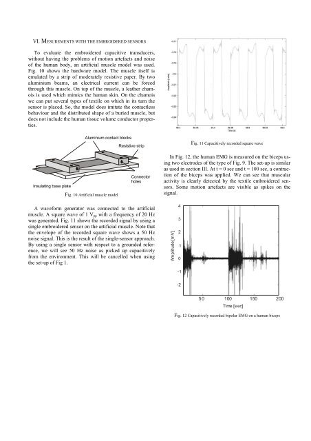
VI. MESUREMENTS WITH THE EMBROIDERED SENSORS<br />
To evaluate the <strong>embroidered</strong> capacitive transducers,<br />
without having the problems of motion artefacts and noise<br />
of the human body, an artificial muscle model was used.<br />
Fig. 10 shows the hardware model. The muscle itself is<br />
emulated by a strip of moderately resistive paper. By two<br />
aluminium beams, an electrical current can be forced<br />
through this muscle. On top of the muscle, a leather chamois<br />
is used which mimics the human skin. On the chamois<br />
we can put several types of <strong>textile</strong> on which in its turn the<br />
sensor is placed. So, the model does imitate the contactless<br />
behaviour and the distributed shape of a buried muscle, but<br />
does not include the human tissue volume conductor properties.<br />
Insulating base plate<br />
Aluminium contact blocks<br />
Fig. 10 Artificial muscle model<br />
Resistive strip<br />
Connector<br />
holes<br />
A waveform generator was connected to the artificial<br />
muscle. A square wave of 1 Vpp with a frequency of 20 Hz<br />
was generated. Fig. 11 shows the recorded signal by using a<br />
single <strong>embroidered</strong> sensor on the artificial muscle. Note that<br />
the envelope of the recorded square wave shows a 50 Hz<br />
noise signal. This is the result of the single-sensor approach.<br />
By using a single sensor with respect to a grounded reference,<br />
we will see 50 Hz noise as picked up capacitively<br />
from the environment. This will be cancelled when using<br />
the set-up of Fig 1.<br />
Fig. 11 Capacitively recorded square wave<br />
In Fig. 12, the human <strong>EMG</strong> is measured on the biceps using<br />
two electrodes of the type of Fig. 9. The set-up is similar<br />
as used in section III. At t = 0 sec and t = 100 sec, a contraction<br />
of the biceps was applied. We can see that muscular<br />
activity is clearly detected by the <strong>textile</strong> <strong>embroidered</strong> <strong>sensors</strong>.<br />
Some motion artefacts are visible as spikes on the<br />
signal.<br />
Amplitude [mV]<br />
4<br />
3<br />
2<br />
1<br />
0<br />
-1<br />
-2<br />
50 100<br />
Time [s ec]<br />
150 200<br />
Fig. 12 Capacitively recorded bipolar <strong>EMG</strong> on a human biceps