coloration - Marine Biological Laboratory
coloration - Marine Biological Laboratory
coloration - Marine Biological Laboratory

You also want an ePaper? Increase the reach of your titles
YUMPU automatically turns print PDFs into web optimized ePapers that Google loves.
REVIEW<br />
Mechanisms and behavioural functions of<br />
structural <strong>coloration</strong> in cephalopods<br />
Lydia M. Mäthger 1,2,3, * ,† , Eric J. Denton 3,‡ , N. Justin Marshall 2<br />
and Roger T. Hanlon 1<br />
1 <strong>Marine</strong> <strong>Biological</strong> <strong>Laboratory</strong>, Woods Hole, MA 02543, USA<br />
2 Sensory Neurobiology Group, School of Biomedical Sciences, University of Queensland,<br />
Brisbane, Queensland 4072, Australia<br />
3 <strong>Marine</strong> <strong>Biological</strong> Association of the UK, Citadel Hill, Plymouth PL1 2PB, UK<br />
Octopus, squid and cuttlefish are renowned for rapid adaptive <strong>coloration</strong> that is used for a wide<br />
range of communication and camouflage. Structural <strong>coloration</strong> plays a key role in augmenting<br />
the skin patterning that is produced largely by neurally controlled pigmented chromatophore<br />
organs. While most iridescence and white scattering is produced by passive reflectance or<br />
diffusion, some iridophores in squid are actively controlled via a unique cholinergic, non-synaptic<br />
neural system. We review the recent anatomical and experimental evidence regarding the<br />
mechanisms of reflection and diffusion of light by the different cell types (iridophores and<br />
leucophores) of various cephalopod species. The structures that are responsible for the optical<br />
effects of some iridophores and leucophores have recently been shown to be proteins. Optical<br />
interactions with the overlying pigmented chromatophores are complex, and the recent<br />
measurements are presented and synthesized. Polarized light reflected from iridophores can be<br />
passed through the chromatophores, thus enabling the use of a discrete communication channel,<br />
because cephalopods are especially sensitive to polarized light. We illustrate how structural<br />
<strong>coloration</strong> contributes to the overall appearance of the cephalopods during intra- and<br />
interspecific behavioural interactions including camouflage.<br />
1. INTRODUCTION<br />
Keywords: iridescence; multilayer reflector; light diffusion; leucophore<br />
Animals can be extraordinarily colourful. There are<br />
innumerable examples in both the vertebrate and<br />
invertebrate worlds, and, while in some animals these<br />
colours function to camouflage their owners, in others<br />
they have clear functions in signalling, and some may<br />
even perform both functions simultaneously.<br />
Colour is produced by either a structural or<br />
pigmentary medium. Structural <strong>coloration</strong> involves<br />
materials that are themselves colourless—the colours<br />
are created by coherent scattering. A pigmented<br />
material has selective absorbance properties that<br />
determine the spectral reflectance of the incident light.<br />
Cephalopods (squid, cuttlefish, octopus) show an<br />
impressive repertoire of body patterns for camouflage<br />
and signalling, despite their apparent colour blindness<br />
*Author and address for correspondence: <strong>Marine</strong> <strong>Biological</strong> <strong>Laboratory</strong>,<br />
Woods Hole, MA 02543, USA (lmathger@mbl.edu).<br />
† Some of the presented material was part of L.M.M.’s postdoctoral<br />
research, Sensory Neurobiology Group, School of Biomedical<br />
Sciences, University of Queensland, Brisbane, Queensland 4072,<br />
Australia, and PhD, <strong>Marine</strong> <strong>Biological</strong> Association of the UK, Citadel<br />
Hill, Plymouth PL1 2PB, UK.<br />
‡ Deceased (2 January 2007).<br />
J. R. Soc. Interface<br />
doi:10.1098/rsif.2008.0366.focus<br />
Published online<br />
(Holmes 1940; Brown & Brown 1958; Packard &<br />
Hochberg 1977; Hanlon 1982; Moynihan 1985; Hanlon<br />
& Messenger 1988, 1996; Marshall & Messenger 1996;<br />
Hanlon & Shashar 2003; Mäthger et al. 2006; but see<br />
examples of cephalopod with colour vision, e.g. Kito<br />
et al. 1992; Michinomae et al. 1994). What is even more<br />
impressive is their ability to almost instantaneously<br />
change colour and pattern. This is mediated by the dual<br />
action of thousands of chromatophores, which are small<br />
pigmented organs (grouped into two or three colour<br />
classes depending on the species: red; yellow/orange;<br />
and brown/black), and structural reflector cells (iridophores<br />
and leucophores) (Williams 1909; Schäfer 1937;<br />
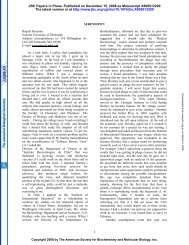
Cloney & Brocco 1983; Messenger 2001; figure 1). The<br />
appearance of the animal thus depends on which skin<br />
elements affect the light incident on the skin. Light may<br />
be reflected by either chromatophores or structural<br />
reflectors, or a combination of both, and it is the<br />
physiological changeability of the chromatophores and<br />
iridophores that enables these animals to produce such<br />
a wide repertoire of optical effects.<br />
A chromatophore consists of a pigment-containing<br />
sac that has dozens of radial muscles attached to its<br />
periphery. These muscles are innervated directly by<br />
Received 28 August 2008<br />
Accepted 19 November 2008 1 This journal is q 2008 The Royal Society
2 Review. Structural <strong>coloration</strong> in cephalopods L. M. Mäthger et al.<br />
(a) (b)<br />
(c)<br />
(e)<br />
ir.<br />
the brain, and by contracting and relaxing the chromatophore<br />
muscles the pigment sac increases or decreases<br />
in area (Florey 1969) in less than a second (Hill &<br />
Solandt 1935). Chromatophore size varies among<br />
cephalopods. In squid, such as Loligo plei, an expanded<br />
chromatophore may be up to 1.5 mm in diameter, while<br />
ch.<br />
leuc.<br />
( f ) (g)<br />
(h) (i)<br />
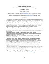
Figure 1. (a) Iridescent spots in the squid Loligo pealeii. (b) Blue–green iridescence and white scattering leucophore stripes in<br />
cuttlefish (Sepia apama). (c) Camouflaged S. apama with pink iridescent arms and white markings caused by leucophores.<br />
(d ) White leucophores in S. apama. (e) Skin in cross section showing the location of chromatophores (ch.) and structural<br />
reflectors (ir., iridophores; leuc., leucophores) in cephalopods. (f ) Close-up of cuttlefish skin (Sepia officinalis) showing<br />
chromatophores (yellow, expanded; dark brown, partially retracted; orange, retracted) and white leucophores. Scale bar, 1 mm.<br />
(g) Brown, red and yellow chromatophores of squid (L. pealeii ). Scale bar, 1 mm. (h) Combination of chromatophores and<br />
iridophores to illustrate the range of colours. Scale bar, 1 mm. (i) Electron micrograph showing iridophore plates (ir.) and<br />
spherical leucophores (leuc.) of cuttlefish (S. officinalis) skin. Scale bar, 1 mm (image courtesy of Alan Kuzirian).<br />
J. R. Soc. Interface<br />
(d)<br />
leuc.<br />
a retracted chromatophore may be barely visible to<br />
the naked eye, measuring as little as a tenth of a<br />
millimetre (Hanlon 1982; Mäthger & Hanlon 2007).<br />
By selectively expanding and retracting distinct<br />
groups of chromatophores, cephalopods can produce<br />
an array of patterns, such as bands, stripes and spots.<br />
ir.
In this review, we deal mainly with the other optical<br />
system that interacts with light—the different types of<br />
structural reflectors. Various cell arrangements have<br />
been identified and described in the cephalopod<br />
literature: (i) those that produce iridescence, a working<br />
definition of which would be the production of rainbowlike<br />
colours or a metallic sheen, and (ii) those that<br />
produce scattering broadband diffuse reflectance, the<br />
most striking of which are the distinct white body<br />
markings of many cephalopod species.<br />
2. IRIDESCENCE<br />
There has been some confusion about the various terms<br />
that have been introduced in the literature to describe<br />
cephalopod skin structures that produce iridescence.<br />
For example, Cloney & Brocco (1983) used the term<br />
‘reflector cell’ (containing plates that are arranged into<br />
distinct groups, called reflectosomes) to describe the<br />
cells that produce iridescence by thin-film interference,<br />
whereas ‘iridocytes’ were described as cells that<br />
function by diffracting light. Iridescent cells whose<br />
optical mechanisms were unclear were termed ‘iridophores’<br />
following these authors’ definition; iridophores<br />
contain a number of iridosomal plates that are grouped<br />
into iridosomes. These plates (along with the spaces<br />
separating them) are ultimately responsible for the<br />
observed iridescence. Since the 1980s, there have been<br />
several studies looking at the ultrastructure, optical<br />
mechanisms and composition of iridescent cells in<br />
cephalopods, some of which have revealed inconsistencies<br />
with the definitions of Cloney & Brocco (1983). For<br />
example, in their paper, the iridescent cells of squid<br />
were described as iridocytes, implying that they<br />
function as diffraction gratings. This has not been<br />
confirmed—on the contrary, all existing papers show<br />
evidence that squid reflectors are multilayer reflectors,<br />
i.e. they reflect light by thin-film interference, which<br />
would warrant the term reflector cell. However, in the<br />
more recent literature, these cells are consistently<br />
referred to as iridophores. This review will be consistent<br />
with these recent publications insofar as iridophores are<br />
defined as the cells that produce iridescence.<br />
In this paper, we deal mainly with multilayer<br />
reflectance, rather than diffraction, because to date,<br />
there is no convincing evidence that diffraction is<br />
responsible for iridescence in cephalopod skin. Diffraction<br />
gratings differ from multilayer reflectors, in that<br />
the structures producing iridescence are oriented in the<br />
same plane as the surface. They are characterized by an<br />
orderly array of ridges that are ‘engraved’ into the<br />
surface. Diffraction gratings produce spectra on the left<br />
and right sides of the zero order (the direction of the<br />
incident light beam) and have been shown to be the<br />
basis of iridescence in some beetles, wasps, ostracod<br />
crustaceans and butterflies (Hinton & Gibbs 1969;<br />
Parker 1995; Brink & Lee 1999). Certainly, considering<br />
the large number of modern cephalopod species (over<br />
700) (Hanlon & Messenger 1996), it may nevertheless<br />
be possible that both diffraction gratings and multilayer<br />
reflectors play a role in creating iridescence, but<br />
evidence thus far suggests that only multilayer reflectors<br />
are involved.<br />
J. R. Soc. Interface<br />
Review. Structural <strong>coloration</strong> in cephalopods L. M. Mäthger et al. 3<br />
2.1. ‘Spectral’ iridescence<br />
Cephalopods have iridophores in many parts of the<br />
body and they have precise arrangements that may<br />
signify specific functions. Squid generally have only<br />
iridophores, i.e. they do not have the broadband<br />
reflecting leucophores found in octopus and cuttlefish<br />
(some species of the squid genus Sepioteuthis may be an<br />
exception; see §3). In most parts of the body (e.g.<br />
mantle and head), the iridophores are located in a<br />
distinct layer beneath the pigmented chromatophores<br />
(Williams 1909; Schäfer 1937; Mirow 1972a,b; Hanlon<br />
1982; Cloney & Brocco 1983). Iridophores are colourless<br />
cells that vary in size but are generally smaller than<br />
1 mm (Mirow 1972b; Cooper et al. 1990). They contain<br />
stacks of thin plates that reflect light by thin-film<br />
interference (Denton & Land 1971; Land 1972;<br />
Mäthger & Denton 2001; Mäthger et al. 2004). Light<br />
reflected from a multilayer reflector is almost always<br />
coloured, as long as two prerequisites are met: (i) there<br />
is a difference in the refractive index between the plates<br />
and the spaces separating them, and (ii) the plates and<br />
spaces have a specific thickness for the constructive<br />
interference of light to occur. The mechanism of<br />
reflectance is the same as that of coloured soap bubbles.<br />
If the soap film (or multilayer plate) is very thin,<br />
shorter wavelengths are reflected, e.g. blue light; if it is<br />
thicker, longer wavelengths, such as yellow and red, are<br />
reflected (Boys 1959; Huxley 1968).<br />
Multilayer reflectors have distinct optical features,<br />
the most obvious of which is the effect of changing the<br />
angle of illumination/observation on the spectrum of<br />
the reflected light. The more oblique the angle, the<br />
shorter the peak wavelengths of the reflected light, i.e. a<br />
multilayer reflector that appears red at near-normal<br />
viewing angles will appear first yellow, then green and<br />
blue at increasingly oblique angles (figure 2a). This may<br />
seem counterintuitive, and, for the interested reader,<br />
we point out that the book by Boys (1959) has a<br />
beautifully written section on this optical phenomenon.<br />
Furthermore, at around Brewster’s angle (angle at<br />
which maximum linear polarization occurs), the<br />
reflected light is highly polarized (figure 2a), an<br />
interesting property that may have some behavioural<br />
function because cephalopods have the ability to detect<br />
polarized light (see below).<br />
Some species of squid (such as Loligo vulgaris, Loligo<br />
forbesii, Loligo pealeii, Alloteuthis subulata, Sepioteuthis<br />
australis, Loliolus noctiluca, Euprymna tasmanica,<br />
Todaropsis eblanae and probably others) appear to<br />
have prominent red-reflecting iridophore stripes<br />
arranged longitudinally on each side of the mantle<br />
(figure 2a,b). These are arranged in either distinct<br />
stripes or ‘splotches’. Mäthger & Denton (2001)<br />
suggested that these iridophores may aid in communication<br />
between individuals of a school. Although the<br />
availability of daylight in the red parts of the spectrum is<br />
much reduced at the depths at which these squid live,<br />
the majority of the reflective plates of these iridophores<br />
are oriented approximately parallel to the skin surface,<br />
which means that shorter wavelengths (green and blue)<br />
are reflected in a horizontal direction. This would make<br />
the stripes highly visible when viewed horizontally,
4 Review. Structural <strong>coloration</strong> in cephalopods L. M. Mäthger et al.<br />
(a)<br />
reflectance (arb. units)<br />
(b)<br />
(c)<br />
reflectance (arb. units)<br />
100<br />
80<br />
60<br />
40<br />
20<br />
0<br />
400 500 600<br />
wavelength (nm)<br />
700 800<br />
(i)<br />
(ii)<br />
(iii)<br />
100<br />
80<br />
60<br />
40<br />
20<br />
45°<br />
40°<br />
±30°<br />
±30°<br />
10°<br />
±30°<br />
on<br />
off<br />
0<br />
400 500 600 700 800 900 1000<br />
wavelength (nm)<br />
Figure 2. (a) Spectral reflectance of iridophores (L. pealeii ) at<br />
different angles of incidence and planes of polarization,<br />
showing that with increasing angle of incidence (i.e. 408 and<br />
458) the reflected light shifts towards the shorter end of the<br />
spectrum and becomes polarized. Two reflectance spectra are<br />
shown for each angle of incidence: the spectrum reflected in<br />
the plane parallel to the plane of incidence and the<br />
perpendicular plane of incidence. At oblique angles (408 and<br />
458), the spectral reflectance in the perpendicular plane is<br />
much reduced in comparison with the parallel plane,<br />
indicating that the reflected light is linearly polarized.<br />
(b) The visibility of the ‘red’ stripe of squid from different<br />
orientations taking into account the light distribution in the<br />
J. R. Soc. Interface<br />
because green and blue are the most prominent<br />
wavelengths found in the light environments these<br />
animals inhabit (such as the example given in<br />
figure 2b, calculated for a depth of 19 m; Mäthger &<br />
Denton 2001). Furthermore, the patterns change<br />
dramatically with direction and movement of the<br />
squid, so that they may be used by squid to coordinate<br />
the movements of individuals of a school.<br />
Squid are able to change their iridescence depending<br />
on behavioural context, showing iridescence especially<br />
during agonistic encounters (Hanlon 1982). In vitro,<br />
iridescence is changed by the topical application of<br />
acetylcholine (ACh) acting on muscarinic cholinergic<br />
receptors (Hanlon 1982; Cooper & Hanlon 1986;<br />
Cooper et al. 1990; Hanlon et al. 1990; Mäthger et al.<br />
2004). However, in contrast to the chromatophores that<br />
can change within a fraction of a second, iridophore<br />
reflectance changes take longer, e.g. several seconds to<br />
minutes. In L. pealeii, the reflected wavelengths have<br />
been shown to shift by over 100 nm (Mäthger & Hanlon<br />
2007), from non-reflective to red and orange after the<br />
application of ACh. The reflectance changes in vitro<br />
typically take several seconds (up to 1–2 min). Interestingly,<br />
in L. pealeii, the collar region (anterior end) of<br />
the mantle has highly reflective iridophores in a<br />
tightly packed arrangement, and this group has been<br />
shown to reflect in the IR parts of the spectrum<br />
(approx. 800 nm) when they appear non-reflective to<br />
the human eye (figure 2c).<br />
To date, it is not known exactly how this wavelength<br />
change is achieved. Recent evidence has provided<br />
confirmation that the plates of squid iridophores are<br />
made up of proteins called reflectins (Crookes et al.<br />
2004); see also Cooper et al. (1990), who pointed out<br />
that a protein state change (affecting refractive index)<br />
combined with a change in the thickness of plates could<br />
explain the observed changes in reflectance.<br />
The squid reflectin protein has received interest from<br />
researchers in the fields of materials science and optical<br />
nanotechnology. For example, Kramer et al. (2007)<br />
showed that reflectin proteins have self-assembling<br />
properties and that they can be processed into thin<br />
films, photonic grating structures and fibres that could<br />
find various applications in society.<br />
Another way of changing iridescence is by selectively<br />
expanding and retracting the overlying chromatophores.<br />
Since chromatophores are innervated directly<br />
from the brain, this effect can be immediate. Chromatophores<br />
are generally located in a distinct layer above<br />
the iridophores and, by selective expansion, they can<br />
either change the reflected spectrum of the iridescence<br />
sea. (i) An observer looking down on a squid will not see<br />
any iridescence. (ii) An observer looking down at a squid<br />
from a 458 angle will see iridescence from the most anterior<br />
and posterior ends of the stripe. (iii) An observer looking<br />
directly from the side will see strong iridescence from the<br />
entire stripe. See Mäthger & Denton (2001) for more details<br />
(modified from Mäthger & Denton 2001). (c) Acetylcholine<br />
(1 mM) changes iridescence from non-reflective (black lines,<br />
reflectance in IR) through various IR steps (black and grey<br />
lines) to red reflective in the squid L. pealeii. Measurements<br />
taken at 15 s intervals.
(e) 100<br />
(f )<br />
reflectance<br />
(arb. units)<br />
75<br />
50<br />
25<br />
0<br />
(g) 10<br />
(h)<br />
reflectance<br />
(%)<br />
5<br />
(a) (b)<br />
(c) (d)<br />
green<br />
iridescence<br />
yellow chromatophore<br />
over green iridescence<br />
0<br />
400 450 500 550 600 650 700<br />
wavelength (nm)<br />
(figure 3a–e; see also Mäthger & Hanlon 2007), create<br />
contrast against which iridescence is viewed (see, for<br />
example, the blue iridescent rings of the blue-ringed<br />
octopus; figure 4b) or block it altogether, such as a<br />
brown chromatophore expanding over reflective iridophores<br />
(figure 3f ).<br />
Some of the aforementioned squid (L. vulgaris,<br />
L. pealeii, A. subulata and L. noctiluca) also have a<br />
blue reflective stripe along the side of the mantle (see<br />
Mäthger & Denton 2001). These iridophores are best<br />
seen when observed and illuminated from above the<br />
animal (rather than from a horizontal direction where<br />
the ‘red’ stripes are visible), and we speculate that they<br />
may function in ways similar to the red stripes<br />
described above: since the reflectance pattern and<br />
red<br />
iridescence<br />
brown chromatophore<br />
over red iridescence<br />
400 450 500 550 600 650 700<br />
wavelength (nm)<br />
Figure 3. (a–d ) Close-up images of L. pealeii skin showing chromatophores and iridophores. Chromatophores can be used to<br />
modulate iridescence. Light reflected from iridophores filtered through (e) a yellow and (f ) a brown chromatophore. Reflectance<br />
spectra of (g) yellow and (h) brown chromatophore.<br />
J. R. Soc. Interface<br />
Review. Structural <strong>coloration</strong> in cephalopods L. M. Mäthger et al. 5<br />
intensity change with the movements of the animal,<br />
other members of a school may be able to use this<br />
information to coordinate their movements.<br />
In all pelagic squid studied so far (namely L. vulgaris,<br />
L. pealeii and L. noctiluca), the ventral iridophores<br />
reflect red light, when viewed and illuminated from the<br />
side (see Mäthger & Denton 2001). The iridophores<br />
making up the ventral side are densely packed and their<br />
flat surfaces make an angle of approximately 608 with<br />
the skin surface. Owing to this orientation, when the<br />
iridophores are viewed at oblique angles of incidence<br />
(such as from below), the light reflected from the<br />
iridophores comes from the inside of the mantle (see<br />
fig. 16 in Mäthger & Denton 2001). This may have an<br />
interesting function. Midwater animals are most visible
6 Review. Structural <strong>coloration</strong> in cephalopods L. M. Mäthger et al.<br />
(a)<br />
(b)<br />
(c)<br />
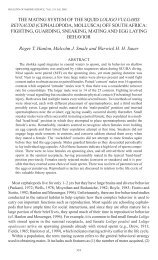
Figure 4. (a) Blue-ringed octopus, Hapalochlaena lunulata,<br />
well camouflaged in a laboratory tank. Note the muted<br />
iridescent blue rings. (b) Bright blue iridescence is typically<br />
seen when a blue-ringed octopus flashes its rings. (c) Electron<br />
micrograph of the blue rings, showing closely packed<br />
iridophore plates (scale bar, 1 mm).<br />
when seen from below against the background of the<br />
strongest radiance in the sea. Any downwelling light<br />
that is absorbed by the animal cannot be replaced by<br />
the reflections of light from any other direction<br />
(bioluminescence is the only solution to this problem).<br />
Being transparent makes a midwater animal less likely<br />
to be detected by the predators lurking below, and in<br />
comparison with other muscular animals, such as fishes,<br />
cephalopods can be very transparent. However, total<br />
transparency is impossible because the internal organs<br />
(e.g. ink sac, digestive tracts) cannot be made<br />
transparent. The dorsal mantle iridophores transmit a<br />
great portion of the incoming light into the mantle<br />
cavity. The light that enters the mantle cavity then<br />
J. R. Soc. Interface<br />
falls on the ventral iridophores obliquely and is<br />
channelled downwards. This may help mitigate shadows<br />
cast to an observer below the animal (Mäthger &<br />
Denton 2001).<br />
Benthic cephalopods, such as Sepia officinalis and<br />
E. tasmanica, also have densely packed iridophores<br />
lining their ventral mantle, but instead of reflecting red<br />
light as the above squid species, they strongly reflect<br />
green light when viewed and illuminated side-on. In<br />
E. tasmanica, for example, the iridophores lie at an<br />
angle of approximately 508 with the skin surface. Sepia<br />
and Euprymna are much more opaque than most<br />
pelagic cephalopods, and it appears unlikely that any<br />
considerable amount of light enters the mantle cavity to<br />
be channelled downwards in the manner described<br />
above, so their function is unknown.<br />
The blue-ringed octopus is well known for its<br />
potentially deadly bites and flashing blue rings. It was<br />
previously suggested that this blue iridescence may be<br />
the result of the Tyndall scattering (Herring 1994).<br />
However, we found that ultrastructural analysis of the<br />
rings and lines of Hapalochlaena fasciata (the blue-lined<br />
octopus) reveals densely packed reflective plates (up to<br />
approx. 30 in one iridophore) grouped together in a<br />
parallel arrangement, suggesting that the blue iridescence<br />
is caused by multilayer constructive interference<br />
(figure 4), which would warrant the term reflector cell<br />
following Cloney & Brocco’s (1983) definition<br />
(however, see above for the reasoning behind calling<br />
iridescent cells iridophores). Spectrometer measurements<br />
have confirmed this (data not shown). Each<br />
iridophore appears to be oriented at a different angle<br />
relative to the surface of the skin, and this correlates<br />
well with the optical appearance of the rings: the blue<br />
iridescence is visible from a wide range of angles. The<br />
reflective plates have thicknesses of approximately<br />
70 nm. This appears to be the thickness required for<br />
this stack to act as an ideal quarter-wave reflector for<br />
which lmaxZ4nd, where n is the refractive index and d<br />
is the actual thickness of the plate. Assuming a<br />
refractive index of 1.59 (Kramer et al. 2007), the<br />
wavelength of maximum reflectance of the blue-ringed<br />
iridophores would be at 445 nm.<br />
Tyndall scattering may nevertheless be a method of<br />
producing the blue iridescence in some cephalopod<br />
species. For example, Octopus bimaculatus has two<br />
characteristic blue rings (often referred to as ‘ocelli’) on<br />
each side of its head, near the eyes. This has been<br />
attributed to the Tyndall scattering from fine purine<br />
granules (Fox & Vevers 1960; Fox 1976; Packard &<br />
Hochberg 1977). There may also be other squid species<br />
in which the Tyndall scattering produces the blue<br />
iridescence. Herring (1994) suggested that the blue<br />
flashes of the squid Onychia caribbaea are produced by<br />
the expansion of dark chromatophores beneath a bluereflecting<br />
Tyndall scatterer.<br />
2.2. Iridophore polarization and optical<br />
enhancement of chromatophores<br />
For human observers, creating colour is probably the<br />
most notable aspect of cephalopod iridophores for<br />
our eyes. However, when viewed at oblique angles,
cephalopod iridophores also polarize light (Shashar &<br />
Hanlon 1997; Mäthger 2001; Shashar et al. 2001;<br />
Mäthger & Hanlon 2006; Chiou et al. 2007). Light is<br />
polarized most strongly at Brewster’s angle (q), which<br />
is simply defined as follows: tan qZn1/n2, where n1<br />
and n2 are the refractive indices of the two media (e.g.<br />
going from a watery surrounding, nZ1.33, to protein,<br />
nZ1.59, Brewster’s angle is approx. 508).<br />
For cephalopods, this may have a useful behavioural<br />
function. Cephalopods have a rhabdomeric visual<br />
system that allows them to detect linearly polarized<br />
light (Moody & Parriss 1960; Shashar & Cronin 1996;<br />
Shashar et al. 1998, 2002), and it is therefore<br />
conceivable that the polarized aspect of iridescence<br />
may have communication functions (Cronin et al. 2003;<br />
Boal et al. 2004). This has also been termed a ‘hidden<br />
communication channel’ because cephalopod predators,<br />
such as teleost fish, sharks and marine mammals,<br />
are believed not to be polarization sensitive and would<br />
therefore not be able to perceive this sort of visual<br />
information (Shashar et al. 1996; Land & Nilsson 2002;<br />
Boal et al. 2004; Mäthger & Hanlon 2006).<br />
In cephalopod skin, polarization is a ‘side product’ of<br />
iridescence, and iridescence can be very conspicuous.<br />
This is where having a variety of optical structures in<br />
the skin is beneficial. In a study on L. pealeii, it was<br />
shown that the polarized aspect of iridescence is<br />
maintained after passing through the overlying pigmented<br />
chromatophores, which produce the most<br />
visible and dynamically changeable aspect of camouflage<br />
patterns in cephalopods (Mäthger & Hanlon 2006,<br />
2007). Cephalopods are polarization sensitive and can<br />
regulate polarization via skin iridescence (Shashar &<br />
Hanlon 1997; Mäthger & Denton 2001; Chiou et al.<br />
2007). Two behavioural functions come to mind. First,<br />
it is conceivable that they could send polarized signals<br />
to conspecifics while using chromatophores to regulate<br />
the potentially conspicuous iridescence and stay<br />
camouflaged to fishes or mammalian predators, most<br />
of which are probably not polarization sensitive<br />
(Land & Nilsson 2002). Second, they could use<br />
polarization signals during intense intraspecific<br />
interactions (sexual selection); Boal et al. (2004)<br />
found that females showed more polarization signals<br />
than males and also changed their behaviour in<br />
response to polarized patterns. Boal et al.’s results<br />
suggest (but did not prove) that females may use<br />
polarized signals as a source of information about<br />
conspecifics (e.g. species, sex, location, size).<br />
Iridophores are generally located just beneath the<br />
pigmented chromatophores (there are some exceptions<br />
to this) and the final appearance of the animal is<br />
frequently the result of the optical interactions between<br />
iridophores and chromatophores. Chromatophores are<br />
very thin when expanded and can filter light reflected<br />
from the iridophores; or, turning this idea around,<br />
iridophores can enhance the chromatophores’ appearance.<br />
Mäthger & Hanlon (2007) showed evidence that<br />
three colour classes of pigments (yellow, red and brown),<br />
combined with a single type of reflective cell (with<br />
dynamic iridescence), produce colours that encompass<br />
the whole of the visible spectrum, enabling the extreme<br />
visual diversity for dynamic camouflage and signalling.<br />
J. R. Soc. Interface<br />
Review. Structural <strong>coloration</strong> in cephalopods L. M. Mäthger et al. 7<br />
3. BROADBAND REFLECTANCE AND<br />
SCATTERING<br />
3.1. Broadband ‘silvery’ reflectors<br />
Cephalopods have very striking silvery iridescence<br />
around their eyes (figure 5a,b). This was studied in<br />
detail for A. subulata and L. vulgaris and it was shown<br />
that, unlike the iridophores described above, the<br />
reflected wavelengths do not change with angle of<br />
incidence/view (Mäthger 2001) and the reflected light<br />
is not polarized at oblique angles (figure 5c). When<br />
viewed closely, the skin shows some variations in colour<br />
on a scale of a few micrometres (figure 5b). Some parts<br />
reflect red, others green and blue and the reflective<br />
properties of each of these microscopic parts are typical<br />
of multilayer reflectors: with increasing angle of<br />
incidence, the reflected wavelengths move towards the<br />
shorter end of the spectrum and become polarized.<br />
When viewed from a distance, however, the various<br />
coloured parts merge together and the net effect is the<br />
reflectance of unpolarized white light. Ultrastructurally,<br />
we can see that the stacking sequence of the plates<br />
and spaces is responsible for the silvery appearance<br />
(figure 5d ). The plates are long (more than 10 mm) and<br />
wavy with varying thicknesses (ranging from 80 to<br />
130 nm) and irregular spaces between them, which<br />
facilitates this broadband reflectance. This natural<br />
pointillistic approach to creating colour can also be seen<br />
in Ribbonfish (Trichiurus spp.), whose silvery surface<br />
is covered with fine streaks that show a variety of<br />
colours when examined microscopically ( Nicol &<br />
Van Baalen 1968).<br />
The orientation of the reflectors relative to the<br />
surface of the skin offers a clue to the possible function<br />
of this iridescence (figure 5e). These measurements<br />
were obtained following the methods described<br />
originally by Denton & Nicol (1962). A piece of silver<br />
foil is placed adjacent to the reflective area under<br />
investigation, and, using a goniometer, the difference<br />
in angle between the light reflected from the foil<br />
(which equals the skin surface) and the reflector<br />
(which may or may not be angled with respect to the<br />
skin surface) is measured. These measurements show<br />
that the reflective plates are tilted towards the true<br />
vertical. Figure 5e shows that the reflectors in the<br />
dorsal and ventral areas are oriented at an angle with<br />
respect to the skin surface, while those in the anterior<br />
and posterior areas lie parallel. This arrangement<br />
would give close to perfect camouflage, much like that<br />
in silvery fish: the reflectors reflect light as a mirror<br />
suspended vertically in the sea. The orientation of the<br />
reflective plates is such that the light reflected from<br />
almost any angle equals the intensity of the background<br />
light against which the animal is viewed<br />
(Denton & Nicol 1962, 1965, 1966; Denton 1970;<br />
Denton & Land 1971; Denton et al. 1972). The<br />
organization of the plates furthermore minimizes<br />
the polarization of the reflected light, which reduces<br />
the squid’s chances of being detected by the predators<br />
that are polarization sensitive.<br />
Cephalopods have pupils that respond to the<br />
changes in light levels as well as under specific<br />
behavioural contexts. In squid (L. vulgaris, L. pealeii,
8 Review. Structural <strong>coloration</strong> in cephalopods L. M. Mäthger et al.<br />
(c)<br />
reflectance<br />
( f )<br />
1<br />
(a)<br />
0<br />
400 450 500 550 600 650 700 750<br />
wavelength (nm)<br />
(d)<br />
1 µm<br />
(e)<br />
lens<br />
dorsal<br />
ventral<br />
dorsal<br />
pupil<br />
ventral<br />
ch.<br />
anterior<br />
and<br />
posterior<br />
parts<br />
ir.<br />
1 µm<br />
Figure 5. (a) Silvery iridescence around the eyes of squid<br />
(L. pealeii ). (b) Close-up of the section highlighted in<br />
(a) showing variations in spectral reflectance. (c) Spectral<br />
reflectance on both planes of polarization at 458 incidence<br />
(black and grey lines), showing that the light reflected from<br />
silvery reflectors is not polarized. (d ) Electron micrograph<br />
of silvery reflectors in cross section, showing wavy arrangement<br />
of reflective plates. (e) Orientation of the reflective plates of<br />
silvery eyes obtained using the techniques described in the text.<br />
The reflective plates (short, thick black lines) are tilted towards<br />
J. R. Soc. Interface<br />
(g)<br />
(b)<br />
A. subulata), these pupils are lined with silvery<br />
iridophores that are tilted towards the horizontal.<br />
The iridophores are weakly reflective when the pupil<br />
covers the eye but reflect strongly when the pupil is<br />
open, for example during low light levels or aggressive<br />
encounters. In such situations, the iridophores may<br />
function to prevent strong downwelling light from<br />
entering the eye. The pupil of Octopus vulgaris is also<br />
lined with silvery iridophores (Froesch & Messenger<br />
1978), although the plates are much shorter (less than<br />
3 mm in length) and do not have the wavy structure as<br />
those of squid.<br />
Longley (1917) found that the internal organs of<br />
some transparent fishes (e.g. Coralliozetus cardonae)<br />
are camouflaged by the principle of countershading.<br />
This is also true of the ink sac of cephalopods: the dorsal<br />
area is black; the sides are silvery; and the ventral area<br />
is white (Denton 1970; Denton & Land 1971; Mäthger<br />
2001). In A. subulata, the silvery iridophores of the ink<br />
sac reflect light in a way similar to that of the<br />
iridophores around the eyes and their structural<br />
arrangement is also similar, with plates ranging in<br />
thickness from 85 to 120 nm with irregular spaces<br />
between the wavy plates (Mäthger 2001).<br />
At least some cephalopods from the families<br />
Ommastrephidae, Sepiolidae and Pyroteuthidae (e.g.<br />
T. eblanae, Sthenoteuthis pteropus, Ommastrephes<br />
bartrami, Heteroteuthis dispar and Pterygioteuthis<br />
microlampas) have a very striking silvery golden stripe<br />
that runs along each side of the mantle and head. As is<br />
common in silvery fish, the iridophores of this stripe are<br />
tilted towards the vertical (figure 5f ), suggesting an<br />
obvious role in camouflage. The iridophores of this<br />
stripe are very densely packed and, unlike other<br />
iridophores, no single iridophore cell can be distinguished<br />
by eye. Figure 5g shows that the plates are the<br />
thickest (approx. 140 nm) nearer the skin surface and<br />
become thinner farther from the skin surface (to as thin<br />
as 70 nm) (Mäthger 2001).<br />
3.2. Broadband reflectance from<br />
photophore reflectors<br />
In habitats with ample supply of daylight, such as the<br />
coastal areas and the upper layers of the sea, camouflage<br />
and communication signals can be created by reflecting<br />
light. However, at night and at greater depths to which<br />
daylight does not penetrate, this is nearly impossible,<br />
and, instead, many animals, such as crustaceans,<br />
cephalopods and fishes, use luminescence for camouflage<br />
and communication, either through intracellular<br />
mechanisms, extracellular excretions or symbiosis with<br />
bioluminescent bacteria (e.g. Nicol 1960; Herring 1988,<br />
2000). Bioluminescence is particularly widespread in the<br />
the vertical, much like the reflectors on the scales of the silvery<br />
fish. (f ) Orientation of the reflective plates of golden silvery<br />
stripe along the lateral side of the oceanic squid Todaropsis.<br />
Reflective plates are also oriented towards the vertical.<br />
(g) Electron micrograph of Todaropsis golden silvery stripe,<br />
showing a chromatophore (chr.) and iridophore plates (ir.).
marine environment, but it is by no means restricted to<br />
it. Some terrestrial organisms, such as some fungi,<br />
bacteria and insects emit light, although their luminescent<br />
structures are generally quite simple, lacking the<br />
additional optical components (such as reflectors, lenses<br />
and filters) that control the optical characteristics of the<br />
more complex light-emitting photophores in some<br />
cephalopods and fishes (Harvey 1952; Lloyd 1975,<br />
1978; Lall et al. 1980; Herring 1988, 2000; Wilson &<br />
Hastings 1998). However, particularly in insects, luminescence<br />
behaviour may be very elaborate: glow-worms<br />
and fireflies are known to point their light organs in<br />
specific directions to attract mates or lure prey. This<br />
makes up for the lack of directionality in their light<br />
organs. At the same time, male/female signalling<br />
dialogues may be extremely complex (Lloyd 1975, 1978).<br />
Cephalopods and fishes have many photophores that<br />
can be highly directional and have very complex<br />
structures (e.g. Chun 1910; Nicol 1960; Robison &<br />
Young 1981, Herring 1988, 2000; Young & Bennett<br />
1988; Johnsen et al. 1999a,b). Although there is an<br />
impressive anatomical variety in cephalopod photophores,<br />
especially with respect to the occurrence and<br />
position of lenses, filters and reflectors, the common<br />
theme among cephalopods is a thick layer of reflective<br />
plates at the base of the photophore. Acting together, in<br />
ventral photophores, these optical structures help to<br />
channel and direct light downwards for effective<br />
countershading (e.g. when viewed from below against<br />
the more brightly lit downwelling light; see Arnold et al.<br />
1974; Herring et al. 1981; Herring 1985, 1988, 2000;<br />
McFall-Ngai & Montgomery 1990). Cephalopod photophore<br />
reflectors differ in their composition and appearance<br />
from diffuse to highly specular. In the midwater<br />
squid Abralia and Watasenia, both of which have<br />
several different types of photophore, the simplest ones<br />
have only a basal concave reflector, formed of a highly<br />
organized multilayer of collagen rods that reflect<br />
specific wavelengths (Young & Arnold 1982; Young &<br />
Bennett 1988; see also reflectin protein in Euprymna<br />
photophores, Crookes et al. 2004). Collagen fibres also<br />
provide the main reflector in the photophores of<br />
Stauroteuthis syrtensis, Vampyroteuthis infernalis and<br />
Spirula spirula, as well as being primarily responsible<br />
for the silvery appearance of the main body wall of the<br />
last species (Herring et al. 1981, 1994; Johnsen et al.<br />
1999b). Some of the more complex photophores (lensed<br />
and filtered) have additional areas with reflective<br />
tissue, such as light guides and filters (Herring 1988;<br />
Young & Bennett 1988). Photophores often have a<br />
pearly iridescent appearance that can result from<br />
irregularity in thickness and organization of reflective<br />
plates, similar to the silvery reflectors around the eyes<br />
of cephalopods (described above). In the shallow-water<br />
Hawaian bobtail squid Euprymna, for example, the<br />
reflective plates appear to be of various thicknesses<br />
(average 100 nm) and orientations (McFall-Ngai &<br />
Montgomery 1990). Some mesopelagic squid, such as<br />
Liocranchia, have highly wavy reflective plates; in<br />
Sandalops and Megalocranchia, the reflective plates are<br />
highly organized into stacks but the overall orientation<br />
differs between stacks (Herring et al. 2002). Both<br />
arrangements could be the cause of the photophore’s<br />
J. R. Soc. Interface<br />
Review. Structural <strong>coloration</strong> in cephalopods L. M. Mäthger et al. 9<br />
‘pearly’ appearance. These basal reflectors reflect light<br />
back through the photophore for more efficient light<br />
emission. Reflectors can also play a role in tuning the<br />
wavelength of the emitted light. In the squid Pterygioteuthis,<br />
the reflective plates are spaced and oriented so<br />
that they reflect only blue light vertically downwards<br />
(Arnold et al. 1974). The spectral characteristics of<br />
light production are determined by the luminescence<br />
chemistry, but the photophore emission can be<br />
modified by spectrally selective filters and reflectors<br />
that alter the bandwidth. Interestingly, some squid are<br />
able to regulate their bioluminescence emission spectra<br />
to match the spectral composition of downwelling light.<br />
Young & Mencher (1980) showed that, in the squid<br />
Abraliopsis and Abralia, the changes in water temperature<br />
experienced during diel vertical migration alter the<br />
spectral emission of light from their photophores,<br />
resulting in a match with the downwelling light<br />
spectrum, which is different during day and night.<br />
This may be achieved in the following way: the<br />
photophore iridophores are surrounded by muscle<br />
tissue, and the tensional forces of the muscles could<br />
affect the spacing of the plates, which in turn could<br />
affect the spectral composition of the emitted light<br />
(Arnold et al. 1974; Young & Arnold 1982). There<br />
appears to be no evidence to suggest that the<br />
photophore iridophores are physiologically active<br />
as the iridophores in the mantle skin of many<br />
squid (see above), but to our knowledge this has not<br />
been tested.<br />
3.3. Diffuse white scattering<br />
The body patterns that cephalopods use for camouflage<br />
and communication show a wide variety of contrast and<br />
brightness levels. In addition to iridophores, cuttlefish<br />
and octopus have another structural reflector type that<br />
creates bright white patterns, shapes and spots. These<br />
reflectors are called leucophores (‘white cells’) and they<br />
are made up of spherical assemblages called leucosomes<br />
(Froesch & Messenger 1978; Cloney & Brocco 1983).<br />
Leucophores reflect the ambient wavelengths of light<br />
(i.e. they look white in white light, red in red light, blue<br />
in blue light, etc.) and are found in high density in<br />
specific skin areas (zebra stripes, fin spots, fin line and<br />
various other skin components) that can be bright<br />
white depending on the body patterning that is shown<br />
(figure 6a). Squid generally do not have leucophores<br />
(Sepioteuthis lessoniana and Sepioteuthis sepioidea<br />
may be exceptions).<br />
Mäthger et al. (submitted) show that the leucophores<br />
of cuttlefish, S. officinalis, have the optical<br />
properties of a perfect diffuser, appearing equally bright<br />
from all angles of view and that they reflect light at<br />
consistently high levels (up to 70% at wavelengths from<br />
300 to 900 nm). The light-scattering elements of<br />
leucophores are spheres (leucosomes) that range in<br />
size from 250 to 1250 nm in diameter (figure 6b) and are<br />
composed of mucoproteins—highly sulphated and<br />
weakly acidic mucopolysaccharides (they stain negatively<br />
with antibodies to reflectin, which is the lightreflecting<br />
protein in squid skin).
10 Review. Structural <strong>coloration</strong> in cephalopods L. M. Mäthger et al.<br />
4. CONCLUSIONS<br />
(a) (b)<br />
(c) (d)<br />
(e) ( f )<br />
In cephalopods, colour change is mediated by two types of<br />
skin components: pigmented chromatophores and structurally<br />
reflecting cells (iridophores and leucophores).<br />
Iridophores are the cells that are made up of stacks of thin<br />
protein plates that function as multilayer reflectors,<br />
whereas leucophores contain spherical protein assemblages<br />
that scatter light equally well throughout the<br />
visible, IR and UV parts of the spectrum.<br />
In this paper, we have shown that iridescence in<br />
cephalopods spans the whole of the visible spectrum,<br />
including the near-IR. <strong>Biological</strong>ly, IR reflectance most<br />
likely plays little or no role in cephalopod behaviour<br />
because IR is absorbed rapidly by sea water. Furthermore,<br />
most cephalopods studied thus far appear to be<br />
colour blind, having only one visual pigment, with the<br />
absorbance peak generally in the blue–green part of the<br />
spectrum (Brown & Brown 1958; Bellingham et al.<br />
1998; but see Kito et al. 1992; Michinomae et al. 1994).<br />
Therefore, reflectance in the IR is most likely irrelevant<br />
for vision in these animals. Interestingly, in the<br />
cephalopod species discussed in this paper, there has<br />
so far been no evidence for iridescence in the UV parts<br />
of the spectrum, but considering the diversity of<br />
0035 15 KV 10 µm WD33<br />
Figure 6. Whiteness in cuttlefish is created by scattering of light from leucophores. (a) Disruptive pattern, (b) zebra pattern,<br />
(c) close-up of white square, (d ) close-up of zebra pattern, (e) close-up of white finspot, created by leucophores. Note the<br />
chromatophores in the superficial layer; chromatophores can modulate whiteness. (f ) Scanning electron micrograph of<br />
leucophores showing spherical arrangement of leucosomes (courtesy of Alan Kuzirian).<br />
J. R. Soc. Interface<br />
cephalopod skin <strong>coloration</strong>, and the fact that some<br />
fish predators can see UV, this possibility should not be<br />
ruled out.<br />
Leucophores create the white skin areas that are part<br />
of various camouflage and signalling body patterns of<br />
cuttlefish and octopus. They provide a backdrop against<br />
which chromatophores (and iridophores) can create<br />
highly contrasting patterns, such as those typical of the<br />
light skin components of disruptive <strong>coloration</strong> in cuttlefish<br />
(Hanlon & Messenger 1988; Chiao & Hanlon 2001;<br />
Barbosa et al. 2007; Kelman et al. 2007; figure 6a).<br />
Leucophores are not physiologically active (as some<br />
iridophores are), they do not polarize light and they look<br />
equally bright from all angles of view. Leucophores reflect<br />
the ambient wavelengths of light (they look red in red<br />
light, blue in blue light, white in white, etc.), which may<br />
aid both wavelength and intensity matching at least at a<br />
localized level in the skin (Froesch & Messenger 1978).<br />
This may be particularly useful in the habitats in which<br />
shorter (blue and green) wavelengths predominate,<br />
primarily those of greater depths (Jerlov 1976).<br />
Two clear functions of structural <strong>coloration</strong> in<br />
cephalopods emerge: (i) camouflage and (ii) signalling/<br />
communication.
4.1. Camouflage<br />
Many iridophores are oriented in precise ways to<br />
facilitate reflectance in specific directions. Iridophores<br />
provide a range of wavelengths that complement the<br />
yellow, red and brown pigments in the chromatophores,<br />
so that camouflage can encompass the entire visible<br />
spectrum. Additionally, some squid are able to regulate<br />
the intensity of light reflected from iridophores, and<br />
matching the intensities of the ambient light field is a key<br />
step towards achieving effective camouflage. Furthermore,<br />
the silvery iridescence that is found around the<br />
eyes, the ink sac and the sides of the mantle suggests a<br />
role in camouflage by acting as vertically oriented<br />
mirrors. The reflective plates are tilted towards the<br />
vertical and they maximally reflect the incident light,<br />
much in the same way as silvery fish (e.g. Denton & Nicol<br />
1962; Denton 1970; Denton & Land 1971).<br />
Leucophores also play a role in camouflage: they<br />
provide light areas that may facilitate both background<br />
matching (by resembling specific light objects in the<br />
background) and disruptive <strong>coloration</strong> (by visually<br />
breaking the body into distinct objects of highcontrasting<br />
patches) (Cott 1940; Hanlon & Messenger<br />
1988; Hanlon et al. in press).<br />
4.2. Signalling/communication<br />
The optical properties of the structural reflectors<br />
provide cephalopods with an excellent means of<br />
communication. Optical signals, both intra- and<br />
interspecific, can vary dramatically from the zebra<br />
stripes of cuttlefish (the light stripes are created by<br />
leucophores; figure 6b) to the flashing blue iridescent<br />
rings of the blue-ringed octopus (figure 4). Some<br />
iridescent areas are strongly polarized at certain angles<br />
(e.g. Shashar & Hanlon 1997; Mäthger & Hanlon 2006;<br />
Chiou et al. 2007) and, since cephalopods have the<br />
ability to detect polarized light, this may have<br />
behavioural functions (e.g. Cronin et al. 2003; Boal<br />
et al. 2004). It has been shown that cuttlefish take<br />
advantage of their polarization vision when hunting for<br />
silvery fish whose scales polarize light (Shashar et al.<br />
2000), so that it is conceivable that polarization may be<br />
used in various signalling aspects of cephalopod<br />
behaviour (Boal et al. 2004).<br />
Squid are commonly found in large schools. The<br />
communication of the movements of individuals in a<br />
school is crucial for maintaining the integrity of a school<br />
and iridescence may play such a role. The various<br />
iridescent stripes and splotches in squid (e.g. Hanlon<br />
1982; Hanlon et al. 1999; Mäthger & Denton 2001) have<br />
characteristic optical features that differ depending on<br />
where the onlooker is positioned, so that it is possible<br />
that the squid may use their iridescent markings to<br />
communicate changes in swimming direction, etc.<br />
(Mäthger & Denton 2001).<br />
Thus far, there is no optical evidence that cephalopod<br />
iridescence may be caused by diffraction gratings.<br />
In a short communication, Hanlon et al. (1983), showed<br />
that the iridophore plates of some squid are oriented<br />
‘edge on’, i.e. with their short ends facing the<br />
skin surface, in some ways resembling the face of<br />
J. R. Soc. Interface<br />
Review. Structural <strong>coloration</strong> in cephalopods L. M. Mäthger et al. 11<br />
a diffraction grating. By contrast, Mäthger & Denton<br />
(2001) subsequently showed that iridophores with<br />
reflective plates of this orientation, as commonly<br />
observed in the iridophores of the lateral and ventral<br />
mantle skin, nevertheless act as multilayer reflectors.<br />
However, considering how few species have been<br />
studied in detail, compared with the diversity of the<br />
class Cephalopoda, it should not be ruled out that<br />
diffraction gratings may be a mechanism of iridescence<br />
in some cephalopod species.<br />
Iridescence is widespread in the animal kingdom and<br />
its function is not always associated with signalling or<br />
camouflage (e.g. mother of pearl, in which the<br />
iridescent layer on the interior of the mollusc shell<br />
provides structural strength; Barthelat et al. 2007) but<br />
for many species it is. Some of the most exotic iridescent<br />
patterns can be seen in a great number of butterfly<br />
species (e.g. Ghiradella 1991; Vukusic et al. 1999, 2000;<br />
Sweeney et al. 2003; Vukusic & Sambles 2003; Stavenga<br />
et al. 2004) and the plumage and skin of many birds<br />
(Prum et al. 1998; Vorobyev et al. 1998; Cuthill et al.<br />
1999; Osorio & Ham 2002; Prum & Torres 2003). The<br />
feathers of the male peacocks and birds of paradise<br />
(Frith & Beehler 1998; Zi et al. 2003) are superb<br />
examples of how nature has built complex optical<br />
devices for achieving special visual effects.<br />
Many marine vertebrate and invertebrate species—<br />
mantis shrimp and coral reef fish are particularly<br />
remarkable—have incorporated iridescent structures<br />
into their body markings whose function it is to attract<br />
females, warn intruders or aid in blending into the<br />
visual background (Kasukawa et al. 1987; Fujii et al.<br />
1989; Lythgoe & Shand 1989a; Fujii 1993; Herring<br />
1994; Marshall 2000; Marshall et al. 2003; Mazel et al.<br />
2004; Siebeck 2004; Chiou et al. 2005; R. Caldwell 2008,<br />
personal communication). Arachnids also have iridescent<br />
markings, some of which are most prominent in the<br />
UV parts of the spectrum (Oxford & Gillespie 1998;<br />
Lim & Li 2007; Taylor & McGraw 2007).<br />
Most animals have available to them only one body<br />
pattern that may undergo seasonal or ontogenetic<br />
changes but more often stays the same throughout the<br />
animal’s life (Cott 1940; Edmunds 1974; Booth 1990).<br />
Changeable iridescence is not common in the animal<br />
kingdom, presumably because of the physical challenge<br />
of creating such devices. Squid are one of a few known<br />
animals that have changeable iridescence. In<br />
vertebrates, iridescence changes occur: most have not<br />
been well quantified, and those that have been generally<br />
appear to take minutes, hours or even days (e.g. fishes:<br />
Lythgoe & Shand 1982, 1989b, Kasukawa et al. 1987,<br />
Fujii et al. 1989; lizards: Hadley & Oldman 1969,<br />
Taylor & Hadley 1970, Morrison et al. 1996; tree frogs:<br />
Stegen et al. 2004). One known exception may be the<br />
paradise whiptail (Pentapodus paradiseus), a tropical<br />
fish whose reflective changes are very fast (i.e. fraction<br />
of a second (Mäthger et al. 2003). Billfish are also<br />
reported to have fast reflective changes, but this has not<br />
been quantified to the best of our knowledge (e.g. Davie<br />
1990; Fritsches et al. 2000).<br />
By studying animal structural <strong>coloration</strong>, we gain<br />
insight into how nature has solved the problem of<br />
creating optical devices with specific functions in
12 Review. Structural <strong>coloration</strong> in cephalopods L. M. Mäthger et al.<br />
ecological contexts. The impressive repertoire of<br />
cephalopod colour change reaches far beyond the field<br />
of biology. In recent years, cephalopod iridophores and<br />
chromatophores have received interest from materials<br />
scientists who aim to model the optical properties of<br />
these structures and create synthetic materials with<br />
similar characteristics for various applications in<br />
optical nanotechnology (e.g. Crookes et al. 2004;<br />
Kramer et al. 2007; Sutherland et al. 2008a,b; Vaia &<br />
Baur 2008).<br />
In summary, studying animal structural <strong>coloration</strong> is<br />
mesmerizing not only because of the sheer beauty that<br />
is created by the microscopic assemblies of reflective<br />
materials with highly precise arrangements and orientations,<br />
but also because of what we can learn from<br />
these biophotonic structures in our efforts to produce<br />
electronic visual displays as well as various kinds of<br />
paints and coatings (e.g. Vaia & Baur 2008). Despite<br />
the recent attention to cephalopod structural <strong>coloration</strong>,<br />
many questions remain unanswered. For<br />
example, what are the behavioural functions of<br />
iridescent signals? What other structural light reflectors<br />
are present in the diverse class Cephalopoda?<br />
Cephalopods have made their way into all the world’s<br />
oceans, including the tropics and polar waters, and even<br />
to the depths exceeding 3000 m (Hanlon & Messenger<br />
1996), so we are likely to find more fascinating<br />
structural reflectors in the future.<br />
We thank Malcolm Clarke for providing some of the specimens<br />
that L.M.M. used during her PhD. Sir E. J. Denton was<br />
L.M.M.’s PhD mentor and he was involved in much of the<br />
characterization of iridescence of squid presented in this<br />
paper. We would also like to thank Morley Stone for<br />
continuous stimulating discussions of these topics. L.M.M.<br />
gratefully acknowledges funding from the Gottlieb Daimlerand<br />
Karl Benz-Foundation (PhD) and the Royal Society<br />
(postdoctoral fellowship in Australia). We are also grateful to<br />
Peter Herring and an anonymous reviewer for their constructive<br />
comments that greatly improved this manuscript.<br />
REFERENCES<br />
Arnold, J. M., Young, R. E. & King, M. V. 1974<br />
Ultrastructure of a cephalopod photophore. II. Iridophores<br />
as reflectors and transmitters. Biol. Bull. 147, 522–534.<br />
(doi:10.2307/1540737)<br />
Barbosa, A., Mäthger, L. M., Chubb, C., Florio, C., Chiao,<br />
C.-C. & Hanlon, R. T. 2007 Disruptive <strong>coloration</strong> in<br />
cuttlefish: a visual perception mechanism that regulates<br />
ontogenetic adjustment of skin patterning. J. Exp. Biol.<br />
210, 1139–1147. (doi:10.1242/jeb.02741)<br />
Barthelat, F., Tang, H., Zavattieri, P. D., Li, C.-M. &<br />
Espinosa, H. D. 2007 On the mechanics of mother-of-pearl:<br />
a key feature in the material hierarchical structure.<br />
J. Mech. Phys. Solids 55, 306–337. (doi:10.1016/j.jmps.<br />
2006.07.007)<br />
Bellingham, J., Morris, A. G. & Hunt, D. M. 1998 The<br />
rhodopsin gene of the cuttlefish Sepia officinalis: sequence<br />
and spectral tuning. J. Exp. Biol. 201, 2299–2306.<br />
Boal, J. G., Shashar, N., Grable, M. M., Vaughan, K. H.,<br />
Loew, E. R. & Hanlon, R. T. 2004 Behavioral evidence for<br />
intraspecific signaling with achromatic and polarized light<br />
by cuttlefish (Mollusca: Cephalopoda). Behaviour 141,<br />
837–861. (doi:10.1163/1568539042265662)<br />
J. R. Soc. Interface<br />
Booth, C. L. 1990 Evolutionary significance of ontogenetic<br />
colour change in animals. Biol. J. Linn. Soc. 40, 125–163.<br />
(doi:10.1111/j.1095-8312.1990.tb01973.x)<br />
Boys, C. V. 1959 Soap bubbles—their colors and forces which<br />
mold them. New York, NY: Dover Publications, Inc.<br />
Brink, D. J. & Lee, M. E. 1999 Confined blue iridescence by a<br />
diffracting microstructure: an optical investigation of the<br />
Cynandra Opis butterfly. Appl. Opt. 38, 5282–5289.<br />
(doi:10.1364/AO.38.005282)<br />
Brown, P. K. & Brown, P. S. 1958 Visual pigments of the<br />
octopus and cuttlefish. Nature 182, 1288–1290. (doi:10.<br />
1038/1821288a0)<br />
Chiao, C.-C. & Hanlon, R. T. 2001 Cuttlefish camouflage:<br />
visual perception of size, contrast and number of white<br />
squares on artificial checkerboard substrata initiates<br />
disruptive <strong>coloration</strong>. J. Exp. Biol. 204, 2119–2125.<br />
Chiou, T.-H., Cronin, T. W., Caldwell, R. L. & Marshall, N. J.<br />
2005 <strong>Biological</strong> polarized light reflectors in stomatopod<br />
crustaceans. Proc. Soc. Photo Opt. Instrum. Eng. 5888,<br />
58881B. (doi:10.1117/12.613117)<br />
Chiou, T.-H., Mäthger, L. M., Hanlon, R. T. & Cronin, T. W.<br />
2007 Spectral and spatial properties of polarized light<br />
reflections from the arms of squid (Loligo pealeii ) and<br />
cuttlefish (Sepia officinalis L.). J. Exp. Biol. 210,<br />
3624–3635. (doi:10.1242/jeb.006932)<br />
Chun, C. 1910 Die Cephalopoden. Wiss Ergebn. Dt. Tiefsee-<br />
Exped Valdivia 18, 1–552.<br />
Cloney, R. A. & Brocco, S. L. 1983 Chromatophore organs,<br />
reflector cells, iridocytes and leucophores in cephalopods.<br />
Am. Zool. 23, 581–592. (doi:10.1093/icb/23.3.581)<br />
Cooper, K. M. & Hanlon, R. T. 1986 Correlation of iridescence<br />
with changes in iridophore platelet ultrastructure in the<br />
squid Lolliguncula brevis. J. Exp. Biol. 121, 451–455.<br />
Cooper, K. M., Hanlon, R. T. & Budelmann, B. U. 1990<br />
Physiological color-change in squid iridophores. II. Ultrastructural<br />
mechanisms in Lolliguncula brevis. Cell Tissue<br />
Res. 259, 15–24. (doi:10.1007/BF00571425)<br />
Cott, H. B. 1940 Adaptive <strong>coloration</strong> in animals. London, UK:<br />
Methuen & Co., Ltd.<br />
Cronin, T. W., Shashar, N., Caldwell, R. L., Marshall, N. J.,<br />
Cheroske, A. G. & Chiou, T.-H. 2003 Polarization vision<br />
and its role in biological signaling. Integr. Comp. Biol. 43,<br />
549–558. (doi:10.1093/icb/43.4.549)<br />
Crookes, W. J., Ding, L., Huang, Q. L., Kimbell, J. R.,<br />
Horwitz, J. & McFall-Ngai, M. J. 2004 Reflectins: the<br />
unusual proteins of squid reflective tissues. Science 303,<br />
235–238. (doi:10.1126/science.1091288)<br />
Cuthill, I. C., Bennett, A. T. D., Partridge, J. C. & Maier,<br />
E. J. 1999 Plumage reflectance and the objective assessment<br />
of avian sexual dichromatism. Am. Nat. 160,<br />
183–200. (doi:10.1086/303160)<br />
Davie, P. S. 1990 Pacific marlins: anatomy and physiology.<br />
Palmerston North, New Zealand: Massey University.<br />
Denton, E. J. 1970 On the organization of reflecting surfaces<br />
in some marine animals. Phil. Trans. R. Soc. B 258,<br />
285–313. (doi:10.1098/rstb.1970.0037)<br />
Denton, E. J. & Land, M. F. 1971 Mechanism of reflexion in<br />
silvery layers of fish and cephalopods. Proc. R. Soc. Lond.<br />
A 178, 43–61. (doi:10.1098/rspb.1971.0051)<br />
Denton, E. J. & Nicol, J. A. C. 1962 Why fishes have silvery<br />
sides; and a method of measuring reflectivity. J. Physiol.<br />
Lond. 165, 13P–15P.<br />
Denton, E. J. & Nicol, J. A. C. 1965 Studies on reflexion of<br />
light from silvery surfaces of fishes, with special reference<br />
to the bleak, Alburnus alburnus. J. Mar. Biol. Assoc. UK<br />
45, 683–703.<br />
Denton, E. J. & Nicol, J. A. C. 1966 A survey of reflectivity in<br />
silvery teleosts. J. Mar. Biol. Assoc. UK 46, 685–722.
Denton, E. J., Gilpin-Brown, J. B. & Wright, P. G. 1972 The<br />
angular distribution of the light produced by some<br />
mesopelagic fish in relation to their camouflage. Proc.<br />
R. Soc. Lond. B 182, 145–158. (doi:10.1098/rspb.1972.<br />
0071)<br />
Edmunds, M. 1974 Defence in animals—a survey of antipredator<br />
defences. Harlow, UK: Longman Group Ltd.<br />
Florey, E. 1969 Ultrastructure and function of cephalopod<br />
chromatophores. Am. Zool. 9, 429–442. (doi:10.1093/icb/<br />
9.2.429)<br />
Fox, D. L. 1976 Animal biochromes and structural colours.<br />
Berkeley, CA: University of California Press.<br />
Fox, H. M. & Vevers, G. 1960 The nature of animal colours.<br />
London, UK: Sidgwick and Jackson.<br />
Frith, C. B. & Beehler, B. M. 1998 The birds of paradise.<br />
Oxford, UK: Oxford University Press.<br />
Fritsches, K. A., Partridge, J. C., Pettigrew, J. D. &<br />
Marshall, N. J. 2000 Colour vision in billfish. Phil. Trans.<br />
R. Soc. B 355, 1253–1256. (doi:10.1098/rstb.2000.0678)<br />
Froesch, D. & Messenger, J. B. 1978 On leucophores and the<br />
chromatic unit of Octopus vulgaris. J. Zool. Lond. 186,<br />
163–173.<br />
Fujii, R. 1993 Coloration and chromatophores. In The<br />
physiology of fishes (ed. D. H. Evans), pp. 535–562. Boca<br />
Raton, FL: CRC Press.<br />
Fujii, R., Kasukawa, H. & Miyaji, K. 1989 Mechanisms of skin<br />
<strong>coloration</strong> and its changes in the blue-green damselfish,<br />
Chromis viridis. Zool. Sci. 6, 477–486.<br />
Ghiradella, H. 1991 Light and colour of the wing: structural<br />
colours in butterflies and moths. Appl. Opt. 30, 3492–3500.<br />
Hadley, M. E. & Oldman, J. M. G. 1969 Physiological color<br />
changes in reptiles. Am. Zool. 9, 489–504. (doi:10.1093/<br />
icb/9.2.489)<br />
Hanlon, R. T. 1982 The functional organization of chromatophores<br />
and iridescent cells in the body patterning of<br />
Loligo plei (Cephalopoda: Myopsida). Malacologia 23,<br />
89–119.<br />
Hanlon, R. T. & Messenger, J. B. 1988 Adaptive <strong>coloration</strong> in<br />
young cuttlefish (Sepia officinalis L.): the morphology and<br />
development of body patterns and their relation to<br />
behaviour. Phil. Trans. R. Soc. B 320, 437–487. (doi:10.<br />
1098/rstb.1988.0087)<br />
Hanlon, R. T. & Messenger, J. B. 1996 Cephalopod behaviour.<br />
Cambridge, UK: Cambridge University Press.<br />
Hanlon, R. T. & Shashar, N. 2003 Aspects of the sensory<br />
ecology of cephalopods. In Sensory processing in the<br />
aquatic environment (eds S. P. Collin & N. J. Marshall),<br />
pp. 266–282. Heidelberg, Germany: Springer.<br />
Hanlon, R. T., Cooper, K. M. & Cloney, R. A. 1983<br />
Do iridophores of the squid mantle reflect light or diffract<br />
light in the production of structural colors? Am. Malacol.<br />
Bull. 2, 91.<br />
Hanlon, R. T., Cooper, K. M., Budelmann, B. U. & Pappas,<br />
T. C. 1990 Physiological color-change in squid iridophores.<br />
I. Behavior, morphology and pharmacology in Lolliguncula<br />
brevis. Cell Tissue Res. 259, 3–14. (doi:10.1007/<br />
BF00571424)<br />
Hanlon, R. T., Maxwell, M. R., Shashar, N., Loew, E. R. &<br />
Boyle, K. L. 1999 An ethogram of body patterning<br />
behavior in the biomedically and commercially valuable<br />
squid Loligo pealei off Cape Cod, Massachusetts. Biol.<br />
Bull. 197, 49–62. (doi:10.2307/1542996)<br />
Hanlon, R. T., Chiao, C., Mäthger, L., Barbosa, A., Buresch, K.<br />
C. & Chubb, C. In press. Cephalopod dynamic camouflage:<br />
bridging the continuum between background matching and<br />
disruptive <strong>coloration</strong>. Phil. Trans. R. Soc. B. (doi:10.1098/<br />
rstb.2008.0270)<br />
Harvey, E. N. 1952 Bioluminescence. New York, NY:<br />
Academic Press.<br />
J. R. Soc. Interface<br />
Review. Structural <strong>coloration</strong> in cephalopods L. M. Mäthger et al. 13<br />
Herring, P. J. 1985 How to survive in the dark: bioluminescence<br />
in the deep sea. Symp. Soc. Exp. Biol. 39, 323–350.<br />
Herring, P. J. 1988 Luminescent organs. The mollusca, form<br />
and function, vol. 11, pp. 449–489. San Diego, CA:<br />
Academic Press.<br />
Herring, P. J. 1994 Reflective systems in aquatic animals.<br />
Comp. Biochem. Physiol. A 109, 513–546. (doi:10.1016/<br />
0300-9629(94)90192-9)<br />
Herring, P. J. 2000 Bioluminescent signals and the role of<br />
reflectors. J. Opt. A Pure Appl. Opt. 2, R29–R38. (doi:10.<br />
1088/1464-4258/2/6/202)<br />
Herring, P. J., Clarke, M. R., Boletzky, S. V. & Ryan, K. P.<br />
1981 The light organs of Sepiola atlantica and Spirula<br />
spirula (mollusca: Cephalopoda): bacterial and intrinsic<br />
systems in the order Sepioidea. J. Mar. Biol. Assoc. UK 61,<br />
901–916.<br />
Herring, P. J., Dilly, P. N. & Cope, C. 1994 The bioluminescent<br />
organs of the deep-sea cephalopod Vampyroteuthis infernalis<br />
(Cephalopoda: Vampyromorpha). J. Zool. Lond. 233, 45–55.<br />
Herring, P. J., Dilly, P. N. & Cope, C. 2002 The photophores of<br />
the squid family Cranchiidae (Cephalopoda: Oegopsida).<br />
J. Zool. Lond. 258, 73–90.<br />
Hill, A. V. & Solandt, D. Y. 1935 Myograms from the<br />
chromatophores of Sepia. J. Physiol. Lond. 83, 13–14.<br />
Hinton, H. E. & Gibbs, D. F. 1969 Diffraction gratings in<br />
phalacrid beetles. Nature 221, 953–954. (doi:10.1038/<br />
221953a0)<br />
Holmes, W. 1940 The colour changes and colour patterns of<br />
Sepia officinalis L. Proc. Zool. Soc. Lond. A 110, 2–35.<br />
Huxley, A. F. 1968 A theoretical treatment of the reflexion of<br />
light by multi-layer structures. J. Exp. Biol. 48, 227–245.<br />
Jerlov, N. G. 1976 <strong>Marine</strong> optics. Amsterdam, The Netherlands:<br />
Elsevier.<br />
Johnsen, S., Balser, E. J., Fisher, E. D. & Widder, E. A. 1999a<br />
Bioluminescence in the deep-sea cirrate octopod Stauroteuthis<br />
syrtensis Verrill (Mollusca: Cephalopoda). Biol.<br />
Bull. 197, 26–39. (doi:10.2307/1542994)<br />
Johnsen, S., Balser, E. J. & Widder, E. A. 1999b Lightemitting<br />
suckers in an octopus. Nature 398, 113–114.<br />
(doi:10.1038/18131)<br />
Kasukawa, H., Oshima, N. & Fujii, R. 1987 Mechanism of<br />
light reflection in blue damselfish motile iridophore. Zool.<br />
Sci. 4, 243–257.<br />
Kelman, E. J., Baddeley, R. J., Shohet, A. J. & Osorio, D. 2007<br />
Perception of visual texture and the expression of disruptive<br />
camouflage by the cuttlefish, Sepia officinalis. Proc. R. Soc.<br />
B 274, 1369–1375. (doi:10.1098/rspb.2007.0240)<br />
Kito, Y., Narita, K., Seidou, M., Michinomae, M., Yoshihara,<br />
K., Partridge, J. C. & Herring, P. J. 1992 A blue-sensitive<br />
visual pigment based on 4-hydroxyretinal is found widely<br />
in mesopelagic cephalopods. In Structures and functions of<br />
retinal proteins (ed. J. L. Rigaud), vol. 221, pp. 411–414.<br />
Montrouge, France: Colloque INSERM/Jhon Libbey<br />
Eurotext Ltd.<br />
Kramer, R. M., Crookes-Goodson, W. J. & Naik, R. R. 2007<br />
The self-organizing properties of squid reflectin protein.<br />
Nat. Mater. 6, 533–538. (doi:10.1038/nmat1930)<br />
Lall, A. B., Seliger, H. H., Biggley, W. H. & Lloyd, J. E. 1980<br />
Ecology and colors of firefly bioluminescence. Science 210,<br />
560–562. (doi:10.1126/science.210.4469.560)<br />
Land, M. F. 1972 The physics and biology of animal reflectors.<br />
Prog. Biophys. Mol. Biol. 24, 75–106. (doi:10.1016/0079-<br />
6107(72)90004-1)<br />
Land, M. F. & Nilsson, D. E. 2002 Animal eyes. Oxford, UK:<br />
Oxford University Press.<br />
Lim, M. L. M. & Li, D. 2007 Effects of age and feeding history<br />
on structure-based UV ornaments of a jumping spider<br />
(Araneae: Salticidae). Proc. R. Soc. B 274, 569–575.<br />
(doi:10.1098/rspb.2006.0020)
14 Review. Structural <strong>coloration</strong> in cephalopods L. M. Mäthger et al.<br />
Lloyd, J. E. 1975 Aggressive mimicry in Photuris fireflies:<br />
signal repertoires by femmes fatales. Science 187, 588–591.<br />
(doi:10.1126/science.187.4175.452)<br />
Lloyd, J. E. 1978 Insect bioluminescence. In Bioluminescence<br />
in action (ed. P. J. Herring), pp. 241–272. London, UK:<br />
Academic Press.<br />
Longley, W. H. 1917 Studies upon the biological significance<br />
of animal <strong>coloration</strong>. I. The colors and color changes of<br />
West Indian reef-fishes. J. Exp. Biol. 1, 533–601.<br />
Lythgoe, J. N. & Shand, J. 1982 Changes in spectral reflexions<br />
from the iridophores of the neon tetra. J. Physiol. Lond.<br />
325, 23–34.<br />
Lythgoe, J. N. & Shand, J. 1989a The structural basis for<br />
iridescent colour changes in dermal and corneal iridophores<br />
in fish. J. Exp. Biol. 141, 313–325.<br />
Lythgoe, J. N. & Shand, J. 1989b The structural basis for<br />
iridescent colour changes in dermal and corneal iridophores<br />
in fish. J. Exp. Biol. 141, 313–325.<br />
Marshall, N. J. 2000 Communication and camouflage with the<br />
same ‘bright’ colours in reef fishes. Phil. Trans. R. Soc. B<br />
355, 1243–1248. (doi:10.1098/rstb.2000.0676)<br />
Marshall, N. J. & Messenger, J. B. 1996 Colour-blind<br />
camouflage. Nature 382, 408–409. (doi:10.1038/382408b0)<br />
Marshall, N. J., Jennings, K. J., McFarland, W. N.,<br />
Loew, E. R. & Losey, G. S. 2003 Visual biology of<br />
Hawaiian coral reef fishes. II. Colors of Hawaiian coral reef<br />
fish. Copeia 3, 455–466. (doi:10.1643/01-055)<br />
Mäthger, L. M. 2001 A study of the properties and functions<br />
of light reflectors in squid. PhD, University of Sheffield,<br />
Sheffield, UK.<br />
Mäthger, L. M. & Denton, E. J. 2001 Reflective properties of<br />
iridophores and fluorescent ‘eyespots’ in the loliginid squid<br />
Alloteuthis subulata and Loligo vulgaris. J. Exp. Biol. 204,<br />
2103–2118.<br />
Mäthger, L. M. & Hanlon, R. T. 2006 Anatomical basis for<br />
camouflaged polarized light communication in squid. Biol.<br />
Lett. 2, 494–496. (doi:10.1098/rsbl.2006.0542)<br />
Mäthger, L. M. & Hanlon, R. T. 2007 Malleable skin <strong>coloration</strong> in<br />
cephalopods: selective reflectance, transmission and absorbance<br />
of light by chromatophores and iridophores.Cell Tissue<br />
Res. 329, 179–186. (doi:10.1007/s00441-007-0384-8)<br />
Mäthger, L. M., Land, M. F., Siebeck, U. E. & Marshall, N. J.<br />
2003 Rapid colour changes in multilayer reflecting stripes<br />
in the Paradise Whiptail, Pentapodus paradiseus. J. Exp.<br />
Biol. 206, 3607–3613. (doi:10.1242/jeb.00599)<br />
Mäthger, L. M., Collins, T. F. T. & Lima, P. A. 2004 The role<br />
of muscarinic receptors and intracellular Ca 2C in the<br />
spectral reflectivity changes of squid iridophores. J. Exp.<br />
Biol. 207, 1759–1769. (doi:10.1242/jeb.00955)<br />
Mäthger, L. M., Barbosa, A., Miner, S. & Hanlon, R. T. 2006<br />
Color blindness and contrast perception in cuttlefish<br />
(Sepia officinalis) determined by a visual sensorimotor<br />
assay. Vision Res. 46, 1746–1753. (doi:10.1016/j.visres.<br />
2005.09.035)<br />
Mäthger, L. M., Kuzirian, A. M. & Hanlon, R. T. Submitted.<br />
Exceptional bright white diffusion by cuttlefish skin.<br />
Mazel, C. H., Cronin, T. W., Caldwell, R. L. & Marshall, N. J.<br />
2004 Fluorescent enhancement of signaling in a mantis<br />
shrimp. Science 303, 51. (doi:10.1126/science.1089803)<br />
McFall-Ngai, M. J. & Montgomery, M. K. 1990 The anatomy<br />
and morphology of the adult bacterial light organ of<br />
Euprymna scolopes Berry (Cephalopoda: Sepiolidae). Biol.<br />
Bull. 179, 332–339. (doi:10.2307/1542325)<br />
Messenger, J. B. 2001 Cephalopod chromatophores: neurobiology<br />
and natural history. Biol. Rev. 76, 473–528.<br />
Michinomae, M., Masuda, H., Seidou, M. & Kito, Y. 1994<br />
Structural basis for wavelength discrimination in the<br />
banked retina of the firefly squid Watasenia scintillans.<br />
J. Exp. Biol. 193, 1–12.<br />
J. R. Soc. Interface<br />
Mirow, S. 1972a Skin color in the squids Loligo pealii and<br />
Loligo opalescens. I. Chromatophores. Z. Zellforsch. 125,<br />
143–175. (doi:10.1007/BF00306786)<br />
Mirow, S. 1972b Skin color in the squids Loligo pealii and<br />
Loligo opalescens. II. Iridophores. Z. Zellforsch. 125,<br />
176–190. (doi:10.1007/BF00306787)<br />
Moody, M. F. & Parriss, J. R. 1960 The visual system of<br />
octopus. Discrimination of polarized light by octopus.<br />
Nature 186, 839–840. (doi:10.1038/186839a0)<br />
Morrison, R. L., Sherbrooke, W. C. & Frost-Mason, S. K.<br />
1996 Temperature-sensitive, physiologically active iridophores<br />
in the lizard Urosaurus ornatus: an ultrastructural<br />
analysis of color change. Copeia 1996, 804–812. (doi:10.<br />
2307/1447641)<br />
Moynihan, M. 1985 Communication and noncommunication<br />
by cephalopods. Bloomington, IN: Indiana University<br />
Press.<br />
Nicol, J. A. C. 1960 The regulation of light emission in<br />
animals. Biol. Rev. 35, 1–42. (doi:10.1111/j.1469-185X.<br />
1960.tb0321.x)<br />
Nicol, J. A. C. & Van Baalen, C. 1968 Studies on the reflective<br />
layers of fishes. Contrib. Mar. Sci. 13, 65–88.<br />
Osorio, D. & Ham, A. D. 2002 Spectral reflectance and<br />
directional properties of structural <strong>coloration</strong> in bird<br />
plumage. J. Exp. Biol. 205, 2017–2027.<br />
Oxford, G. S. & Gillespie, R. G. 1998 Evolution and ecology of<br />
spider <strong>coloration</strong>. Annu. Rev. Entomol. 41, 619–643.<br />
(doi:10.1146/annurev.ento.43.1.619)<br />
Packard, A. & Hochberg, F. G. 1977 Skin patterning in<br />
Octopus and other genera. Symp. Zool. Soc. Lond. 38,<br />
191–231.<br />
Parker, A. R. 1995 Discovery of functional iridescence and its<br />
coevolution with eyes in the phylogeny of Ostracoda<br />
(Crustacea). Proc. R. Soc. B 262, 349–355. (doi:10.1098/<br />
rspb.1995.0216)<br />
Prum, R. O. & Torres, R. H. 2003 Structural colouration of<br />
avian skin: convergent evolution of coherently scattering<br />
dermal collagen arrays. J. Exp. Biol. 206, 2409–2429.<br />
(doi:10.1242/jeb.00431)<br />
Prum, R. O., Torres, R. H., Williamson, S. & Dyck, J. 1998<br />
Coherent light scattering by blue feather barbs. Nature<br />
396, 28–29. (doi:10.1038/23838)<br />
Robison, B. H. & Young, R. E. 1981 Bioluminescence in<br />
pelagic octopods. Pac. Sci. 35, 39–44.<br />
Schäfer, W. 1937 Bau, Entwicklung und Farbentstehung bei<br />
den Flitterzellen von Sepia officinalis. Z. Zellforsch 27,<br />
222–245. (doi:10.1007/BF01880083)<br />
Shashar, N. & Cronin, T. W. 1996 Polarization contrast<br />
vision in octopus. J. Exp. Biol. 199, 999–1004.<br />
Shashar, N. & Hanlon, R. T. 1997 Squids (Loligo pealei and<br />
Euprymna scolopes) can exhibit polarized light patterns<br />
produced by their skin. Biol. Bull. 193, 207–208.<br />
Shashar, N., Rutledge, P. S. & Cronin, T. W. 1996<br />
Polarization vision in cuttlefish—a concealed communication<br />
channel? J. Exp. Biol. 199, 2077–2084.<br />
Shashar, N., Hanlon, R. T. & Petz, A. D. 1998 Polarization<br />
vision helps detect transparent prey. Nature 393, 222–223.<br />
(doi:10.1038/30380)<br />
Shashar, N., Hagan, R., Boal, J. G. & Hanlon, R. T. 2000<br />
Cuttlefish use polarization sensitivity in predation on<br />
silvery fish. Vision Res. 40, 71–75. (doi:10.1016/S0042-<br />
6989(99)00158-3)<br />
Shashar, N., Borst, D. T., Ament, S. A., Saidel, W. M.,<br />
Smolowitz, R. M. & Hanlon, R. T. 2001 Polarization<br />
reflecting iridophores in the arms of the squid Loligo<br />
pealeii. Biol. Bull. 201, 267–268. (doi:10.2307/1543358)<br />
Shashar, N., Milbury, C. A. & Hanlon, R. T. 2002<br />
Polarization vision in cephalopods: neuroanatomical and
ehavioral features that illustrate aspects of form and<br />
function. Mar. Freshw. Behav. Physiol. 35, 57–68. (doi:10.<br />
1080/10236240290025617)<br />
Siebeck, U. E. 2004 Communication in coral reef fish: the role<br />
of ultraviolet colour patterns in damselfish territorial<br />
behaviour. Anim. Behav. 68, 273–282. (doi:10.1016/<br />
j.anbehav.2003.11.010)<br />
Stavenga, D. G., Stowe, S., Siebke, K., Zeil, J. & Arikawa, K.<br />
2004 Butterfly wing colours: scale beads make white pierid<br />
wings brighter. Proc. R. Soc. B 271, 1577–1584. (doi:10.<br />
1098/rspb.2004.2781)<br />
Stegen, J. C., Gienger, C. M. & Sun, L. 2004 The control of<br />
color change in the Pacific tree frog, Hyla regilla. Can.<br />
J. Zool. 82, 889–896. (doi:10.1139/z04-068)<br />
Sutherland, R. L., Mäthger, L. M., Hanlon, R. T., Urbas, A. M.<br />
& Stone, M. O. 2008a Cephalopod <strong>coloration</strong> model. I. Squid<br />
chromatophores and iridophores. J. Opt. Soc. Am. A. 25,<br />
588–599. (doi:10.1364/JOSAA.25.000588)<br />
Sutherland, R. L., Mäthger, L. M., Hanlon, R. T., Urbas,<br />
A. M. & Stone, M. O. 2008b Cephalopod <strong>coloration</strong> model.<br />
II. Multiple layer skin effects. J. Opt. Soc. Am. A. 25,<br />
2044–2054. (doi:10.1364/JOSAA.25.002044)<br />
Sweeney, A., Jiggins, C. & Johnsen, S. 2003 Polarized light as<br />
a butterfly mating signal. Nature 423, 31–32. (doi:10.1038/<br />
423031a)<br />
Taylor, J. D. & Hadley, M. E. 1970 Chromatophores and color<br />
change in the lizard, Anolis carolinensis. Cell Tissue Res.<br />
104, 282–294. (doi:10.1007/BF00309737)<br />
Taylor, L. A. & McGraw, K. J. 2007 Animal <strong>coloration</strong>: sexy<br />
spider scales. Curr. Biol. 17, R592–R593. (doi:10.1016/<br />
j.cub.2007.05.064)<br />
Vaia, R. & Baur, J. 2008 Adaptive composites. Science 319,<br />
420–421. (doi:10.1126/science.1152931)<br />
J. R. Soc. Interface<br />
Review. Structural <strong>coloration</strong> in cephalopods L. M. Mäthger et al. 15<br />
Vorobyev, M., Osorio, D., Bennett, A. T. D., Marshall, N. J.<br />
& Cuthill, I. C. 1998 Tetrachromacy, oil droplets and bird<br />
plumage colours. J. Comp. Physiol. A 183, 621–633.<br />
(doi:10.1007/s003590050286)<br />
Vukusic, P. & Sambles, J. R. 2003 Photonic structures in<br />
biology. Nature 424, 852–855. (doi:10.1038/nature01941)<br />
Vukusic, P., Sambles, J. R., Lawrence, C. R. & Wootton, R. J.<br />
1999 Quantified interference and diffraction in single<br />
Morpho butterfly scales. Proc. R. Soc. B 266, 1403–1411.<br />
(doi:10.1098/rspb.1999.0794)<br />
Vukusic, P., Sambles, J. R. & Lawrence, C. R. 2000 Colour<br />
mixing in wing scales of a butterfly. Nature 404, 457.<br />
(doi:10.1038/35006561)<br />
Williams, L. 1909 The anatomy of the common squid, Loligo<br />
pealii. Leiden, Holland: E.J. Brill.<br />
Wilson, T. & Hastings, J. W. 1998 Bioluminescence. Annu.<br />
Rev. Cell Dev. Biol. 14, 197–230. (doi:10.1146/annurev.<br />
cellbio.14.1.197)<br />
Young, R. E. & Arnold, J. M. 1982 The functional<br />
morphology of a ventral photophore from the mesopelagic<br />
squid Abralia trigonura. Malacologia 23, 135–163.<br />
Young, R. E. & Bennett, T. M. 1988 Photophore structure<br />
and evolution within the Enoploteuthinae (Cephalopoda).<br />
The mollusca, paleontology and neontology of cephalopods,<br />
vol. 12, pp. 241–251. San Diego, CA: Academic Press.<br />
Young, R. E. & Mencher, F. M. 1980 Bioluminescence in<br />
mesopelagic squid: diel color change during counterillumination.<br />
Science 208, 1286–1288. (doi:10.1126/<br />
science.208.4449.1286)<br />
Zi, J., Yu, X., Li, Y., Hu, X., Xu, C., Wang, X., Liu, X. & Fu,<br />
R. 2003 Coloration strategies on peacock feathers. Proc.<br />
Natl Acad. Sci. USA 100, 12 576–12 578. (doi:10.1073/<br />
pnas.2133313100)