Malleable skin coloration in cephalopods: selective reflectance ...
Malleable skin coloration in cephalopods: selective reflectance ...
Malleable skin coloration in cephalopods: selective reflectance ...

Create successful ePaper yourself
Turn your PDF publications into a flip-book with our unique Google optimized e-Paper software.
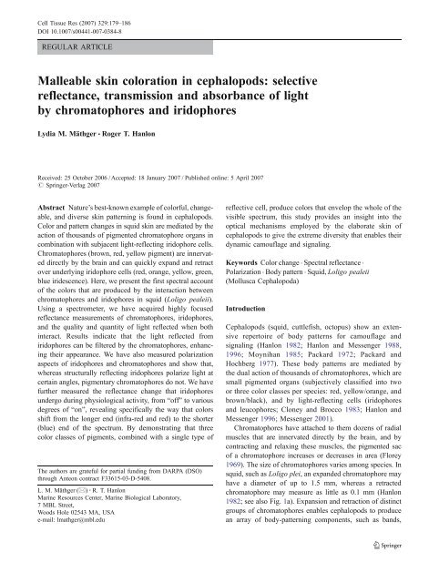
Cell Tissue Res (2007) 329:179–186<br />
DOI 10.1007/s00441-007-0384-8<br />
REGULAR ARTICLE<br />
<strong>Malleable</strong> <strong>sk<strong>in</strong></strong> <strong>coloration</strong> <strong>in</strong> <strong>cephalopods</strong>: <strong>selective</strong><br />
<strong>reflectance</strong>, transmission and absorbance of light<br />
by chromatophores and iridophores<br />
Lydia M. Mäthger & Roger T. Hanlon<br />
Received: 25 October 2006 /Accepted: 18 January 2007 / Published onl<strong>in</strong>e: 5 April 2007<br />
# Spr<strong>in</strong>ger-Verlag 2007<br />
Abstract Nature’s best-known example of colorful, changeable,<br />
and diverse <strong>sk<strong>in</strong></strong> pattern<strong>in</strong>g is found <strong>in</strong> <strong>cephalopods</strong>.<br />
Color and pattern changes <strong>in</strong> squid <strong>sk<strong>in</strong></strong> are mediated by the<br />
action of thousands of pigmented chromatophore organs <strong>in</strong><br />
comb<strong>in</strong>ation with subjacent light-reflect<strong>in</strong>g iridophore cells.<br />
Chromatophores (brown, red, yellow pigment) are <strong>in</strong>nervated<br />
directly by the bra<strong>in</strong> and can quickly expand and retract<br />
over underly<strong>in</strong>g iridophore cells (red, orange, yellow, green,<br />
blue iridescence). Here, we present the first spectral account<br />
of the colors that are produced by the <strong>in</strong>teraction between<br />
chromatophores and iridophores <strong>in</strong> squid (Loligo pealeii).<br />
Us<strong>in</strong>g a spectrometer, we have acquired highly focused<br />
<strong>reflectance</strong> measurements of chromatophores, iridophores,<br />
and the quality and quantity of light reflected when both<br />
<strong>in</strong>teract. Results <strong>in</strong>dicate that the light reflected from<br />
iridophores can be filtered by the chromatophores, enhanc<strong>in</strong>g<br />
their appearance. We have also measured polarization<br />
aspects of iridophores and chromatophores and show that,<br />
whereas structurally reflect<strong>in</strong>g iridophores polarize light at<br />
certa<strong>in</strong> angles, pigmentary chromatophores do not. We have<br />
further measured the <strong>reflectance</strong> change that iridophores<br />
undergo dur<strong>in</strong>g physiological activity, from “off” to various<br />
degrees of “on”, reveal<strong>in</strong>g specifically the way that colors<br />
shift from the longer end (<strong>in</strong>fra-red and red) to the shorter<br />
(blue) end of the spectrum. By demonstrat<strong>in</strong>g that three<br />
color classes of pigments, comb<strong>in</strong>ed with a s<strong>in</strong>gle type of<br />
The authors are grateful for partial fund<strong>in</strong>g from DARPA (DSO)<br />
through Anteon contract F33615-03-D-5408.<br />
L. M. Mäthger (*) : R. T. Hanlon<br />
Mar<strong>in</strong>e Resources Center, Mar<strong>in</strong>e Biological Laboratory,<br />
7 MBL Street,<br />
Woods Hole 02543 MA, USA<br />
e-mail: lmathger@mbl.edu<br />
reflective cell, produce colors that envelop the whole of the<br />
visible spectrum, this study provides an <strong>in</strong>sight <strong>in</strong>to the<br />
optical mechanisms employed by the elaborate <strong>sk<strong>in</strong></strong> of<br />
<strong>cephalopods</strong> to give the extreme diversity that enables their<br />
dynamic camouflage and signal<strong>in</strong>g.<br />
Keywords Color change . Spectral <strong>reflectance</strong> .<br />
Polarization . Body pattern . Squid, Loligo pealeii<br />
(Mollusca Cephalopoda)<br />
Introduction<br />
Cephalopods (squid, cuttlefish, octopus) show an extensive<br />
repertoire of body patterns for camouflage and<br />
signal<strong>in</strong>g (Hanlon 1982; Hanlon and Messenger 1988,<br />
1996; Moynihan 1985; Packard 1972; Packard and<br />
Hochberg 1977). These body patterns are mediated by<br />
the dual action of thousands of chromatophores, which are<br />
small pigmented organs (subjectively classified <strong>in</strong>to two<br />
or three color classes per species: red, yellow/orange, and<br />
brown/black), and by light-reflect<strong>in</strong>g cells (iridophores<br />
and leucophores; Cloney and Brocco 1983; Hanlon and<br />
Messenger 1996; Messenger 2001).<br />
Chromatophores have attached to them dozens of radial<br />
muscles that are <strong>in</strong>nervated directly by the bra<strong>in</strong>, and by<br />
contract<strong>in</strong>g and relax<strong>in</strong>g these muscles, the pigmented sac<br />
of a chromatophore <strong>in</strong>creases or decreases <strong>in</strong> area (Florey<br />
1969). The size of chromatophores varies among species. In<br />
squid, such as Loligo plei, an expanded chromatophore may<br />
have a diameter of up to 1.5 mm, whereas a retracted<br />
chromatophore may measure as little as 0.1 mm (Hanlon<br />
1982; see also Fig. 1a). Expansion and retraction of dist<strong>in</strong>ct<br />
groups of chromatophores enables <strong>cephalopods</strong> to produce<br />
an array of body-pattern<strong>in</strong>g components, such as bands,
180 Cell Tissue Res (2007) 329:179–186<br />
stripes and spots (see, for example, the repertoire of the<br />
subject of this study, Loligo pealeii (Hanlon et al. 1999); an<br />
image of the squid is shown <strong>in</strong> Fig. 1a).<br />
Importantly, however, cephalopod <strong>sk<strong>in</strong></strong> has another<br />
system that <strong>in</strong>teracts with light: the various types of<br />
structural reflectors that lie subjacent to the pigmented<br />
chromatophore organs (Cloney and Brocco 1983). Squid<br />
generally only have iridophores. These are colorless cells of<br />
variable sizes, generally smaller than 1 mm (Mirow 1972b).<br />
They are made up of stacks of th<strong>in</strong> plates that reflect light<br />
by th<strong>in</strong>-film <strong>in</strong>terference (Denton and Land 1971; Land<br />
1972; Mäthger et al. 2004; Mäthger and Denton 2001). The<br />
light reflected from a multilayer reflector of this k<strong>in</strong>d is<br />
almost always colored. Two pre-requisites are that (1) there<br />
is a difference <strong>in</strong> refractive <strong>in</strong>dex between the plates and the<br />
spaces separat<strong>in</strong>g them, and (2) the plates and spaces have<br />
thicknesses comparable to the wavelength of light. The<br />
mechanism of <strong>reflectance</strong> is the same as that of colored<br />
soap bubbles (Boys 1959).<br />
Light <strong>reflectance</strong> from iridophores can be changed by<br />
applications of acetylchol<strong>in</strong>e (ACh) act<strong>in</strong>g on muscar<strong>in</strong>ic<br />
chol<strong>in</strong>ergic receptors, and changeable <strong>reflectance</strong> has been<br />
observed <strong>in</strong> liv<strong>in</strong>g squid (Cooper and Hanlon 1986;<br />
Cooper et al. 1990; Hanlon 1982; Hanlon et al. 1990;<br />
Mäthger et al. 2004). However, <strong>in</strong> contrast to chromatophores<br />
that can change with<strong>in</strong> a fraction of a second,<br />
changes <strong>in</strong> iridophore <strong>reflectance</strong> take longer, e.g., several<br />
seconds to m<strong>in</strong>utes. In the lolig<strong>in</strong>id squid Lolliguncula<br />
brevis, Cooper and Hanlon (1986) have reported that the<br />
reflected wavelengths shift from the long (red) end of the<br />
visible spectrum to the shorter (blue/ultraviolet [UV]) end<br />
with <strong>in</strong>creas<strong>in</strong>g concentrations of ACh. To date, the way<br />
that this wavelength change is achieved rema<strong>in</strong>s unknown.<br />
Recent evidence has provided confirmation that the plates<br />
of squid iridophores are made up of prote<strong>in</strong>s called<br />
reflect<strong>in</strong>s (Crookes et al. 2004; see also Cooper et al.<br />
1990). Cooper et al. (1990) have po<strong>in</strong>ted out that a prote<strong>in</strong><br />
state change (affect<strong>in</strong>g refractive <strong>in</strong>dex) comb<strong>in</strong>ed with a<br />
change <strong>in</strong> the thickness of plates could expla<strong>in</strong> the<br />
observed changes <strong>in</strong> <strong>reflectance</strong>.<br />
Iridophores are located <strong>in</strong> a dist<strong>in</strong>ct layer beneath the<br />
chromatophores (Hanlon and Messenger 1996; Williams<br />
1909). The appearance of the animal thus depends on<br />
which <strong>sk<strong>in</strong></strong> elements affect the light <strong>in</strong>cident on the <strong>sk<strong>in</strong></strong>.<br />
Light may be reflected by either chromatophores or<br />
iridophores, or a comb<strong>in</strong>ation of both, and the physiological<br />
changeability of chromatophores and iridophores thereby<br />
enables the <strong>sk<strong>in</strong></strong> to produce such impressive optical<br />
malleability.<br />
We present the first quantified measurements of different<br />
wavelengths that can be recorded at the surface of squid<br />
<strong>sk<strong>in</strong></strong>. In previous publications, we, and others, have looked<br />
at some aspects of squid <strong>coloration</strong> caused by either<br />
iridophores or chromatophores, or otherwise on a grosser<br />
scale of observation (Cooper and Hanlon 1986; Cornwell et<br />
al. 1997; Hanlon 1982; Hanlon et al. 1990, 1999; Mäthger<br />
and Denton 2001; Mirow 1972a, b). However, to date, no
Cell Tissue Res (2007) 329:179–186 181<br />
documentation exists regard<strong>in</strong>g the detailed nature of the<br />
optical <strong>in</strong>teractions between the pigmentary and structurally<br />
reflect<strong>in</strong>g <strong>sk<strong>in</strong></strong> elements.<br />
Materials and methods<br />
Specimen preparation<br />
Squid (Loligo pealeii), found <strong>in</strong> coastal waters around Cape<br />
Cod, were caught by trawls and held <strong>in</strong> an open sea water<br />
system at the Mar<strong>in</strong>e Resources Center (MBL, Woods<br />
Hole, USA). For <strong>sk<strong>in</strong></strong> measurements, squid were killed by<br />
decapitation. Small specimens of mantle tissue with <strong>in</strong>tact<br />
<strong>sk<strong>in</strong></strong> were dissected and p<strong>in</strong>ned onto the Sylgard covered<br />
dish of a goniometer, thereby allow<strong>in</strong>g 3-dimensional<br />
position<strong>in</strong>g of the tissue.<br />
Squid <strong>sk<strong>in</strong></strong> is highly elastic. Hence, if the mantle tissue is<br />
left attached to the <strong>sk<strong>in</strong></strong>, it ensures that the <strong>sk<strong>in</strong></strong> elements<br />
reta<strong>in</strong> their natural orientations with<strong>in</strong> the tissue.<br />
Spectral measurements of <strong>sk<strong>in</strong></strong> components<br />
Spectral <strong>reflectance</strong> and transmission measurements were<br />
obta<strong>in</strong>ed by us<strong>in</strong>g a fiber optic spectrometer (USB2000,<br />
Ocean Optics, USA; spectra recorded on PC, us<strong>in</strong>g<br />
OOIBase 32 software, which automatically plotted <strong>reflectance</strong>)<br />
connected, via a 1-mm-diameter fiber, to the c-mount<br />
of a dissect<strong>in</strong>g microscope (Zeiss; angle of acceptance:<br />
13°). At the highest magnification of the microscope, the<br />
area of the measured field was approximately 0.3 mm <strong>in</strong><br />
diameter. Illum<strong>in</strong>ation was provided by a Schott fiber-optic<br />
microscope-light source. The spectral range of measurements<br />
was limited from 400 nm to 800 nm, ma<strong>in</strong>ly because<br />
the microscope optics absorbed wavebands outside this<br />
range. Measurements <strong>in</strong> the UV and <strong>in</strong>fra-red (IR) part of<br />
the spectrum were taken without use of the microscope.<br />
Instead, optical fibers were held <strong>in</strong> place by micromanipulators.<br />
To take measurements <strong>in</strong> the UV part of the<br />
spectrum, we illum<strong>in</strong>ated <strong>sk<strong>in</strong></strong> samples with a xenon light<br />
source (PX-1, Ocean Optics, range 200–750 nm). Illum<strong>in</strong>ation<br />
for IR measurements was provided by a tungsten<br />
halogen light source (LS-1, Ocean Optics, range 360–<br />
Fig. 1 a (i) Squid (Loligo pealeii) chromatophores and reflective<br />
iridophores. (ii) Brown (B), red (R), and yellow (Y) chromatophores <strong>in</strong><br />
transmitted light. b, c Spectral <strong>reflectance</strong> (%) and transmission (%)<br />
measurements, respectively, of yellow, red, and brown chromatophores<br />
(n=8 for each color class). Bars <strong>in</strong>dicate SE of the mean. d<br />
Spectral measurements of reflected light <strong>in</strong> both planes of polarization<br />
(90 degree angle to each other) for a red chromatophore, show<strong>in</strong>g that<br />
reflected light is unpolarized, i.e., light is reflected equally well <strong>in</strong> both<br />
planes of polarization<br />
2000 nm). A diffuse reflection standard (WS-1, Ocean<br />
Optics) was used to standardize measurements.<br />
For transmission measurements of chromatophores,<br />
chromatophore layers were dissected, p<strong>in</strong>ned out to orig<strong>in</strong>al<br />
size <strong>in</strong> a Petri-dish filled with Sylgard, and placed over a<br />
diffuse glass plate and <strong>in</strong>to a beam of light reflected directly<br />
<strong>in</strong>to the microscope by us<strong>in</strong>g a mirror. Brown and red<br />
chromatophores (approximately 1–1.5 mm diameter fully<br />
expanded) were larger than yellow chromatophores. Therefore,<br />
we could measure s<strong>in</strong>gle brown or red chromatophores<br />
but were not always able to measure s<strong>in</strong>gle yellow<br />
chromatophores. Instead, groups of 2–3 yellow chromatophores<br />
were measured. We ensured that only yellow<br />
chromatophores were expanded, and that the overlap<br />
between chromatophores was m<strong>in</strong>imal (less than 10% of<br />
area), so that the effects on <strong>reflectance</strong> and transmission<br />
were negligible.<br />
For polarization measurements, a l<strong>in</strong>ear polarizer<br />
(Jessop, UK) was placed under the microscope and turned<br />
to analyze polarization from the specimen.<br />
Results<br />
Chromatophore <strong>reflectance</strong> and transmission properties<br />
In Loligo pealeii, as <strong>in</strong> most other lolig<strong>in</strong>id squid, there are<br />
three long-wavelength classes of chromatophores (previously<br />
described as brown, red, and yellow; Fig. 1a,b).<br />
Spectral measurements of <strong>reflectance</strong> properties of expanded<br />
chromatophores are shown <strong>in</strong> Fig. 1b. In the longwavelength<br />
band (above 550 nm), all three color classes of<br />
chromatophores reflect less than 5% of <strong>in</strong>com<strong>in</strong>g light. In<br />
the shorter wavelengths (below 550 nm), light is heavily<br />
absorbed; <strong>reflectance</strong> is as low as 1% (Fig. 1b). Most of the<br />
light <strong>in</strong>cident on chromatophores is transmitted through the<br />
pigment to the underly<strong>in</strong>g iridophores. For yellow and red<br />
chromatophores, transmission between 550 nm to 700 nm<br />
may be as high as 70%–90%. Brown chromatophores<br />
transmit less light <strong>in</strong> comparison; between 700–750 nm,<br />
this may be as little as 20%–30% (Fig. 1c). The amount of<br />
light reflected or transmitted depends critically on the<br />
relative expansion of the chromatophore pigment sac. Light<br />
reflected from all three color classes of chromatophores is<br />
unpolarized, i.e., light is reflected equally well <strong>in</strong> both<br />
planes of polarization (Fig. 1d for red chromatophore; data<br />
for yellow and brown not shown).<br />
Only chromatophores considered to be expanded have<br />
been measured <strong>in</strong> this study. When chromatophores are<br />
retracted, more unfiltered light is transmitted through the<br />
<strong>sk<strong>in</strong></strong> to the underly<strong>in</strong>g iridophores. So far, we have been<br />
unable to take spectral measurements of chromatophores <strong>in</strong><br />
a retracted state.
182 Cell Tissue Res (2007) 329:179–186<br />
Iridophore <strong>reflectance</strong> properties<br />
One of the key features <strong>in</strong> squid <strong>sk<strong>in</strong></strong> <strong>coloration</strong> is the<br />
iridescence produced by light-reflect<strong>in</strong>g iridophores. These<br />
are found <strong>in</strong> a dist<strong>in</strong>ct layer all over the body and may<br />
appear dimly or brightly iridescent red, green, or blue. All<br />
iridophores have similar optical properties. (1) With<br />
<strong>in</strong>creas<strong>in</strong>g angle of <strong>in</strong>cidence, the wavelengths of the<br />
reflected light shift toward the shorter (blue/UV) end of<br />
the spectrum (Fig. 2a,b). (2) At oblique angles of <strong>in</strong>cidence,<br />
the reflected light is l<strong>in</strong>early polarized. Polarization is<br />
maximal at 45–50° <strong>in</strong>cidence (as predicted by Brewster’s<br />
law; i.e., angle of maximal polarization; Fig. 2b).<br />
The wavelengths reflected by iridophores vary widely.<br />
For example, <strong>in</strong> L. pealeii, “splotches” of reddish p<strong>in</strong>k<br />
(viewed at normal <strong>in</strong>cidence) occur on the dorsal mantle:<br />
however, spectrometer measurements reveal variations from<br />
deep red (above 700 nm) to orange and almost yellow<br />
(approximately 600 nm). As has been shown before,<br />
iridophore <strong>reflectance</strong> is physiologically active, with ACh<br />
act<strong>in</strong>g on muscar<strong>in</strong>ic chol<strong>in</strong>ergic receptors (Hanlon et al.<br />
1990; Mäthger et al. 2004). In Fig. 2c,e,f, we provide the<br />
first spectral measurements of this change. Images of this<br />
change are illustrated <strong>in</strong> Fig. 2d, <strong>in</strong> which a non-reflective<br />
iridophore patch (i) turns reflective after applications of<br />
1 mM ACh: first red (ii), then orange (iii), and f<strong>in</strong>ally<br />
yellow (iv). Figure 2c shows an iridophore reflect<strong>in</strong>g red<br />
light (approximately 640 nm), <strong>in</strong> the process of “turn<strong>in</strong>g<br />
off” of its own accord; this <strong>in</strong>volves a shift <strong>in</strong> reflected<br />
wavelengths toward the longer (IR) end of the spectrum<br />
and a subsequent decrease <strong>in</strong> overall <strong>reflectance</strong>. The<br />
iridophores reflect wavelengths around 705 nm before<br />
turn<strong>in</strong>g off completely.<br />
In the example shown <strong>in</strong> Fig. 2e, applications of 1 mM<br />
ACh cause a splotch of red-reflect<strong>in</strong>g iridophores to<br />
undergo an approximately 60-nm shift toward the shorter<br />
end of the spectrum and a doubl<strong>in</strong>g of absolute <strong>reflectance</strong>.<br />
Iridophores of the collar region reflect weakly <strong>in</strong> the IR part<br />
of the spectrum when visually “turned off”. Applications of<br />
ACh cause these iridophores to “turn on” with wavelengths<br />
shift<strong>in</strong>g from approximately 800 nm to 670 nm (Fig. 2f).<br />
Iridophores appear not to reflect <strong>in</strong> the UV part of the<br />
spectrum.<br />
Chromatophore-iridophore <strong>in</strong>teractions<br />
Chromatophores have the ability to expand over underly<strong>in</strong>g<br />
iridophores thereby chang<strong>in</strong>g the optical appearance of the<br />
<strong>sk<strong>in</strong></strong>. Follow<strong>in</strong>g <strong>reflectance</strong> from an iridophore, light<br />
pass<strong>in</strong>g upward through a chromatophore is filtered (i.e.,<br />
partially absorbed), thus alter<strong>in</strong>g the <strong>reflectance</strong> spectrum of<br />
the iridophore and add<strong>in</strong>g to the light reflected from the<br />
chromatophore. This <strong>in</strong>teraction makes the chromatophore<br />
appear brighter, <strong>in</strong> some <strong>in</strong>stances giv<strong>in</strong>g rise to a color that<br />
neither chromatophore nor iridophore alone is capable of<br />
produc<strong>in</strong>g. As shown above, because of their structural<br />
nature, iridophores can reflect almost any waveband of the<br />
visible spectrum from deep red to near-UV. Three color<br />
classes of chromatophores cover the body of a squid, and<br />
thus a variety of iridophore-chromatophore comb<strong>in</strong>ations<br />
are possible, but only a few examples are given here.<br />
Figure 3a illustrates one comb<strong>in</strong>ation of a yellow<br />
chromatophore cover<strong>in</strong>g greenish-blue iridescent iridophores.<br />
L<strong>in</strong>e 1 (peak 530 nm) shows the <strong>reflectance</strong> of<br />
the iridophores alone. L<strong>in</strong>e 2 represents a yellow<br />
chromatophore alone (peak 575 nm). L<strong>in</strong>e 3 demonstrates<br />
the <strong>in</strong>teractive effects: the spectrum of the iridophore<br />
shifts toward that of the chromatophore (peak 595 nm),<br />
greatly enhanc<strong>in</strong>g its appearance. In Fig. 3b, a red<br />
chromatophore (l<strong>in</strong>e 2) cover<strong>in</strong>g an orange-reflect<strong>in</strong>g<br />
iridophore (peak at 630 nm; l<strong>in</strong>e 1) is shown. Although<br />
not as pronounced as the previous example, the spectral<br />
change (peak at 670 nm; l<strong>in</strong>e 3) is nevertheless noticeable.<br />
In Fig. 3c, a yellow chromatophore (l<strong>in</strong>e 2) covers a red<br />
iridophore (peak at 680 nm; l<strong>in</strong>e 1). Here, the peak<br />
wavelength is not shifted as the chromatophore covers the<br />
iridophore, but the spectrum becomes broader, and the<br />
color appears as orange. Figure 3d shows an <strong>in</strong>terest<strong>in</strong>g<br />
color comb<strong>in</strong>ation that is not possible to produce by<br />
iridophores or chromatophores alone. A red chromatophore<br />
cover<strong>in</strong>g a blue iridophore (red iridophore at 50°<br />
<strong>in</strong>cidence, see Fig. 2a,b) results <strong>in</strong> the appearance of a<br />
purple color. The peak wavelength of the blue iridescence<br />
(l<strong>in</strong>e 1, 470 nm) is greatly reduced, whereas the long<br />
wavelength part of the <strong>reflectance</strong> (630 nm–750 nm) is<br />
reta<strong>in</strong>ed.<br />
Brown chromatophores transmit little to no light.<br />
Therefore, their function may be to block, rather than<br />
modify, <strong>reflectance</strong> from iridophores.<br />
Discussion<br />
The rapid color change found <strong>in</strong> <strong>cephalopods</strong> is unique <strong>in</strong><br />
the animal k<strong>in</strong>gdom. Most animals only have one available<br />
body pattern; this may undergo seasonal or ontogenetic<br />
changes but more often stays the same throughout the life<br />
of the animal (Cott 1940; Edmunds 1974; Booth 1990).<br />
Although most animal body patterns may not be changeable,<br />
some are highly reflective, such as those of many<br />
birds (Cuthill et al. 1999; Osorio and Ham 2002; Vorobyev<br />
et al. 1998) and reef fishes (Marshall 2000). In most<br />
animals with changeable body patterns, these changes<br />
generally take several seconds, hours, or even days (e.g.,<br />
fish: Fujii 1993; Kasukawa et al. 1987; Lythgoe and Shand<br />
1989; lizards: Hadley and Oldman 1969; Taylor and Hadley
Cell Tissue Res (2007) 329:179–186 183<br />
Fig. 2 a Iridophore “splotch” of the dorsal mantle viewed <strong>in</strong> white light<br />
at near-normal <strong>in</strong>cidence (left, red) and at approximately 50° <strong>in</strong>cidence<br />
(right, blue). b Reflective measurements for both planes of polarization<br />
of a dorsal iridophore splotch, reflect<strong>in</strong>g red at near normal <strong>in</strong>cidence<br />
(10°). At 40° <strong>in</strong>cidence, the color is greenish-yellow at a peak<br />
wavelength of approximately 550 nm; at 50°, the color is blue-green<br />
at a peak wavelength of approximately 500 nm (thick l<strong>in</strong>e parallel plane,<br />
th<strong>in</strong> l<strong>in</strong>e perpendicular plane of polarization). At near normal <strong>in</strong>cidence<br />
(10°), reflected light is almost unpolarized; at oblique angles, polarization<br />
is strong. c Spectral measurements of an iridophore patch <strong>in</strong> the act<br />
of “turn<strong>in</strong>g off”. Peak wavelength at the beg<strong>in</strong>n<strong>in</strong>g of the measurement<br />
is 640 nm. Measurements were taken at 0 m<strong>in</strong> (highest <strong>reflectance</strong>), 30 s,<br />
1 m<strong>in</strong>, 1 m<strong>in</strong> 30 s, 2 m<strong>in</strong>, 2 m<strong>in</strong> 30 s, 3 m<strong>in</strong>, 3 m<strong>in</strong> 30 s, 4 m<strong>in</strong>, and<br />
1970; tree frogs: Stegen et al. 2004). One known exception<br />
may be the paradise whiptail (Pentapodus paradiseus), a<br />
tropical fish whose reflective changes are fast (i.e., fractions<br />
of a second; Mäthger et al. 2003).<br />
To our knowledge, this is the first study that exam<strong>in</strong>es, at<br />
a microscopic level, the <strong>in</strong>teractions between pigments and<br />
structural reflectors of any animal. In fish, <strong>coloration</strong> and<br />
pattern<strong>in</strong>g are also produced by several structures <strong>in</strong> the<br />
<strong>sk<strong>in</strong></strong> (chromatophores, leucophores, iridophores); a descriptive<br />
account of these <strong>in</strong>teractions can be found <strong>in</strong> Fujii<br />
(1993). However, no spectral measurements are given.<br />
5 m<strong>in</strong> (lowest <strong>reflectance</strong>). d In vitro iridophore color change after<br />
application of 1 mM acetylchol<strong>in</strong>e (ACh). Images were taken at 20-s<br />
<strong>in</strong>tervals. Iridophores change from non-reflective (i) to orange (iv) with<strong>in</strong><br />
1 m<strong>in</strong>. e Reflective changes result<strong>in</strong>g from ACh application. Measurements<br />
taken at 15-s <strong>in</strong>tervals (curve 1 before ACh, curve 2 15 s after<br />
ACh application, curve 3 30 s after ACh application, curve 4 45 s after<br />
ACh application, curve 5 1 m<strong>in</strong> after ACh application; cf. c). f Reflective<br />
changes result<strong>in</strong>g from ACh application (1 mM). Before ACh<br />
application, iridophores are non-reflective (black l<strong>in</strong>e <strong>in</strong>fra-red <strong>reflectance</strong><br />
<strong>in</strong>visible to the human eye). Iridophores then move through the<br />
<strong>in</strong>fra-red part of the spectrum (800–750 nm) before reflect<strong>in</strong>g red light<br />
visible to the human eye. Measurements taken at 30-s <strong>in</strong>tervals. Highest<br />
<strong>reflectance</strong> measured after 5 m<strong>in</strong>: 670 nm<br />
Several authors have tackled the optical properties of<br />
cephalopod iridophores and chromatophores <strong>in</strong>dividually,<br />
but we stress that the dynamically chang<strong>in</strong>g <strong>in</strong>teractions of<br />
pigmentary and structural color are the keys to understand<strong>in</strong>g<br />
the optical malleability of cephalopod <strong>sk<strong>in</strong></strong>.<br />
First, we have confirmed previous f<strong>in</strong>d<strong>in</strong>gs that squid<br />
iridophores act as multilayer reflectors, as described <strong>in</strong><br />
Land (1972) and Mäthger and Denton (2001). Because of<br />
their structural nature, iridophores can reflect almost any<br />
waveband of the visible spectrum from deep red to near-<br />
UV. Moreover, the wavelengths of iridescence can be
184 Cell Tissue Res (2007) 329:179–186<br />
Fig. 3 Spectral measurements<br />
and correspond<strong>in</strong>g images of<br />
chromatophores cover<strong>in</strong>g iridophores,<br />
show<strong>in</strong>g the resultant<br />
spectral shift (curve 1 reflective<br />
spectrum of iridophores only,<br />
curve 2 chromatophore only,<br />
curve 3 chromatophore over<br />
iridophores, arrows location of<br />
measurement). a Yellow chromatophore<br />
over green iridophores.<br />
b Red chromatophore<br />
over p<strong>in</strong>k iridophores. c Yellow<br />
chromatophore over p<strong>in</strong>k iridophores.<br />
d Red chromatophore<br />
over blue iridophores<br />
changed swiftly under physiological control and can also be<br />
turned on and off; such delicate changes nevertheless<br />
greatly affect the gross appearance of the <strong>sk<strong>in</strong></strong>. Three color<br />
classes of chromatophores cover the body of L. pealeii<br />
(yellow, red, and brown). Thus, a variety of iridophorechromatophore<br />
color comb<strong>in</strong>ations are possible, even<br />
though we have only given a few examples here. Generally,<br />
red and yellow chromatophores transmit much of the<br />
<strong>in</strong>com<strong>in</strong>g light (Fig. 1b,c) and therefore play an important<br />
role <strong>in</strong> modulat<strong>in</strong>g iridescence. Brown chromatophores<br />
transmit less light <strong>in</strong> comparison, especially when only<br />
partially expanded, and their function may be (1) to block
Cell Tissue Res (2007) 329:179–186 185<br />
iridescence and polarization and (2) to produce the darkest<br />
aspect of gross pattern<strong>in</strong>g <strong>in</strong> body <strong>coloration</strong>. From our<br />
results, we conclude that the comb<strong>in</strong>ation of the three<br />
pigmentary colors (red, yellow, and brown), comb<strong>in</strong>ed with<br />
multilayer reflectors, enables squid to reflect any wavelength<br />
of visible light. Reflectance <strong>in</strong> the IR part of the<br />
spectrum has only been found <strong>in</strong> the collar region of the<br />
mantle. The biological function that this may have is<br />
unknown, s<strong>in</strong>ce squid eyes are not sensitive <strong>in</strong> the IR<br />
(Morris et al. 1993). Furthermore, IR light is absorbed<br />
heavily by water (Jerlov 1976) and may not be available for<br />
<strong>reflectance</strong>.<br />
Interest<strong>in</strong>gly, polarized light reflected from iridophores<br />
is not depolarized when it passes through the<br />
pigmented chromatophores, a recent f<strong>in</strong>d<strong>in</strong>g (Mäthger<br />
and Hanlon 2006) suggest<strong>in</strong>g that this may be a private<br />
communication channel that might reach an <strong>in</strong>tended<br />
receiver while the animal is camouflaged by us<strong>in</strong>g<br />
pigmented chromatophores.<br />
We have used squid as a model to study the <strong>in</strong>teractions<br />
between chromatophores and iridophores, primarily for<br />
reasons of simplicity. L. pealeii, like other Loligo spp.<br />
(Hanlon 1982), have large chromatophores whose density<br />
<strong>in</strong> the <strong>sk<strong>in</strong></strong> is low; furthermore, the dermal layers of<br />
chromatophores and iridophores are easily separated by<br />
dissection. The <strong>sk<strong>in</strong></strong> of octopus and cuttlefish is similar,<br />
although more complicated <strong>in</strong> structure, because the density<br />
of chromatophores and various reflect<strong>in</strong>g cells is much<br />
greater (Hanlon 1988; Packard and Hochberg 1977). The<br />
layers are not structured as clearly, and <strong>in</strong> addition, octopus<br />
and cuttlefish have light-scatter<strong>in</strong>g leucophores that produce<br />
whiteness <strong>in</strong> the <strong>sk<strong>in</strong></strong> (Cloney and Brocco 1983;<br />
Froesch and Messenger 1978; Hanlon and Messenger<br />
1988). To date, no spectral data are available for the<br />
<strong>in</strong>teraction between the <strong>sk<strong>in</strong></strong> components of cuttlefish and<br />
octopus, but the general pr<strong>in</strong>ciples that we report here for L.<br />
pealeii probably also apply to cuttlefish and octopus.<br />
At shallow depths of water, where daylight penetrates<br />
without much loss of the red and UV ends of the spectrum,<br />
the natural environment can be colorful (Jerlov 1976;<br />
Marshall et al. 2003). Hav<strong>in</strong>g the ability to modulate <strong>sk<strong>in</strong></strong><br />
<strong>coloration</strong> to such a f<strong>in</strong>e degree makes color match<strong>in</strong>g for<br />
camouflage purposes necessary, s<strong>in</strong>ce many predators of<br />
<strong>cephalopods</strong> (teleost fishes, for example) have color vision<br />
(Fritsches et al. 2000; Losey et al. 2003; Partridge 1990).<br />
However, no data are available to show whether squid, or<br />
any cephalopod, match background colors. Evidence thus<br />
far suggests that squid, cuttlefish, and octopus are colorbl<strong>in</strong>d<br />
(Brown and Brown 1958; Marshall and Messenger<br />
1996; Mäthger et al. 2006; Messenger 1973; Morris et al.<br />
1993). Therefore, the concept of color match<strong>in</strong>g is<br />
especially <strong>in</strong>trigu<strong>in</strong>g consider<strong>in</strong>g that these animals have<br />
no color perception. At greater depths, the question of color<br />
match<strong>in</strong>g becomes less important, s<strong>in</strong>ce daylight is reduced<br />
to the blue and green parts of the spectrum (Jerlov 1976).<br />
At such depths, squid <strong>sk<strong>in</strong></strong> will lose its colorful appearance<br />
and <strong>in</strong>stead will appear as more or less bright shades of the<br />
prevalent waveband. Here, squid <strong>sk<strong>in</strong></strong> may be adapted to<br />
<strong>in</strong>tensity and contrast match<strong>in</strong>g by controll<strong>in</strong>g iridescence<br />
and modulat<strong>in</strong>g it us<strong>in</strong>g overly<strong>in</strong>g pigmented chromatophores.<br />
Further <strong>in</strong>vestigations <strong>in</strong>to the nature of the optical<br />
<strong>in</strong>teractions between chromatophores and iridophores <strong>in</strong><br />
cephalopod <strong>sk<strong>in</strong></strong> should help us understand better the<br />
mechanisms of camouflage and signal<strong>in</strong>g <strong>in</strong> these animals<br />
and to broaden our knowledge of animal visual ecology.<br />
Acknowledgments Many thanks to Phil McFadden (U. Oregon),<br />
Peter J. S. Smith (MBL) and two anonymous reviewers for constructive<br />
comments.<br />
References<br />
Booth CL (1990) Evolutionary significance of ontogenetic colour<br />
change <strong>in</strong> animals. Biol J L<strong>in</strong>n Soc 40:125–163<br />
Boys CV (1959) Soap bubbles - their colors and forces which mold<br />
them. Dover, New York<br />
Brown PK, Brown PS (1958) Visual pigments of the octopus and<br />
cuttlefish. Nature 182:1288–1290<br />
Cloney RA, Brocco SL (1983) Chromatophore organs, reflector cells,<br />
iridocytes and leucophores <strong>in</strong> <strong>cephalopods</strong>. Am Zool 23:581–<br />
592<br />
Cooper KM, Hanlon RT (1986) Correlation of iridescence with<br />
changes <strong>in</strong> iridophore platelet ultrastructure <strong>in</strong> the squid<br />
Lolliguncula brevis. J Exp Biol 121:451–455<br />
Cooper KM, Hanlon RT, Budelmann BU (1990) Physiological colorchange<br />
<strong>in</strong> squid iridophores. II. Ultrastructural mechanisms <strong>in</strong><br />
Lolliguncula brevis. Cell Tissue Res 259:15–24<br />
Cornwell CJ, Messenger JB, Hanlon RT (1997) Chromatophores and<br />
body pattern<strong>in</strong>g <strong>in</strong> the squid Alloteuthis subulata. J Mar Biol<br />
Assoc UK 77:1243–1246<br />
Cott HB (1940) Adaptive colouration <strong>in</strong> animals. Methuen, London<br />
Crookes WJ, D<strong>in</strong>g L, Huang QL, Kimbell JR, Horwitz J, McFall-Ngai<br />
MJ (2004) Reflect<strong>in</strong>s: the unusual prote<strong>in</strong>s of squid reflective<br />
tissues. Science 303:235–238<br />
Cuthill IC, Bennett ATD, Partridge JC, Maier EJ (1999) Plumage<br />
<strong>reflectance</strong> and the objective assessment of avian sexual<br />
dichromatism. Am Nat 160:183–200<br />
Denton EJ, Land MF (1971) Mechanism of reflexion <strong>in</strong> silvery layers<br />
of fish and <strong>cephalopods</strong>. Proc R Soc Lond [A] 178:43–61<br />
Edmunds M (1974) Defence <strong>in</strong> animals—a survey of anti-predator<br />
defences. Longman, Harlow, UK<br />
Florey E (1969) Ultrastructure and function of cephalopod chromatophores.<br />
Am Zool 9:429–442<br />
Fujii R (1993) Coloration and chromatophores. In: Evans DH (ed) The<br />
physiology of fishes. CRC Press, Boca Raton, pp 535–562<br />
Fritsches KA, Partridge J, Pettigrew JD, Marshall NJ (2000) Colour<br />
vision <strong>in</strong> billfish. Philos Trans R Soc Lond Biol 355:1253–1256<br />
Froesch D, Messenger JB (1978) On leucophores and the chromatic<br />
unit of Octopus vulgaris. J Zool (Lond) 186:163–173<br />
Hadley ME, Oldman JMG (1969) Physiological color changes <strong>in</strong><br />
reptiles. Am Zool 9:489–504
186 Cell Tissue Res (2007) 329:179–186<br />
Hanlon RT (1982) The functional organization of chromatophores and<br />
iridescent cells <strong>in</strong> the body pattern<strong>in</strong>g of Loligo plei (Cephalopoda:<br />
Myopsida). Malacologia 23:89–119<br />
Hanlon RT (1988) Behavioral and body pattern<strong>in</strong>g characters useful <strong>in</strong><br />
taxonomy of field identification of <strong>cephalopods</strong>. Malacologia<br />
29:247–264<br />
Hanlon RT, Messenger JB (1988) Adaptive <strong>coloration</strong> <strong>in</strong> young<br />
cuttlefish (Sepia offic<strong>in</strong>alis L.): the morphology and development<br />
of body patterns and their relation to behaviour. Philos Trans R<br />
Soc Lond Biol 320:437–487<br />
Hanlon RT, Messenger JB (1996) Cephalopod behaviour. Cambridge<br />
University Press, Cambridge<br />
Hanlon RT, Cooper KM, Budelmann BU, Pappas TC (1990)<br />
Physiological color-change <strong>in</strong> squid iridophores. I. Behavior,<br />
morphology and pharmacology <strong>in</strong> Lolliguncula brevis. Cell<br />
Tissue Res 259:3–14<br />
Hanlon RT, Maxwell MR, Shashar N, Loew ER, Boyle KL (1999) An<br />
ethogram of body pattern<strong>in</strong>g behavior <strong>in</strong> the biomedically and<br />
commercially valuable squid Loligo pealei off Cape Cod,<br />
Massachusetts. Biol Bull 197:49–62<br />
Jerlov NG (1976) Mar<strong>in</strong>e optics. Elsevier, Amsterdam<br />
Kasukawa H, Oshima N, Fujii R (1987) Mechanism of light reflection<br />
<strong>in</strong> blue damselfish motile iridophore. Zool Sci 4:243–257<br />
Land MF (1972) The physics and biology of animal reflectors. Progr<br />
Biophys Mol Biol 24:75–106<br />
Losey GS, McFarland WN, Loew ER, Zamzow JP, Nelson PA,<br />
Marshall NJ (2003) Visual biology of Hawaiian coral reef fishes.<br />
I. Ocular transmission and visual pigments. Copeia 3:433–454<br />
Lythgoe JN, Shand J (1989) The structural basis for iridescent colour<br />
changes <strong>in</strong> dermal and corneal iridophores <strong>in</strong> fish. J Exp Biol<br />
141:313–325<br />
Marshall NJ (2000) Communication and camouflage with the same<br />
“bright” colours <strong>in</strong> reef fishes. Philos Trans R Soc Lond Biol<br />
355:1243–1248<br />
Marshall NJ, Messenger JB (1996) Colour-bl<strong>in</strong>d camouflage. Nature<br />
382:408–409<br />
Marshall NJ, Jenn<strong>in</strong>gs KJ, McFarland WN, Loew ER, Losey GS<br />
(2003) Visual biology of Hawaiian coral reef fishes. III.<br />
Environmental light and an <strong>in</strong>tegrated approach to the ecology<br />
of reef fish vision. Copeia 3:467–480<br />
Mäthger LM, Denton EJ (2001) Reflective properties of iridophores<br />
and fluorescent “eyespots” <strong>in</strong> the lolig<strong>in</strong>id squid Alloteuthis<br />
subulata and Loligo vulgaris. J Exp Biol 204:2103–2118<br />
Mäthger LM, Hanlon RT (2006) Anatomical basis for camouflaged<br />
polarized light communication <strong>in</strong> squid. Biol Lett 2:494–496<br />
Mäthger LM, Land MF, Siebeck UE, Marshall NJ (2003)<br />
Rapid colour changes <strong>in</strong> multilayer reflect<strong>in</strong>g stripes <strong>in</strong> the<br />
paradise whiptail, Pentapodus paradiseus. J Exp Biol<br />
206:3607–3613<br />
Mäthger LM, Coll<strong>in</strong>s TFT, Lima PA (2004) The role of<br />
muscar<strong>in</strong>ic receptors and <strong>in</strong>tracellular Ca 2+ <strong>in</strong> the spectral<br />
reflectivity changes of squid iridophores. J Exp Biol<br />
207:1759–1769<br />
Mäthger LM, Barbosa A, M<strong>in</strong>er S, Hanlon RT (2006) Color<br />
bl<strong>in</strong>dness and contrast perception <strong>in</strong> cuttlefish (Sepia offic<strong>in</strong>alis)<br />
determ<strong>in</strong>ed by a visual sensorimotor assay. Vision Res 46:1746–<br />
1753<br />
Messenger JB (1973) Some evidence for colour-bl<strong>in</strong>dness <strong>in</strong> Octopus.<br />
J Exp Biol 59:77–94<br />
Messenger JB (2001) Cephalopod chromatophores: neurobiology and<br />
natural history. Biol Rev 76:473–528<br />
Mirow S (1972a) Sk<strong>in</strong> color <strong>in</strong> the squids Loligo pealii and Loligo<br />
opalescens. I. Chromatophores. Z Zellforsch 125:143–175<br />
Mirow S (1972b) Sk<strong>in</strong> color <strong>in</strong> the squids Loligo pealii and Loligo<br />
opalescens. II. Iridophores. Z Zellforsch 125:176–190<br />
Morris A, Bowmaker JK, Hunt DM (1993) The molecular basis of a<br />
spectral shift <strong>in</strong> the rhodops<strong>in</strong>s of two species of squid from<br />
different photic environments. Proc R Soc Lond [Biol] 254:233–<br />
240<br />
Moynihan M (1985) Communication and noncomm<strong>in</strong>ucation by<br />
<strong>cephalopods</strong>. Indiana University Press, Bloom<strong>in</strong>gton<br />
Osorio D, Ham AD (2002) Spectral <strong>reflectance</strong> and directional<br />
properties of structural <strong>coloration</strong> <strong>in</strong> bird plumage. J Exp Biol<br />
205:2017–2027<br />
Packard A (1972) Cephalopods and fish: the limits of convergence.<br />
Biol Rev 47:241–307<br />
Packard A, Hochberg FG (1977) Sk<strong>in</strong> pattern<strong>in</strong>g <strong>in</strong> Octopus and other<br />
genera. Symp Zool Soc Lond 38:191–231<br />
Partridge JC (1990) The colour sensitivity and vision of fishes. In:<br />
Herr<strong>in</strong>g PJ, Campbell AK, Whitfield M, Maddock L (eds) Life<br />
and light <strong>in</strong> the sea. Cambridge University Press, Cambridge,<br />
pp 167–184<br />
Stegen JC, Gienger CM, Sun L (2004) The control of color change <strong>in</strong><br />
the Pacific tree frog, Hyla regilla. Can J Zool 82:889–896<br />
Taylor JD, Hadley ME (1970) Chromatophores and color<br />
change <strong>in</strong> the lizard, Anolis carol<strong>in</strong>ensis. Cell Tissue Res<br />
104:282–294<br />
Vorobyev M, Osorio D, Bennett ATD, Marshall NJ, Cuthill IC (1998)<br />
Tetrachromacy, oil droplets and bird plumage colours. J Comp<br />
Physiol [A] 183:621–633<br />
Williams L (1909) The anatomy of the common squid, Loligo pealii.<br />
Brill, Leiden